Sunday, August 26, 2018

Nonsense Mutation

Nonsense Mutation Definition


A nonsense mutation occurs when the sequence of nucleotides in DNA is changed in a way that stops the normal sequence of amino acids in the final protein. In central dogma of biology, DNA is transposed into RNA, which is then translated into a protein. The protein is a particular sequence of amino acids which confers a particular function onto the cell. The sequence of amino acids determines this role by the properties they contain and the ways they interact.


In the DNA, each amino acid is designated by a series of three nucleotides, called a codon. There are around 21 amino acids which can be designated by this system. There are also two other important signals, “START” and “STOP”. These signals allow the ribosome assembling the protein to know where to begin, and where to end. A nonsense mutation changes the codon for an amino acid into the codon for a “STOP” signal.


This completely changes the structure of the protein, because anything after the “STOP” signal is ignored. The ribosome snips off the incomplete protein, and goes on its way. Without the remainder of the amino acid chain, the protein may function and form completely differently than before. A nonsense mutation can have three basic outcomes.


Outcomes of a Nonsense Mutation


Deleterious


The vast majority of mutations are deleterious, meaning they cause a decrease in the overall fitness and reproductive success of the organism. A nonsense mutation would fall into this category if the mutation affected an important functional protein. Imagine if the nonsense mutation was found in the DNA which coded for an ion channel protein. If this protein was incomplete, it could not function to properly transport ions across the membrane. This would be deleterious to the organism with the nonsense mutation.


Cystic fibrosis is a genetic disorder caused by a nonsense mutation which does exactly that. The protein affected by the nonsense mutation in cystic fibrosis is a regulator protein for ion channels. Without the ability to properly move ions, people with cystic fibrosis often have respiratory problems caused by a mucous buildup due to the unregulated ions in their system. Duchenne muscular dystrophy is another disease cause by a nonsense mutation, and there are many more examples.


Neutral


A neutral mutation occurs when the effects of the mutation go undetected. Imagine that the mutation is found right before the last amino acid in a protein. Further, this final amino acid is really unnecessary for the actual function of the protein within the cell. If this is the case, the nonsense mutation will produce no effect at all. The protein will continue to function, even without the final amino acid. In this case, nothing really changes for the organism.


Beneficial


The least common type of mutation is a beneficial mutation. This is a mutation in which the protein changes in such a way that it increases the fitness and reproductive success of the organism. However, it is extremely unlikely that a nonsense mutation will end up being beneficial. In only the rarest of circumstances, a nonsense mutation may be beneficial if changing the protein it affects somehow provides a benefit to the organism. Imagine if the nonsense mutation affected a protein which inadvertently transports a toxin into cells. In an environment filled with the toxin, a dysfunctional protein might very well be the cure to being constantly bombarded with a toxin. If the protein no longer transported the toxin in, the cells wouldn’t need to worry about it.


In an even more unlikely circumstance, the nonsense mutation may completely alter the function of the protein. In this case, it might alter the protein to not transport the toxin, but rather destroy it or bind to it. This could also be a case in which the nonsense mutation became beneficial. In the most extreme circumstance the nonsense mutation may take a protein used for one process, and create an entirely new active protein by cutting the other one in pieces. Much of this has to do with the exact protein affected and the resulting effects on the organism.


Nonsense Mutation Example


Below is a chart of several point mutations, or mutations of a single nucleotide. A nonsense mutation can be seen in the middle.


Point mutations

Point mutations


In this case, the original codon read “TTC”. This called for an mRNA with the codon “AAG”, which then produced a lysine in the amino acid chain. A nonsense mutation would change the first “T” to an “A”. This makes the first codon “ATC”. The corresponding mRNA segment, “UAG”, is a signal to the ribosome to stop the chain. Unlike any of the other mutations, this ends the chain entirely.


This is likely why nonsense mutations are often noticeable. It is unlikely that these mutations do not affect the resulting protein. Given that all of the amino acids play a role in a protein, dividing it at any point will likely change the way it interacts with the environment. Even if only several amino acids are lost, these could be the crucial external amino acids which attach the protein to the cell membrane or help it interact with other cells.


Quiz


1. What is the difference between a nonsense mutation and a missense mutation?
A. No difference
B. A missense mutation stops the chain of amino acids
C. A nonsense mutation cannot provide the same type of amino acid

Answer to Question #1
C is correct. A nonsense mutation, by definition, is an end to the amino acid chain. In a missense mutation, there is at least still a chance that the amino acid will be replaced with something very similar, and that no loss in protein function will result. A missense mutation also allows the continuation of the chain, which can lessen the impact of the mutation.

2. Which of the following could NOT be caused by a nonsense mutation?
A. A protein controlling glucose intake is disabled, due to the protein being only half formed
B. A protein in jellyfish gains the ability of fluorescence, due to the addition of amino acids
C. A protein used to transport ions is hampered, because several amino acids have been lost

Answer to Question #2
B is correct. The addition of amino acids cannot happen through a nonsense mutation. A nonsense mutation always causes a loss of amino acids. Either A or C could be reasonably caused by this mutation. Remember that they could also be caused by incorrect protein folding and processing after translation. To know that a nonsense mutation occurred, one would have to compare the mutated DNA to the original.

3. Your friend says that because nonsense mutations cause a loss of amino acids, they are always bad. What can you tell him to change his mind?
A. A nonsense mutation on an overactive protein would increase fitness
B. Nonsense mutations can cause functional proteins to stop working
C. All mutations are bad

Answer to Question #3
A is correct. If the protein affected by the mutation is overactive, producing too much of a product, or otherwise hindering fitness through its action, then a nonsense mutation can eliminate that function and provide benefit for the organism. A nonsense mutation could also create a protein with an entirely new beneficial function. Remember that mutation is the driving force behind evolution.

References


  • Hartwell, L. H., Hood, L., Goldberg, M. L., Reynolds, A. E., & Silver, L. M. (2011). Genetics: From Genes to Genomes. Boston: McGraw Hill.

  • Nelson, D. L., & Cox, M. M. (2008). Principles of Biochemistry. New York: W.H. Freeman and Company.

  • Widmaier, E. P., Raff, H., & Strang, K. T. (2008). Vander’s Human Physiology: The Mechanisms of Body Function (11th ed.). Boston: McGraw-Hill Higher Education.


Nonsense Mutation

Virus

Virus Definition


A virus is a chain of nucleic acids (DNA or RNA) which lives in a host cell, uses parts of the cellular machinery to reproduce, and releases the replicated nucleic acid chains to infect more cells. A virus is often housed in a protein coat or protein envelope, a protective covering which allows the virus to survive between hosts.


Virus Structure


A virus can take on a variety of different structures. The smallest virus is only 17 nanometers, barely longer than an average sized protein. The largest virus is nearly a thousand times that size, at 1,500 nanometers. This is really small. A human hair is approximately 20,000 nanometers across. This means that most virus particles are well beyond the capability of a normal light microscope. Below is a scanning electron microscope (SEM) image of the Ebola virus.
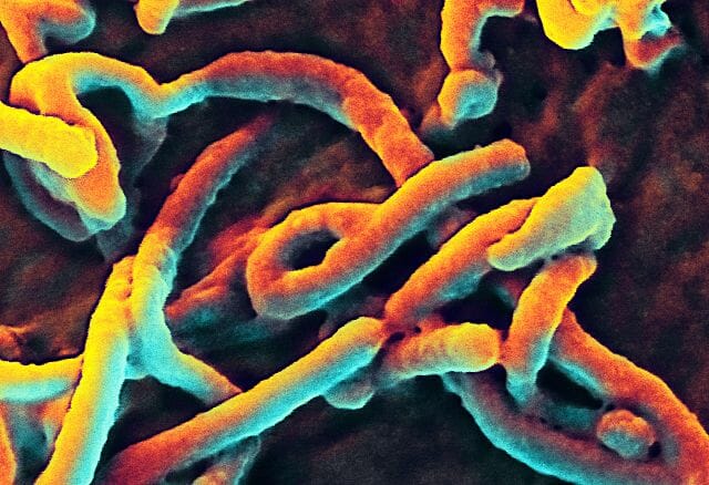

Ebola Virus

Ebola Virus


Here, you can only see the protein coat of the Ebola virus. Each virus looks like a little bent worm. However, these are not cells. Inside of the protein coat is a carefully folded RNA molecule, which contains the information necessary to replicate the protein coat, the RNA molecule, and the components necessary to hijack a cell’s natural processes to complete these tasks.


The exact structure of a virus is dependent upon which species serves as its host. A virus which replicates in mammalian cells will have a protein coat which enables it to attach to and infiltrate mammalian cells. The shape, structure, and function of these proteins changes depending on the species of virus. A typical virus can be seen below.
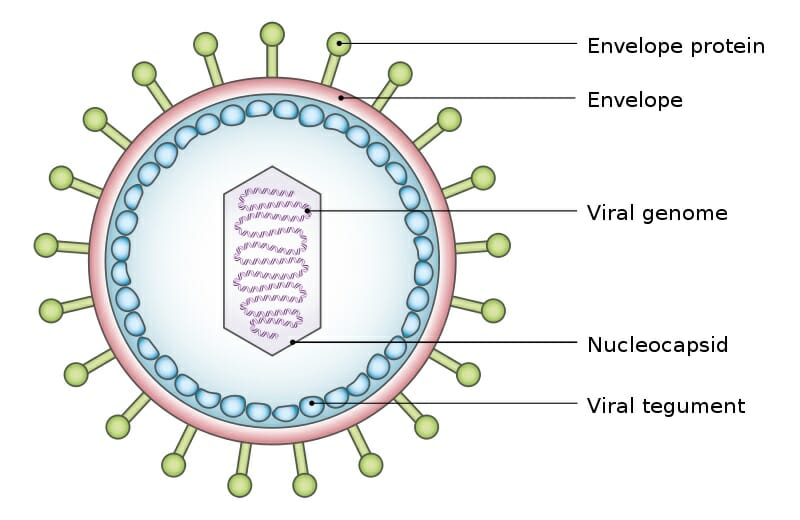

Viral Tegument

Viral Tegument


The above virus shows the typical structure a virus takes, a viral genome surrounded by a shield of proteins. The various envelope proteins will enable the virus to interact with the host cell it finds. Part of the protein coat will then open, puncture through the cell membrane, and deposit the viral genome within the cell. The protein coat can then be discarded, as the viral genome will now replicate within the host cell. The replicated virus molecules will be packaged within their own protein coats, and be released into the environment to find another host. While many virus particles take a simple shape like the one above, some are much more complicated.
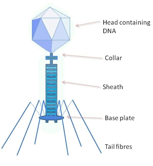

Phage

Phage


The above image shows a phage, a type of virus which specializes on bacterial cells. The protein coat of a phage is much more complex, and has a variety of specialized parts. The head portion contains the viral genome. The collar, sheath, base plate, and tail fibers are part of an intricate system to attach to and inject the genome into a bacterial cell. The tail fibers grasp the bacterial cell, pulling the base plate up to the cell wall or membrane. The sheath and collar compress, puncture the cell, and deposit the DNA into the bacterial cell.


Some virus molecules have no protein coat whatsoever, or have never been identified making on. In some plant virus species, the virus is passed from cell to cell within the plant. When seeds are created within the plant, the virus spreads to the seeds. In this way the virus can live within cells its entire existence, and never need a protein coat to protect it in the environment. Other virus molecules have even larger and more complex protein coats, and specialize on various hosts.


Is a Virus Living?


This is a complicated question. A cell is considered to be living because it contains all the necessary components to replicate its DNA, grow, and divide into new cells. This is the process all life takes, where it is a single-celled organism or a multi-cellular organism. Some people do not consider a virus living because a virus does not contain all of the mechanisms necessary to replicate itself. They would say that a virus, without a host cell, cannot replicate on its own and is therefore not alive.


Yet, by the definition of life laid out before, it seems that when a virus is inside of a host cell it does have all the machinery it needs to survive. The protein coat it exists in outside of a cell is the equivalent of a bacterial spore, a small capsule bacteria form around themselves to survive harsh conditions. Scientists who support a virus being a living organisms note the similarity between a virus in a protein coat and a bacterial spore. Neither organism is active within their protective coat, they only become active when they reach favorable conditions.


In fact, the only reason a virus affects us at all is because it becomes active within our cells. Further, a virus tends to evolve with its host. Most dangerous viruses have just recently jumped to a new species. The biochemistry they evolved to live within the other species is not compatible with the new species, and cell damage and death occur. This causes a number of reactions, depending on which cells were infected. The HIV virus, for instance, attacks immune cells exclusively. This leads to a total loss of immune function in patients. With the virus causing the common cold, the virus attacks respiratory cells and damages them as it does its work.


Yet, not all virus infections will be detrimental to the host. A virus that kills the host will be less successful over time, compared to a virus which doesn’t harm the host. A healthy host increases the number of virus molecules released into the environment, which is the ultimate goal of the virus. In fact, some virus particles may actually benefit the host. A good example is a form of herpes virus, found in mice. This virus, while it is infecting a mouse, provides the mouse with a good defense against the bacteria which carry the plague. While the mechanism is not clear, the virus somehow prevents the bacteria from taking hold in the mouse’s system.


When viewed in this light, it is easy to see how a virus is very similar to a bacteria. The bacteria creates and maintains the tools needed to reproduce DNA, where the virus steals them. This is the only real difference between a virus and a bacteria. Because of this, many scientists consider a virus a living organism. Scientists who study viruses, virologists, note that virus particles (alive or not) have been evolving with life probably as long as the first cells were present. Because of this, there is a virus which specializes on almost every single species on the planet.


Virus Classification


Scientists classify viruses based on how they replicate their genome. Some virus genomes are made of RNA, others are made of DNA. Some viruses use a single strand, others use a double strand. The complexities involved in replicating and packaging these different molecules places viruses into seven different categories.


Class I virus genomes are made of double stranded DNA, the same as the human genome. This makes it easy for these virus molecules to use the cell’s natural machinery to produce proteins from the virus DNA. However, in order for DNA polymerase (the molecule which copies DNA) to be active the cell must be dividing. Some Class I virus molecules include sections of DNA which make the cell actively start dividing. These virus molecules can lead to cancer. Human papilloma virus is a sexually-transmitted Class I virus, and can cause cervical cancer.


A Class II virus contains only a single strand of DNA. Before it can be read by the host’s DNA polymerase enzymes, it must be converted to double stranded DNA. It does this by hijacking the host cell’s histones (DNA proteins) and DNA polymerase. Instead of waiting for the cell to divide or forcing it to, Class II virus DNA contains coding for a protein called Rep. This replication enzyme replicates the original single-stranded virus genome. Other proteins are created from the DNA and used to create protein coats with the cellular machinery. The single-stranded DNA is then packaged into these protein coats, and new virus packages are created.


Class III virus genomes are created from double-stranded RNA. While this is unusual, these virus packages come with their own protein, RNA polymerase. This protein can create messenger RNA (mRNA) from the double-stranded virus RNA. The virus RNA therefore stays within the virus capsule, and only the mRNA enters the cytoplasm of the host. Here, the mRNA is converted into proteins, some of which include more RNA polymerase. This RNA polymerase creates a new double-stranded RNA, which is encapsulated by the proteins and released from the cell.


Class IV viruses are single-stranded RNA, almost identical to mRNA produced by the host cell. With these viruses the entire protein coat is engulfed by an uninfected host cell. The small RNA genome escapes the protein coat, and makes its way into the cytoplasm. This one mRNA-like strand codes for a large polyprotein, which will be created by the hosts ribosomes. The polyprotein naturally breaks into different parts. Some create protein coats, while others read and replicate the original strand of viral RNA. The virus continues to replicate and create new, fully packed virus particles. When the cell is completely full, it ruptures and releases the virus particles into the blood or environment. Up to 10,000 virus particles can be release from a single cell.


The virus genomes in Class V are also single-stranded RNA. However, they run in the opposite direction from normal mRNA. Therefore, the cell’s machinery cannot read them directly. These virus molecules contain a RNA polymerase molecule which can read in reverse. These virus molecules have large capsules, surrounded by cell membrane and proteins. When the virus approaches a cell, its membrane proteins bind with the cell, and it is drawn into the cytoplasm. Here, it breaks apart, releasing the backwards viral RNA and associated proteins. These small complexes produce regular mRNA, which creates new virus complexes. These unfinished complexes move to the cell surface, where they line the cell membrane with proteins they create. When they are finished, they wrap themselves in this membrane, and tear away from the cell.


Class VI virus genomes are the same as Class V, but they use a different method to replicate. Class VI virus particles are known as retroviruses. Instead of creating mRNA from the viral RNA, these virus molecules work with a different protein. Known as reverse transcriptase, this enzyme is able to create DNA from the virus RNA. In doing so, the viral RNA is converted to double-stranded DNA. This DNA then produces new virus. The DNA can incorporate with the host DNA, and in doing so become endogenized. This means that the DNA will remain in the cell as long as the cell lives. If the cell is found in a germ line, such as a sperm or egg, the virus will permanently become a part of the host’s genome. It is estimated that 5-8% of the human genome is left over retrovirus DNA.


The final class, Class VII, includes the pararetroviruses. Similar to Class VI, these virus genomes use reverse transcriptase. However, these virus genomes are package as DNA, not RNA. These viruses insert themselves directly into the host genome, which begins transposing the viral DNA into RNA. Most of this RNA will be mRNA, used to create a polyprotein. Part of the polyprotein is reverse transcriptase. This reverse transcriptase works on pieces of RNA known as pregenome. It reads these RNA molecules and produces the original virus DNA. This is then packaged into viral protein coats. Class VII viruses are often found in plants, and can travel between cells using the plasmodesmata, or they can be carried by herbivorous insects feeding on the plants. Aphids carry many plant diseases, as their proboscis pierces plant cell walls and they drink the cytoplasm.


Examples of a Virus


Polio Virus


The Polio virus, which crippled President Franklin Roosevelt, is a Class III virus. This double-stranded RNA virus encodes for 12 proteins. Like other Class III virus genomes, it reproduces by releasing mRNA strands into the cytosol of host cells, which code for new virus molecules. Interestingly, the polio virus was not deadly, until people started treating their water. Before chlorinated water, polio survived in most water sources. Thus, most infants were exposed to polio right off the bat.


In infants, there are usually no symptoms of polio, and the immune system responds to the virus. However, after chlorinated water was established, most children did not experience polio. However, the disease was not eradicated. Many people were exposed in adulthood to pockets of polio which still persisted. These people suffered greatly from the disease, as the immune system did not react quickly enough to it. Like FDR, they were usually permanently crippled from the effects of the virus on bone health. Luckily the vaccine for polio, one of the first ever created, is easily made from killing live polio virus with heat. The dead protein coats allow the body to develop an immunity to the virus, without cells being infected.


Rabies Virus


The rabies virus is a Class V virus, with a bullet-shaped protein coat. This virus is made of linear, single-stranded RNA. The rabies virus genome codes for five proteins, from 12,000 nucleotides. Interestingly, the symptoms of rabies in many animals include increased aggression. This trait, caused by where the virus attacks and the damage it does, causes animals to bite other animals more often than they normally would. The assembled rabies virus particles accumulate in the saliva. Thus, when an infected animal bites another one the virus is passed to the new animal.


Rabies virus is almost always fatal in humans, if not treated immediately. Yearly, there are nearly 15 million post-exposure vaccinations given for rabies. The vaccine essentially loads the body with the dead virus, allowing a large immune response against the virus. This can stop the virus before it gets established in the system. If this happens, there is little chance of recovery. Dogs are commonly vaccinated pre-exposure, which provides a general protection to their owners on the chance they are bitten by an animal infected with the virus.


Quiz


1. Which of the following classes of virus genome can be reproduced directly by cellular machinery?
A. Class I
B. Class III
C. Class VI

Answer to Question #1
A is correct. Class I virus genomes are made of DNA, and double-stranded at that. This means the viral genome is ready to be copied into mRNA, without intermediate steps found in the other classes of virus.

2. Human Rhinovirus A causes the common cold. The genome of rhinovirus is a single-stranded RNA, similar to mRNAs produced by the host cell. Which class does rhinovirus belong to?
A. Class VII
B. Class II
C. Class IV

Answer to Question #2
C is correct. Class IV includes all of the mRNA-like virus genomes. These viruses can be translated directly by the host’s ribosomes into proteins, skipping the steps other viruses take.

3. Your friend claims that viruses are the same as allergies, as both cause his nose to run. Which of the following will convince your friend otherwise?
A. Only viruses cause an immune reaction
B. A virus not only causes a reaction, it reproduces within your cells
C. Why argue? Your friend is right.

Answer to Question #3
B is correct. Both substances do cause an immune reaction. The immune system is responsible for recognizing self vs other. The difference is that allergens, such as pollen and dust, don’t self-replicate within your cells after taking them over.

References


  • Nelson, D. L., & Cox, M. M. (2008). Principles of Biochemistry. New York: W.H. Freeman and Company.

  • Roossinck, M. J. (2016). Virus. Princeton: Princeton University Press.

  • Widmaier, E. P., Raff, H., & Strang, K. T. (2008). Vander’s Human Physiology: The Mechanisms of Body Function (11th ed.). Boston: McGraw-Hill Higher Education.


Virus

Test Cross

Test Cross Definition


The test cross is an experiment first employed by Gregor Mendel, in his studies of the genetics of traits in pea plants. Mendel’s theory, which holds true today, was that each organism carried two copies of each trait. One was dominant trait, while one could be considered recessive. The dominant trait, if present, would determine the outward appearance of the organism, or the phenotype. Thus, Mendel became interested in the question of determining which organisms with the dominant phenotype had two dominant alleles, and which have one dominant allele and one recessive allele. His answer came in the form of the test cross.


The purpose of the test cross is to determine the genetic makeup of the dominant organism. Mendel wanted to do this so that he could be sure he was working with a dominant organism which was homozygous, or contained only dominant alleles. However, the phenotype alone doesn’t not tell you the genotype of an organism. The organism could be hiding a recessive, non-expressed allele. To find out what this unknown allele was, Mendel developed the technique of breeding this individual with a homozygous recessive individual for the same trait.


The phenotypic results of the offspring then tell you the genetic make-up of the original parents. The recessive phenotype parent is known to have two recessive alleles for the trait, otherwise the dominant trait would show. If the dominant phenotype parent has a recessive allele, this will be given to approximately half of the offspring. These offspring would receive a recessive allele from the other parents, and therefore be homozygous recessive. Thus, if any of the offspring from the test cross have the recessive trait, the dominant phenotype parent was actually heterozygous, having both a dominant and recessive allele.


If, on the other hand, the offspring all show the same dominant phenotype as the unknown parent, then the second allele the dominant phenotype parent has is also dominant. The recessive parent had to donate a recessive allele, either way. Thus, every offspring has at least one recessive allele. If none of the offspring show a recessive phenotype, it means that the dominant parent passed only dominant alleles to the offspring. This would make the unknown parent a homozygous dominant individual for that trait. In other words, the test cross is a genetic test which reveals the unknown genotype of dominant individuals. The test is interpreted through the number and type of offspring. Below are some common examples.


Test Cross Examples


Monohybrid Cross


The typical example of the test cross is the origin experiment Mendel conducted himself, to determine the genotype of a yellow pea. As seen in the image below, the alleles Y and y are used for the yellow and green versions of the allele, respectively. The yellow allele, Y, is dominant over the y allele. Therefore, in an organism with the genotype Yy, only the yellow allele is seen in the phenotype. Mendel had a yellow pea, and he wanted to know whether it was YY or Yy.


This was important to Mendel as it is to many seed producers and farmers today. The quality of a seed is determined by the plant it produces. A YY plant, if self-fertilized, would produce only yellow peas, in all of its offspring. There are many traits which are desirable to reproduce, and a homozygous plant is the obvious choice to do reproduce it with. However, in a dominant/recessive relationship, it is impossible to distinguish between a homozygous dominant plant (YY) and a hybrid, or heterozygous plant (Yy). Both would produce yellow seeds. However, if the Yy plant self-fertilizes, there is a chance of an offspring with the (yy) genotype, which would make green peas. Mendel sought to sort this out once and for all, so he devised the following test cross.


Mendel bred the unknown yellow pea (Y?) with a green pea, being homozygous recessive (yy). The chart below shows the two possible outcomes of the test.


Test Cross


Either the offspring would be all yellow, or around half of them would be green. This is based on the results of the two Punnett squares shown. The top square shows the results if the unknown yellow pea is (YY). In this case, the pea has no recessive allele to pass to the offspring. Therefore, 100% of the offspring receive one Y allele and one y allele, making them all yellow.


In the second case, if the unknown yellow pea has the genotype Yy, half of the offspring will receive this allele. The other allele will be from the green pea, and will also be a green allele (y). In this case, half of the offspring will produce green peas. The test cross itself occurs when the two plants are bred together, by taking pollen from the recessive plant, and carefully placing it on the flowers of the yellow pea plant. Mendel would then carefully rear all of the beans produced (which would be yellow) into plants of their own. The color of peas that these plants produced would determine the genetics of the original plant, which produced the yellow (Y?) seeds.


Dihybrid Test Cross and Beyond


This simple model works well for a single trait, but it can easily be expanded to encompass more traits. The dihybrid cross is a cross which looks at the cross of two separate traits with different alleles. Sticking with the pea color example, we will add a trait to the cross, let’s say shape. Peas can either be round and plump, or wrinkly. Round peas are dominant, created by the (R) allele. Wrinkled peas are only found in homozygous recessive individuals (rr). The following chart shows how to calculate the results of test cross. (Note that wrinkled seeds should have the r allele).


Dihybrid Crosses

Dihybrid Crosses


In the case shown, this is a test cross involving an individual which is homozygous dominant for both traits, with the all recessive test cross individual. This test cross individual will always have all recessive traits, as it allows for immediate detection of the genotype based on the offspring ratio. The image describes using the FOIL method of determining all the possible outcomes. On the first genotype, you would pair the first allele of each gene (RY), then the outside pair (also RY). After carrying this procedure out, you have all the possible gametes formed from each parent. Eliminate the repetitive pairs, and you have the only relevant pairs. In this case, all of the offspring are going to be RrYy. This would tell us that the parent was homozygous dominant for both traits.


If the first parent was heterozygous for both traits, the ratio of phenotypes would look much different. In this case, the first parent would be (RrYy). Using the FOIL method, you arrive at 4 possible gametes from the heterozygous parent: RY, Ry, rY, and ry. Combined with the single gamete type produced by the test cross parent, you can get 4 possible genetic combinations. These are RrYy, Rryy, rrYy, and rryy. The ratio on the bottom would be 1:1:1:1.


Thus, if you had a plant which produced round and yellow peas, but knew nothing else about it, you could put it through a test cross with a wrinkled green plant and know, for certain, the genotype of the original plant. While Mendel was limited in his day, the math of these crosses can be analyzed by computers much faster than humans can fill out Punnett squares. Thus, any number of traits can be analyzed by complex functions, with simple inputs such as color and shape. This has taken much of the guesswork out of genetics. However, many genes do not function by simple dominant/recessive relationships, and are controlled by much more complicated mechanisms.


Quiz


1. What is the purpose of a test cross?
A. Determine the genotype of an unknown plant
B. Produce “true-breeding” offspring
C. Both

Answer to Question #1
C is correct. In this case, Mendel’s goal of understanding plant genetics and the farmer’s goal of producing a steady, consistent crop were aligned.

2. You perform a test cross on some hamsters. You want to know if your brown hamster carries the allele for albinism, a recessive mutation which causes no pigment production. Normal hamsters are BB, and recessive hamsters (bb) have albinism. (Bb) hamsters simply carry the allele, but are still brown. When you breed your hamster (B?) with an albino hamster (bb), you get the following results: 4 brown hamsters and 4 albino hamsters. Does your hamster carry the albino allele?
A. Yes
B. No
C. Impossible to determine

Answer to Question #2
A is correct. To create recessive homozygote offspring, your hamster must have donated a recessive allele. Although he appears brown, he is harboring a recessive, unexpressed allele. It was only seen during the test cross, in the offspring.

3. Someone has claimed you are the offspring of the mailman! To uphold your mother’s nobility, you will use a hypothetical test cross. The mailman is blood type AB. Your mother is blood type O (OO). You are blood type O. Which of the following arguments will set the record straight?
A. The mailman was just being friendly
B. If the mailman is AB, he would have to donate an A or B allele to the offspring
C. See, I’m simply an EXACT replica of my mother!

Answer to Question #3
B is correct. The mailman has two alleles, A and B. Your mother only has one allele to give, O. If you were the mailman’s offspring, you would have received at least one A or B. However, you are blood type O, or OO. If the mailman were blood type A (AO), then he could have passed you an O. But he isn’t.

References


  • Hartwell, L. H., Hood, L., Goldberg, M. L., Reynolds, A. E., & Silver, L. M. (2011).Genetics: From Genes to Genomes. Boston: McGraw Hill.

  • Lodish, H., Berk, A., Kaiser, C. A., Krieger, M., Scott, M. P., Bretscher, A., . . . Matsudaira, P. (2008). Molecular Cell Biology (6th ed.). New York: W.H. Freeman and Company.

  • Widmaier, E. P., Raff, H., & Strang, K. T. (2008). Vander’s Human Physiology: The Mechanisms of Body Function (11th ed.). Boston: McGraw-Hill Higher Education.


Test Cross

Integral Protein

Integral Protein Definition


An integral protein, sometimes referred to as an integral membrane protein, is any protein which has a special functional region for the purpose of securing its position within the cellular membrane. In other words, an integral protein locks itself into the cellular membrane. It does so with regions of specific amino acids which are attracted to the middle of the plasma membrane. A typical integral protein can be seen in the image below.


Transmembrane receptor

Transmembrane receptor


The integral protein seen here crosses the plasma membrane (P) several times. This is not always the case, some integral proteins have only a single region which extends into the hydrophobic internal layer of the plasma membrane. The region of the protein seen in green is also hydrophobic. The positive influence of these non-polar interactions and the negative force of trying to push into a region filled with water keep integral proteins in place. Besides this basic function caused by the similar structure of all integral proteins, a single integral protein can take part in many different reactions.


An integral protein can be compared to a peripheral protein. A peripheral protein is often attached to the plasma membrane, but only to the heads of the phospholipid molecules. Most can detach easily, and are not really bound within the membrane. An integral protein, because of the chemistry of the environment around it, can never leave the plasma membrane. Sometimes a peripheral protein and integral protein will work in conjunction to complete a task.


Integral Protein Function


The basic function of at least one part of every integral protein is to attach the protein to a plasma membrane. This membrane may be the plasma membrane surrounding the mitochondria, or the inner membrane of the mitochondria. They are present on the outermost cell wall, as well as the nuclear envelope, which surround the nucleus and binds the DNA. There is an integral protein associated with every living plasma membrane, and most cells include hundreds, if not thousands of them.


The ultimate function of each integral protein varies by organism, organelle, and even by location along a microscopic piece of plasma membrane. One integral protein may function as messenger, transferring a signal between the extracellular space and the cytosol. Many integral proteins like this are used in the reception of hormones, and the transfer of their messages.


Some integral membrane proteins are part of large complexes of proteins, responsible for a number of reactions which take place across a membrane. ATP synthase, for example, is the multi-protein complex which produces ATP in living organisms from plants to humans. It resides on the inner mitochondrial membrane. Here, the electron transport chain has amassed ions on one side of the membrane, creating a gradient. ATP synthase uses the pressure of this gradient like a hydro-electric dam, and uses the energy provided to produce ATP.


A different integral protein may not extend all the way through the plasma membrane. Instead, these integral proteins may need to be bound to a membrane so that their product is easy to expel. Some of the proteins responsible for producing neurotransmitters operate in this way. This allows the product to be amassed where it is needed most, at the very tips of the neurons where the signal can be released.


Integral Protein Structure


While the structure of an integral protein outside of the plasma membrane binding region can vary widely based on function, there are only three common themes of binding to the plasma membrane within living cells that we currently know of. The first two involve the sequence of amino acids which makes up the protein, and the third involves a modification to the protein after it is created which gives it a lipid-based anchor within the plasma membrane.


The Alpha Helix


The alpha-helix is a shape produced by a certain chain of amino acids which looks exactly as its name implies. The interactions between the amino acids next to each other make a downward and inward bend, creating a structure similar to a spiral staircase. Alpha helices tend to be non-polar, giving them a distinct advantage to staying bound within the hydrophobic tail-region of the membrane. A transmembrane alpha helix spans all the way through the membrane. An integral protein may only have one region of alpha helix, as shown in the far left of the image below.


Transmembrane proteins

Transmembrane proteins


Many other proteins employ several alpha helices, which span the membrane. This allows for the creation of a protein channel, or a hole in the plasma membrane which allows various substances to pass. Common among bacteria is the third image, the beta barrel.


The Beta Barrel


A beta sheet is a complexly folded chain of amino acids which forms a flattened, rigid sheet. Like the alpha helix, it is one of the principle shapes a chain of amino acids can take on. When many beta sheets extend through the membrane, creating a pore, the structure is called a beta barrel. The outsides of the beta sheets have hydrophobic residues, and the integral protein can be locked into the plasma membrane. Like the transmembrane alpha helix, the beta barrel requires the correct sequence of amino acids for the integral protein to maintain contact with the membrane.


The Lipid Anchor


A lipid anchor is a non-polar, hydrophobic attachment to some proteins which allows it to be embedded within the plasma membrane. Instead of being coded into the genetic code of the protein, the protein itself is modified through a different process. Through a biochemical reaction, a fatty acid or other lipid is covalently bonded to the protein itself, usually at one end. The lipid is then used in the constitution of the plasma membrane, where it becomes trapped by its nature with the other lipids of the tail regions of the phospholipids. An integral protein with a lipid anchor is not picture in the above image.


Quiz


1. Which of the following is the defining feature of an integral protein?
A. Portion which binds to the hydrophobic region of the plasma membrane
B. Attaching to the plasma membrane in any way
C. Conducting enzyme reactions near the membrane

Answer to Question #1
A is correct. An integral protein may have an enzyme activity, but it may also just be a structural protein. Part of the name implies that the protein integrates into the plasma membrane, and is not simply attracted to it, as is the case with peripheral proteins.

2. A scientist in the laboratory has learned how to separate integral proteins from the plasma membrane. He simple puts cells in a solution containing detergent, like dish soap, and the proteins are extracted from the membrane. What must detergent be doing to the proteins to extract them whole?
A. Destroying the bonds of their amino acids
B. Replacing the bonds of the plasma membranes with those of the detergent molecules
C. Physically cutting the integral protein from the membrane

Answer to Question #2
B is correct. The integral membrane proteins are being surrounded by detergent molecules, which force their way between the phospholipids. Like the phospholipids, detergent molecules have both polar and non-polar regions. They have a much higher affinity for non-polar interactions, which causes them to surround the integral protein. When all of the bonds between the protein and the membrane are replaced with bonds to the detergent, the integral protein comes free.

3. By only looking at the genetic code, what is one way to distinguish an integral protein from a protein which does not bind to the membrane?
A. There is no way to tell simply by looking at the genetics
B. Look at how many A’s vs T’s there are in the code
C. Look for signs of alpha helices and beta barrels

Answer to Question #3
C is correct. The presence of alpha helices and beta barrels can be detected by simple analysis of the genetic code. Computer simulations are advanced enough, and we know enough about these structures to predict their presence. If their presence is predicted, and the structure suggests they are also hydrophobic residues, it likely means they will be placed or find their way to the nearest plasma membrane as an integral protein.

References


  • Bruice, P. Y. (2011). Organic Chemistry (6th ed.). Boston: Prentice Hall.

  • Nelson, D. L., & Cox, M. M. (2008). Principles of Biochemistry. New York: W.H. Freeman and Company.

  • Widmaier, E. P., Raff, H., & Strang, K. T. (2008). Vander’s Human Physiology: The Mechanisms of Body Function (11th ed.). Boston: McGraw-Hill Higher Education.


Integral Protein

Plant Tissue

Plant Tissue Definition


Plant tissue is a collection of similar cells performing an organized function for the plant. Each plant tissue is specialized for a unique purpose, and can be combined with other tissues to create organs such as leaves, flowers, stems and roots. The following is a brief outline of plant tissues, and their functions within the plant.


Types of Tissue in Plants


Meristematic Tissue


Meristematic plant tissue is different than all other plant tissue, in that it is the main growth tissue of the plant. All cells originate from one meristem or another. The apical meristem is the plant tissue which drives above ground growth, and decides the direction of the plant. Root meristems dig into the soil in search of water and nutrients. Subapical meristems divide the plant and carry leaves in different directions. Intercalary meristems provide growth from the middle of the plant, to extend the leaves upward into the sunlight.


Meristematic plant tissue, at the central point, is undifferentiated and ready to divide into any other type of plant cell. Meristematic cells divide asymmetrically. This means that one plant remains undifferentiated, while the other cell takes on a more specialized form. This cell will then continue to divide and develop into a plant tissue, which can help form a new organ, such as a leaf. In this way meristematic plant tissue is equivalent to animal stem cells. These cells are totipotent or pluripotent, meaning they can divide into many different types of plant tissue.


Simple Plant Tissue


There are several basic forms of plant tissue, formed from mostly identical types of cells. The first is the epidermis. The epidermis in plants serves the same function as it does in animals. It is a plant tissue formed of thin and densely packed cells, meant to separate the inside of the organisms from the outside. The epidermis is often covered in a layer of waxy protection, to stop the plant from burning or drying out in the sun. The epidermis also contains guard cells, which operate small opening called stoma. These stoma control the passage of air and water through the leaves, allowing plants to move water and nutrients up from the soil.


Sometimes, another form of simple plant tissues covers the epidermis, cork. Cork is a plant tissue seen in woody plants, which dies and becomes an outer layer of bark. This tissue is also soaked with a special waxy substance which protects against insects, the sun, and the elements.


As you turn inside the plants, the next plant tissue is parenchyma. This tissue is comprised of thin-walled cells with very large central vacuoles. The turgor pressure of these vacuoles is elevated when they are full of water, which gives structure and support to the plant. Parenchyma plant tissue is found in all parts of the plant, and makes up large portions of the leaves, stems and roots. In the leaves, parenchyma plant tissue is highly involved in the process of photosynthesis. All parenchyma plant tissue is living, and carries out functions continually. Parenchyma tissue, when wounded, can revert back into meristematic plant tissue to regrow damaged areas.


Like cork, sclerenchyma plant tissue is a structural tissue which dies, but the cell wall and structure remain. Sclerenchyma plant tissue forms long, connected fibers called sclereids. These fibers can extend throughout a plant to provide support and strength to various organs. This plant tissue is commonly found in stems, bark, and in the hard shells of some fruits and nuts, such as pears. Collenchyma plant tissue is similar to sclerenchyma, in that it provides support. Often, collenchyma plant tissue is seen in young plants, with a limited number of cells. As such, only a portion of the cell wall in these cells will be thickened for support. This plant tissue is usually found wherever there is new growth and the other structural cells have not set in yet.


Complex Plant Tissue


The complex tissues in a plant deal with moving nutrients and water to the leaves, while removing the products of photosynthesis from the leaves. Photosynthesis produces the sugar glucose. Modified and bound to other 6-carbon sugars, the substance becomes sucrose or a variety of other disaccharides. In this form it can be moved with small amounts of water and can be transported efficiently throughout the plant. The complex tissues of the plant aid in this overall effort to supply the roots with food as they supply the leaves with water and nutrients.


The two main forms of plant tissue used in this process are xylem and phloem. Xylem is a plant tissue specially designed for transporting water and nutrients. This plant tissue can come in several forms, depending on the species. Sometimes, the xylem plant tissue is made up of a long chain of small tubes, called vessels, which interconnect and allow water to travel through unimpeded.


This main tube is supported by other cells, which help pull nutrients from the water and transport it to the cells within the leaves. Starting at the roots, the water is driven by pressure at the bottom and transpiration at the leaves, which sucks the water through the xylem like as straw. It is estimated that up to 95% of the water used by plants is transpired, rather than used in photosynthesis or in the metabolism. This is thought to be necessary to concentrate nutrients found in the soil, a


At certain places, the xylem extends small tubes into the other type of complex plant tissue, the phloem. Like the xylem, the phloem consist of a variety of different cell types which work together to produce a continual interconnected passageway connecting cells of the plant. The phloem, rather than bringing water up from the roots, needs to carry sugar down to the roots and stems. With a little water from the xylem, it can complete this process. It is further aided by companion cells, which surround the actual sieve-tube. The whole structure is then supported by phloem fibers, which give the tube shape and structure.


Other Ways to Classify Plant Tissue


There are other ways to classify the basic plant tissue types, if the above separation seems too complicated. Some choose to classify three types of plant tissue, ground tissue, vascular tissue, and dermal tissue. This is basically the same as above, although it separates the epidermis and related tissue into the dermal category. The remaining tissues which are not vascular, it refers to as ground tissue.


Another way to classify plant tissue is based on its function. Certain tissues are only used for the purposes of photosynthesis and growth. Theses tissues can be referred to as vegetative tissue. The more specialized organs of the plant, such as flowers, fruits, and seeds, are all reproductive tissue. This method of classifying plant tissues is often used by those interested in plant genetics and reproduction, as these forms of the plant are often vastly different, genetically speaking, than the vegetative portions of the plant. Plants have a life-cycle which exhibits the alternation of generations, in which the internal portions of the flower are actually small, multicellular organisms differing genetically from the parent plant. For this reason, some scientists choose to view these tissues as separate.


Quiz


1. Which of the following is not a plant tissue?
A. Parenchyma
B. Cork
C. Leaf

Answer to Question #1
C is correct. A leaf is a plant organ. An organ has many different tissue types and can have different functions. The leaf is the main source of photosynthesis and transpiration for the plant.

2. What is the main different between Parenchyma and Sclerenchyma plant tissues?
A. Parenchyma are protective cells
B. Sclerenchyma plant tissue photosynthesizes
C. Parenchyma cells have thinner walls and remain living

Answer to Question #2
C is correct. Parenchyma cells are sometimes considered the most important plant tissue, because they do much of the work of moving, creating, and storing the products the plants need. However, the other tissues provide the support and strength the plants needs to survive.

3. In your high-tech laboratory, you carefully cut part the epidermis from the top of a plant’s leaf. What will happen to the leaf?
A. It will dry out and die
B. It will keep photosynthesizing, but not regrow the epidermis
C. It will regrow the epidermis and survive

Answer to Question #3
C is correct. This leaf will die, as the water will escape too quickly from the surface of the exposed leaf. The parenchyma cells, when damaged, will become meristematic and begin producing epidermis cells to heal the wound, in a very similar process to how a human wound heals.

References


  • Jones Jr., J. B. (2005). Hydroponics: A Practical Guide for the Soilless Grower (2nd ed.). Boca Raton: CRC Press.

  • McMahon, M. J., Kofranek, A. M., & Rubatzky, V. E. (2011). Plant Science: Growth, Development, and Utilization of Cultivated Plants (5th ed.). Boston: Prentince Hall.

  • Widmaier, E. P., Raff, H., & Strang, K. T. (2008). Vander’s Human Physiology: The Mechanisms of Body Function (11th ed.). Boston: McGraw-Hill Higher Education.


Plant Tissue

Saturday, August 25, 2018

Sucrose

Sucrose Definition


Sucrose, commonly known as “table sugar” or “cane sugar”, is a carbohydrate formed from the combination of glucose and fructose. Glucose is the simple carbohydrate formed as a result of photosynthesis. Fructose is nearly identical, except for the location of a double-bonded oxygen. They are both six-carbon molecules, but fructose has a slightly different configuration. When the two combine, they become sucrose.


Plants use sucrose as a storage molecule. For quick energy, cells may store the sugar for later use. If far too much is accumulated, plants may begin to combine the complex sugars like sucrose into even large and denser molecules, like starches. These molecules, and oily lipids, are the main storage chemicals used by plants. In turn, animals eat these sugars and starches, break them back down into glucose, and use the energy within the bonds of glucose to power our cells.


Sucrose has been an important sugar for humans because it is easy extracted from plants such as sugar cane and sugar beets. These plants tend to store an excess of sugar, and from this we produce the majority of the sugar that we use. Even most “natural” sweeteners, which claim to be healthier than sucrose, are simply a different version of glucose combined in a different manner by plants.


Sucrose Structure


As mentioned above, sucrose is disaccharide, or a molecule made of two monosaccharides. Glucose and fructose are both monosaccharides, but together they make the disaccharide sucrose. This is an important process for the storage and compression of energy. Plants do this to make it easier to transport large amounts of energy, via sucrose. This process can be seen in the following image.


Sucrose condensation

Sucrose condensation


Glucose is seen on the left. Glucose is known as an aldose, meaning the carbonyl group (carbon double bonded to an oxygen) is found at the end of the chain of carbons. When the molecule creates a ring back on itself, it forms a 6-sided ring. Fructose, on the other hand, is a ketose. This means that the carbonyl group is found in the middle of the middle of the molecule. In this case, it forces fructose into a five-sided ring.


In a plant creating sucrose, an enzyme comes along to smash these two rings together, and extract a molecule of water. This process is called a condensation reaction, and forms a glycosidic bond between the two molecules. As you can see in the image, the reaction can also go the other way. To dissolve sucrose into fructose and glucose, a molecule of water can be added back in. This is what happens to sucrose as you digest it.


Sucrose Uses


Sucrose is the most common form of carbohydrate used to transport carbon within a plant. Sucrose is able to be dissolved into water, while maintaining a stable structure. Sucrose can then be exported by plant cells into the phloem, the special vascular tissue designed to transport sugars. From the cells in which it was produces, the sucrose travels through the intercellular spaces within the leaf. It arrives at the vascular bundle, where specialized cells pump it into the phloem. The xylem, or vascular tube which carries water, adds small amounts of water to the phloem to keep the sugar mixture from solidifying. The sucrose mixture then makes its way down the phloem, arriving at cells in the stem and roots which have no chloroplasts and rely on the leaves for energy.


The sucrose is absorbed into these cells, and enzymes begin breaking the sucrose back into its constituent parts. The six-carbon glucose and fructose can be broken down into 3-carbon molecules, which are imported into the mitochondria, where they go through the citric acid cycle (AKA the Krebs Cycle). This process reduces coenzymes, which are then used in oxidative phosphorylation to create ATP. The energy within the bonds of ATP can power many of the reactions these cells need to complete in order to maintain the stem and roots.


Likewise, all other life on Earth is dependent upon sucrose and other carbs produced by plants. Sucrose was one of the first substances to be extracted from plants on a mass-scale, creating the white table sugar we know today. These sugars are extracted and purified from large crops, including sugar cane and sugar beets. To extract the sugar, the plants are usually boiled or heated, releasing the sugar. “Sugar in the Raw” is sugar which has not been treated further, while white table sugar undergoes more purification.


Quiz


1. Which of the following terms describes sucrose?
A. Disaccharide
B. Monosaccharide
C. Ketose

Answer to Question #1
A is correct. Sucrose is a disaccharide, meaning it is made of two monosaccharides, or sugars. You can also think of it as a “complex sugar” or “complex carbohydrate”.

2. Which of the following is NOT a use of sucrose?
A. Sweetener
B. Information storage
C. Carbon-transport

Answer to Question #2
B is correct. Humans use sucrose as a sweetener, while plants use it to transport carbon they have acquired and imbued with energy. Ribose, a five-carbon sugar, is used in the construction of DNA, which does store information. The uses of carbohydrates are truly incredible.

3. The items listed below all store energy within their bonds. Which of the following molecules stores the MOST energy?
A. Glucose
B. Sucrose
C. Starch

Answer to Question #3
C is correct. Remember that sucrose is made of two smaller molecules. Each of these holds a similar number of bonds, meaning sucrose stores twice as much energy as glucose, approximately. Starch is made up of many, many glucose molecules. Therefore, starch stores the most energy of these substances. However, starch is hard to transport throughout the plant, making sucrose the preferred method of transporting energy through a carbon source.

References


  • Jones Jr., J. B. (2005). Hydroponics: A Practical Guide for the Soilless Grower (2nd ed.). Boca Raton: CRC Press.

  • McMahon, M. J., Kofranek, A. M., & Rubatzky, V. E. (2011). Plant Science: Growth, Development, and Utilization of Cultivated Plants (5th ed.). Boston: Prentince Hall.

  • Nelson, D. L., & Cox, M. M. (2008). Principles of Biochemistry. New York: W.H. Freeman and Company.


Sucrose

Leaf Cell

Leaf Cell Definition


A leaf cell, by definition, is any cell found within a leaf. However, there are many different kinds of leaf cell, and each plays an integral role in the overall function of the leaf and the plant itself. A single leaf cell may be designed to simply photosynthesize, or create sugars from the energy in light. Other cells are designed to carry these sugars to the phloem, a specialized tube for transporting the sugars to the rest of the plant. Still other cells are specialized to carry water, which eventually form a rigid tube, the xylem. Another leaf cell is specifically designed to support the xylem and phloem into vascular bundles and transport substances to and from them. Below are several types of leaf cell, and the functions they serve. Their definitions refer to the following image.


Leaf anatomy

Leaf anatomy


Types of Leaf Cell


Epidermis


An epidermal leaf cell is any cell which protects the outside of the leaf. These cells are often short and flattened, much like a square pancake. They form a protective layer over the leaf. They often produce waxy substances which protect the leaf from drying out or being attacked by insects. A leaf cell in the epidermis often lacks chloroplasts, the organelles responsible for creating sugar.


The upper and lower epidermis vary slightly. The upper epidermis, often exposed to direct sunlight, is often a thin layer of translucent cells. Below this are the cells responsible for photosynthesis, so they want to be as close to the light as possible while still being protected. The lower epidermis, on the other hand, is not responsible for protecting the plant from the harmful rays of sunlight. Instead, the lower epidermis has specialized cells for allowing air exchange. These small holes, called stoma, can be opened and closed by a specialized form of leaf cell.


Guard cells, as they are called, react to various condition inside and outside of the leaf, an open and close accordingly. It is through these stoma that the plant can exchange much needed carbon dioxide for the oxygen byproduct it is producing. Another important function of the stoma is transpiration. Through this process, water is passed out of the stoma and sucked up through the roots, bringing vital nutrients to the plant.


Palisade Mesophyll


The palisade mesophyll consists of a type of leaf cell specifically designed to carry out photosynthesis. These cells are absolutely packed with chlorophyll, and simply work their hardest to pump out as much sugar as they can. This sugar they release into the intracellular space, where it works its way to the next type of leaf cell.


Spongy Mesophyll


Spongy mesophyll is exactly what it sounds like: a loose matrix of structural mesophyll cells. These cells are not neatly packed into rows like the palisade cells. Rather, they form networks around bundles of vascular cells, and transport materials to and from the bundles. Like palisade mesophyll leaf cells, they can photosynthesize, but they carry additional functions as well. These two types of leaf cell give the leaf its green color.


Vascular Bundle


The last type of leaf cell is not specific to the leaf, as it travels the entire length of the plant. The cells around the xylem and phloem together make the vascular bundle. These highly specialized cells allow water and minerals to flow up from the roots, while transporting the products of photosynthesis to the entire plant. Like the arteries and veins of a human, they allow the organism to specialize functions in different parts of the body.


References


  • Lodish, H., Berk, A., Kaiser, C. A., Krieger, M., Scott, M. P., Bretscher, A., . . . Matsudaira, P. (2008). Molecular Cell Biology (6th ed.). New York: W.H. Freeman and Company.

  • McMahon, M. J., Kofranek, A. M., & Rubatzky, V. E. (2011). Plant Science: Growth, Development, and Utilization of Cultivated Plants (5th ed.). Boston: Prentince Hall.


Leaf Cell

Wednesday, August 22, 2018

Electron Transport Chain and Oxidative Phosphorylation

Oxidative phosphorylation is a process involving a flow of electrons through the electron transport chain, a series of proteins and electron carriers within the mitochondrial membrane. This flow of electrons allows the electron transport chain to pump protons to one side of the mitochondrial membrane. As the protons build up, they create a proton-motive force, a type of electrochemical pressure. This pressure is relived through specialized protein complexes, which capture the energy of the protons as they flow to the other side of the membrane. The energy is then used to bond a phosphate group to the molecule adenosine diphosphate (ADP), creating adenosine triphosphate (ATP). This completes the process of oxidative phosphorylation.


Steps of Oxidative Phosphorylation


Before the Electron Transport Chain


For the electron transport chain to be able to pump protons to one side of the mitochondrial inner membrane, it must first have a source of those electrons and protons. There are several cellular processes which lead to the oxidation (“burning”) of various cellular food sources. These processes include glycolysis, the citric acid cycle, the fatty acid beta-oxidation metabolism, and the oxidation of amino acids.


All of these processes involve the transfer of electrons and protons to coenzymes. The most common coenzymes are nicotinamide adenine dinucleotide (NAD) and flavin adenine dinucleotide (FAD). NAD can be reduced with electrons and a proton to become NADH, while FAD can take on two protons and four electrons to become FADH2. These coenzymes can bind to the proteins of the electron transport chain, and transfer their electrons and protons. This becomes the first stage in the electron transport chain.


Within the Electron Transport Chain


The electron transport chain consists of four protein complexes, simply named complex I, complex II, complex III, and complex IV. Each complex is designed to receive electrons from a coenzyme or one of the other complexes in the chain. The actions each complex takes can be seen in the image below.


The Electron Transport Chain


Complex I is responsible for relieving NADH of its hydrogen and electrons. The energy received by taking the electrons allows complex I to pump the hydrogen atom through the inner mitochondrial membrane, which concentrates hydrogens in the intermembrane space. The electrons are then passed to coenzyme Q (CoQ). CoQ can take on hydrogens and electrons, and can be reduced to CoQH2. The coenzyme transfers the electrons to complex III.


Meanwhile, complex II is also receiving electrons and protons. These come from FADH2, from the citric acid cycle. Complex II relieves FADH2 of its electrons, and passes them to CoQ. The coenzyme passes them to complex III, which now receives electrons and their energy from two sources. This allows complex III to pump large amounts of hydrogen across the membrane. Cytochrome c (Cyt c) allows the electrons to be passed to complex IV, the final complex in the electron transport chain. This complex passes the electrons to oxygen molecules, where they bind with hydrogens to produce water. With the final bit of energy, another proton is passed through the membrane.


ATP Synthesis


At this point, the electron transport chain has built up a large number of hydrogen ions in the intermembrane space. It did this with the energy it received through passing electrons through a series of energy releasing reactions. The final step of oxidative phosphorylation is the production of ATP, or the process of phosphorylation.


This process takes place in a complex called ATP synthase. This large complex uses the proton-motive force to attach phosphate groups to ADP molecules. Because there are so many protons built up in the intermembrane space, they want to push their way to the other side. ATP synthase uses this energy to undergo a conformational change. In doing so, it forces the ATD and phosphate group together, and reduces the energy they need to bond. ATP can then go on to fuel reactions all over the cell, when it is exported from the mitochondria.


The Electron Transport Chain Within Oxidative Phosphorylation


Oxidative phosphorylation is part of a larger system, cellular respiration. The 4 steps of cellular respiration can be seen in the image below. The first step occurs outside of the mitochondria. This involves the breakdown of glucose, lipids, or amino acids. This step is symbolized here with “Glycolysis” only. Remember that there are other ways to generate pyruvate and intermediates the Krebs cycle (citric acid cycle).


Cellular Respiration


The remaining steps take place within the mitochondria. The yellow lines in the image represent the generation of reduced coenzymes, or molecules which are carrying electrons. While some ATP is generated during glycolysis and the citric acid cycle, the majority is generated through oxidative phosphorylation. The electron transport chain is symbolized by the red staircase, representing the successive release of energy from the electrons. The orange arrows represent ATP synthase, which creates ATP through the proton-motive force.


Oxidative Phosphorylation within Cellular Respiration


Therefore, the electron transport chain is a part of oxidative phosphorylation, which itself is the last stage of cellular respiration. The truly interesting thing about these processes is that they are conserved across evolution. The electron transport chain can be observed in the most basic of organisms. Any eukaryote (cell with organelles), has mitochondria and therefore uses this exact same method to produce ATP. Even plants, which are often considered so different than animals, rely on the same process of oxidative phosphorylation.


Interestingly, the process of photophosphorylation is very similar to oxidative phosphorylation. This process is used in photosynthesis. However, instead of using oxygen to create water, it uses water to create oxygen. Basically the opposite of oxidative phosphorylation, photosynthesis uses an electron transport chain of its own to carry energy from sunlight into the bonds of sugar molecules. The plant can then use these molecules to feed other cells within its body. Just as an animal would, it breaks the glucose into pyruvate, and the pyruvate enters the mitochondria and eventually undergoes oxidative phosphorylation powered by the electron transport chain.


Quiz


1. Which of the following is a true statement?
A. Oxidative phosphorylation and the electron transport chain are unrelated
B. Oxidative phosphorylation drives the electron transport chain
C. Oxidative phosphorylation relies on the electron transport chain

Answer to Question #1
C is correct. The process of ATP synthase attaching phosphate groups to ADP is the process of phosphorylation. The energy used for this process comes from the oxidation of various substances, and the electrons received from doing so. These electrons generate a proton gradient, which drives ATP synthase.

2. What would happen to a cell if there was no electron transport chain?
A. The cell would have no energy
B. The cell would fall apart
C. The cell would have less energy

Answer to Question #2
C is correct. While oxidative phosphorylation does provide a huge supply of energy, there are other pathways cells can take to make energy. Remember that the electron transport chain needs oxygen. Without oxygen, it will stop working. This is when cells have to resort to less efficient methods of energy production such as fermentation.

3. As a scientist in your laboratory, you extract the mitochondria from your own cell, and from the cells of your favorite house plant. You put each mitochondria in a small dish, surrounded with pyruvate. You measure how much ATP each mitochondria makes. What do you expect?
A. The animal mitochondria will make more ATP
B. The plant mitochondria will make more ATP
C. They will produce roughly the same amount of ATP

Answer to Question #3
C is correct. All mitochondria are thought to have arisen from the same bacterial ancestor billions of years ago. Thus, in plants and animals they operate essentially in the same way. A major different between plants and animals may come in the number of mitochondria per cell. An animal may pack their muscle cells with mitochondria to provide energy for contractions, where a plant cell may only need a handful of mitochondria in each cell to provide their energy needs.

References


  • Lodish, H., Berk, A., Kaiser, C. A., Krieger, M., Scott, M. P., Bretscher, A., . . . Matsudaira, P. (2008). Molecular Cell Biology (6th ed.). New York: W.H. Freeman and Company.

  • Nelson, D. L., & Cox, M. M. (2008). Principles of Biochemistry. New York: W.H. Freeman and Company.

  • Widmaier, E. P., Raff, H., & Strang, K. T. (2008). Vander’s Human Physiology: The Mechanisms of Body Function (11th ed.). Boston: McGraw-Hill Higher Education.


Electron Transport Chain and Oxidative Phosphorylation

Peptide Hormones

Peptide Hormones Definition


Peptide hormones are a class of proteins which are bound by receptor proteins and enable or disable a biological pathway. Hormones, in general, are biological molecules used in multicellular organisms to direct and coordinate development, growth, and reproduction. The word peptide refers to peptide bonds between amino acids. A peptide hormone, therefore, is a chain of amino acids which serves the function of a biological communication molecule.


Peptide hormones have a short half-life, meaning they break apart quickly. This allows organisms to use peptide hormones to direct processes quickly and efficiently, without the signal lingering for a long time. This makes peptide hormones ideal candidates for intracellular hormones, operating within cells. However, many peptide hormones are also found in extracellular applications. Peptide hormones can be found in insects, all vertebrates, and many other creatures. Other hormones, such as steroid hormones, must be broken down and excreted through the urine or feces.


Synthesis of Peptide Hormones


Like all proteins, peptide hormones are described in the DNA, translated into the form of a protein, and modified or altered appropriately. A large majority of protein synthesis happens within the endoplasmic reticulum. Large protein complexes known as ribosomes read the messenger RNA and convert the message into a sequence of amino acids. Peptide hormones can be any length, from only a few amino acids to several hundred.


Typically, cells secret peptide hormones via one of two pathways. The first, called regulated secretion works by producing lots of the hormone and storing it in a secretory granule or vesicle. When a signal is given to release the hormone, the granule bursts and hormone is released either into the cell, out of the cell, or into the environment.


Other peptide hormones are released via constitutive secretion. In this form of hormone release something signals the DNA to start producing the peptide hormone. A regulator protein may be removed, or a growth factor may somehow signal enzymes in the nucleus to produce the peptide hormones. As they are produces, they are simultaneously released without being first stored. When the signal is over, the DNA is again protected, and the organism stops producing the peptide hormones.


Peptide Hormones Examples


Insulin


Insulin is one of the most commonly known peptide hormones. Insulin is one of many peptide hormones found in animals which helps regulate the amount of glucose within cells and the blood. Insulin acts on all cells of the body, binding to receptor proteins on the surface of cells and enabling the uptake of glucose. Most importantly, in healthy individuals, insulin is self-regulating because its release is cause by a high level of glucose in the blood. This is shown in the image below.


GSIS in islet beta-cells

GSIS in islet beta-cells


This image represents a cell within the pancreas, the gland responsible for secreting insulin. Within the pancreas specialized islet beta cells have an important receptor on their surface responsible for taking in glucose, the GLUT2 receptor. This protein transports glucose into the cell, where the glucose undergoes the process of glycolysis. Once broken into smaller pieces, it enters the mitochondria where it goes through the Krebs cycle and eventually oxidative phosphorylation to produce ATP.


In the presence of an increase concentration of ATP, the ATP-sensitive potassium channel closes. This means that the potassium ions can no longer escape the cell. Ions on either side of the membrane build up a certain electrical potential, which is completely disrupted by the closing of the potassium channel. This creates a depolarization of the membrane, much like a nerve reaction.


The depolarization travels around the membrane until it reaches voltage-gated calcium channels. The depolarization causes these channels to open, allowing calcium ions to flood the cell. These calcium ions activate secretory vesicles, which carry the insulin. These small sacs fuse with the plasma membrane and dump their pre-made peptide hormones into the bloodstream. There, they can circulate and tell cells to uptake glucose. When the concentration goes back down the ATP within the islet beta-cell will decrease, and the system will reset.


Insulin is one of the peptide hormones which is released by regulated secretion. Long before the signal was received, insulin was transcribed from the DNA and processed by ribosomes. Insulin is one of the longer peptide hormones, at 51 amino acids. The peptide hormone is then passed through the endoplasmic reticulum and the Golgi apparatus before becoming package in secretory vesicles. This arrangement ensures that a lot of insulin can be released in a short period when needed.


Other Human Peptide Hormones


Besides insulin, the human bodily relies on a wide range of other peptide hormones. Among those are prolactin, a hormone responsible for acting on the mammary glands, and growth hormone, which is responsible for controlling many aspects of growth and development. Like insulin, these hormones must be timely and controlled by the DNA. This ensures that the organism develops in the proper fashion.


Peptide Hormones in Other Organisms


Essentially all known organisms use some form of peptide hormones. While hormones in plants have typically been restricted to 5 categories, scientists have recently confirmed the use of peptide hormones within plants. In the animal world, all use some form of peptide hormone. This is likely due to the ease with which peptide hormone pathways would be created evolutionarily. Other hormones, which require entirely novel pathways to create, likely also require more mutations and stable evolution to occur. Peptide hormones could be created through novel interactions of the DNA, a trait caused by mutations anyway.


Quiz


1. Which of the following is a peptide hormone?
A. A molecule made of amino acids used to lower the activation energy of a reaction
B. A molecule made of lipids, used to convey a signal to some part of the cell or organism
C. A molecule made of amino acids used to convey a signal to some part of the cell or organism

Answer to Question #1
C is correct. The first two answers do not describe peptide hormones. Here, Answer A describes any enzyme. Answer B describes a lipid hormone. Remember that protein based hormones are only one class. This leaves the correct description of peptide hormones.

2. Why is it important that insulin is a peptide hormone?
A. It can be eaten in the diet
B. It can be readily synthesized from DNA
C. It lasts a long time

Answer to Question #2
B is correct. Insulin must make a timely and precise signal to an entire organism. Being able to synthesize the hormone from DNA and the normal machinery for proteins allows cells to precisely control the peptide hormones. In addition, it is important that insulin breaks down quickly so that a level of glucose can be maintained in the blood.

3. For people with diabetes, the insulin they receive can be produced by bacteria. How can this be?
A. Magic!
B. The bacteria have been given the gene for insulin
C. The human genome is placed within bacteria

Answer to Question #3
B is correct. While the entire human genome would not fit or function within bacterial cells, one small part of the DNA will. Through the process of transformation scientists can introduce this small DNA sequence into bacterial cells. The machinery of their cells will then naturally produce insulin, which can then be collected and given to patients.

References


  • Colorado State University. (2018, July 6). Hormone Chemistry, Synthesis and Elimination. Retrieved from VIVO Pathophysiology: http://www.vivo.colostate.edu/hbooks/pathphys/endocrine/basics/chem.html

  • Lodish, H., Berk, A., Kaiser, C. A., Krieger, M., Scott, M. P., Bretscher, A., . . . Matsudaira, P. (2008). Molecular Cell Biology (6th ed.). New York: W.H. Freeman and Company.

  • Nelson, D. L., & Cox, M. M. (2008). Principles of Biochemistry. New York: W.H. Freeman and Company.


Peptide Hormones