Monday, July 31, 2017

Geriatrics

Geriatrics Definition


Geriatrics, or geriatric medicine, is a specialty of medicine that focuses on the health care of the elderly. Doctors who practice geriatrics are called geriatricians or geriatric physicians. They work to improve and maintain the health of elderly people by treating and preventing diseases such as dementia, osteoporosis, and heart disease. Geriatrics is different from gerontology, which is the study of the aging process, such as the biological changes that take place in cells.


History of Geriatrics


Ignatz L. Nascher, a physician who was born in Austria and raised in America, was the first to use the term geriatrics in 1909. He was inspired by the Austrian system of caring for elderly people, which was flourishing. Nascher’s views and interest in treating elderly patients differed markedly from his colleagues, and he was initially met with resistance from them. For example, his contemporary William Osler once stated that after age 40, men were relatively useless, and after age 60, men were absolutely useless and should be killed with chloroform. (Osler was in his mid-50s at the time of this speech, and was known for being a jokester, but the devaluation of the elderly was commonly seen in society.) Marjory Warren, a British doctor, was another early leader in geriatrics. In 1935, she was put in charge of the elderly patients at West Middlesex Hospital. She made substantial changes to the way these patients were being treated, including improving the quality of their surroundings, initiating rehabilitation programs, and promoting the motivation and active engagement of older people in their daily lives. She also wrote 27 articles on geriatrics.


Around this time, the field of geriatrics developed more quickly in the United Kingdom than in the United States, possibly because the UK had a greater proportion of elderly people in their population. The American Geriatric Society was founded in 1942, but the first geriatric medicine fellowship in the US was only created in 1966, and it was not until 1982 that the first separate geriatrics department was established in an American university (Mount Sinai School of Medicine). By this time, geriatrics departments in British universities had existed for decades. The Veterans Association was an important organization that contributed to the growth of American geriatrics in the 1970s. It was established as a response to the increase in aging veterans and was responsible for research, education, and patient care. Another crucial turning point happened in 1978, when American doctor Paul E. Beeson, who had taught at Oxford, led a series of Institute of Medicine reports on treating the elderly. The first report was son challenges that doctors faced in treating older patients, and the second emphasized the necessity of training academic leaders in geriatrics, who could then go on to educate others.


After these reports, the field of geriatrics expanded rapidly. However, although the elderly population is increasing in the United States, there is a shortage of geriatricians; in fact, the number of geriatricians is decreasing. This is due to multiple reasons. Geriatrics is newer and less established than other specialties of medicine like cardiology and nephrology, geriatricians are not as well paid, and geriatrics may be seen as less glamorous than other specialties. Nevertheless, the need for geriatricians remains high and will only increase as the population ages.


Common Geriatric Conditions


Common health conditions in elderly patients that geriatricians diagnose, treat, and manage include:


  • Arthritis

  • Cancer

  • Cardiovascular Disease

  • Cataracts

  • Dementia

  • Falls

  • Hearing Loss

  • Incontinence

  • Osteoporosis

  • Sleep Problems

  • Stroke


Geriatrics Careers


With people living longer than they used to and with the older population continuing to increase in number, the demand for geriatricians is growing. In order to become a geriatrician, one must go to college and obtain a bachelor’s degree. Then they must go on to medical school, complete an additional residency after medical school, and become certified to be a doctor. A bachelor’s degree takes about four years, medical school takes an additional four years, and a residency can take from three to seven years, so one who wants to become a geriatrician must be extremely committed to further schooling. A premed student may choose from a variety of different majors as an undergraduate, as long as they are on a premed track that meets the prerequisites for medical school. Usually, this involves taking courses in biology, chemistry, physics, and calculus. Then, in medical school and especially during residency, an individual can begin to specialize in geriatrics.


Geriatricians work long, hard hours, and taking care of the elderly can be especially challenging because of the often debilitating health conditions that are associated with elderly people. But it can be very rewarding to assist patients and directly improve their lives, and geriatricians are aided in their work by a whole team of healthcare professionals. Other jobs that can involve specialization in geriatrics and working closely with geriatricians include being a nurse, psychiatrist, pharmacist, physician assistant, social worker, or physical therapist. Additionally, certain other health professions are not exclusively within geriatrics but may often involve the care of elderly people, such as being an audiologist, podiatrist, or dietitian. Specialized training beyond a bachelor’s degree is required for many of these positions.


Related to but not the same as geriatrics is gerontology, a subfield of biology that studies the changes that take place during aging. Many gerontologists are researchers that work in a laboratory setting, while others are involved in administration and policy. Generally, geriatrics and gerontology complement each other but do not directly overlap; geriatrics involves direct patient care while gerontology has more of an indirect role. However, a geriatrician who takes care of patients and also performs research would be considered both a geriatrician and a gerontologist. People in both of these fields have the same goal: to improve the quality of life for the elderly.


Geriatric nurse

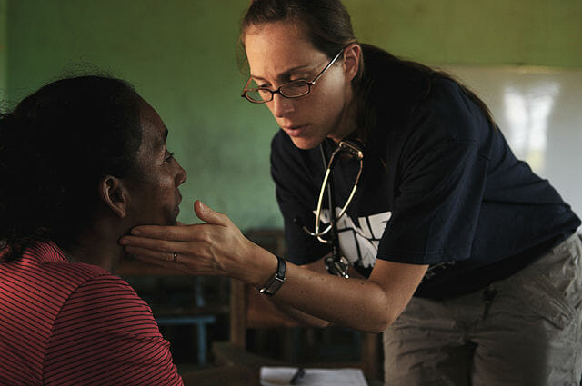
In this image, a geriatric nurse gives a checkup to an elderly patient in Nicaragua.


References


  • n.a. (n.d.). “A Brief History of Geriatrics.” The John A. Hartford Foundation. Retrieved 2017-07-06 from http://www.johnahartford.org/ar2005/2_a_brief_history.html.

  • n.a. (n.d.). “Geriatrics Overview.” Liaison International. Retrieved 2017-07-10 from https://explorehealthcareers.org/field/geriatrics/.

  • n.a. (n.d.). “The History of Geriatric Medicine.” IPC / Senior Care of Colorado. Retrieved 2017-07-06 from http://www.seniorcareofcolorado.com/index.php?option=com_content&view=article&id=152&Itemid=151.

  • n.a. (2015-12). “A Guide to Geriatric Syndromes: Common and Often Related Medical Conditions in Older Adults.” Health in Aging. Retrieved 2017-07-07 from http://www.healthinaging.org/resources/resource:guide-to-geriatric-syndromes-part-i/.

  • Morley, John E. (2004). “A Brief History of Geriatrics.” Journal of Gerontology: Medical Sciences 59A(11): 1132-1152.


Geriatrics

Biophysics

Biophysics Definition


Biophysics is a branch of science that uses the methods of physics to study biological processes. Physics uses mathematical laws to explain the natural world, and it can be applied to biological organisms and systems to gain insight into their workings. Research in biophysics has helped prevent and treat disease, advance drug development, and create technology to allow humans to live more sustainably and protect the changing environment.


History of Biophysics


Biophysics is a relatively young branch of science; it arose as a definite subfield in the early to mid-20th Century. However, the foundations for the study of biophysics were laid down much earlier, in the 19th Century, by a group of physiologists in Berlin. The Berlin school of physiologists included Hermann von Helmholtz, Emil DuBois-Reymond, Ernst von Brücke, and Carl Ludwig. In 1856, Adolf Fick, one of Ludwig’s students, even published the first biophysics textbook. But technology in physics had not sufficiently advanced at this time to study lifeforms in a detailed way, such as at the molecular level.


In the first half of the 20th Century, German scientists dominated the biophysics. They studied electromagnetic fields and light, and they became mainly concerned with studying the effects of radiation on living things. The popularity of biophysics rose when the Austrian physicist Erwin Schrödinger published the book What is Life? in 1944. This book was based on a series of public lectures that Schrödinger gave on explaining the processes of living things through physics and chemistry. In it, he proposed the idea that there was a molecule in living things that contained genetic information in covalent bonds. This inspired scientists such as James Watson and Francis Crick to search for and characterize the genetic molecule, and with the aid of Rosalind Franklin’s x-ray crystallography research, they discovered the double helix structure of DNA in 1953.


By the mid-20th century, biophysics programs had sprung up and gained popularity in other countries, and from 1950-1970, biophysics research occurred at a faster rate than ever before. In addition to the discovery of DNA and its structure, biophysics techniques were also used to create vaccines, develop imaging techniques such as MRI and CAT scans to help doctors diagnose diseases, and create new treatment methods such as dialysis, radiation therapy, and pacemakers. Currently, biophysics has also begun to focus on issues related to the Earth’s changing climate. For example, some biophysicists are working on developing biofuels from living microorganisms that could replace gasoline as a fuel.


Areas of Biophysics


Biophysics is incorporated into many diverse areas of biology. Some research topics in biophysics or involving biophysics include:


  • Membrane biophysics—the study of the structure and function of cell membranes, including the ion channels, proteins, and receptors embedded within them.

  • Computational/theoretical biophysics—using mathematical modeling to study biological systems.

  • Protein engineering—creating and modifying proteins to advance synthetic biology. Often used to advance human health in the form of new disease treatments.

  • Molecular structures—biophysics studies the molecular structures of biological molecules including proteins, nucleic acids, and lipids.

  • Mechanisms—using physical mechanisms to explain the occurrence of biological processes. Some physical mechanisms include energy transduction in membranes, protein folding and structure leading to specific functions, cell movement, and the electrical behavior of cells.


Biophysics Lab

Here, a biophysicist in a U.S. Food and Drug Administration lab is studying the electrical activity of the heart as related to pacemaker and defibrillator use.


Biophysics Major


Some universities offer undergraduate Bachelor of Arts or Bachelor of Science degrees in Biophysics, while others only offer a Biophysics degree at the graduate level (i.e., a master’s and/or doctorate degree). Biophysics degrees are heavily focused on physics and biophysics courses, and usually those who major in biophysics are required to take numerous math and chemistry classes as well. At the undergraduate level, one can expect to take courses in general and organic chemistry, calculus, mechanics, linear algebra, and biochemistry. Other possible courses include cell biology, genetics, molecular biology, statistics, and computational biology, among others. Another important component of many biophysics degrees is research; some programs require research in a laboratory to be done for a certain number of semesters, culminating in a senior research project. The specific courses offered in a biophysics major program can vary from university to university, but majoring in biophysics will adequately prepare a student to begin their career in biophysics research.


If a student is interested in biophysics but their school does not offer a biophysics degree, there are often comparable programs found in other majors that include much of the same courses. Majoring in physics is another good option, and one may consider adding another major or minor in biochemistry, chemistry, or biology depending on research interests and the programs offered.


Biophysics Careers


The most common career options for biophysicists include research, teaching, or a combination of both. A master’s degree is generally needed to become a biophysics teacher, lab manager or research associate, while a PhD is necessary in order to be the principal investigator of a research laboratory. Principal investigators design experiments and oversee all of the research being done in a lab, while lab managers and research associates have a more supporting role and assist the principal investigator in carrying out their research. Those with bachelor’s degrees may obtain positions as research technicians, which are also important in the laboratory. Research technicians carry out a lot of the benchwork of scientific experiments, allowing the principal investigator time to write scientific papers, research proposals, and grants.


References


  • n.a. (n.d.). “Biophysical Mechanisms.” Biophysical Society. Retrieved 2017-07-03 from http://www.biophysics.org/Education/SelectedTopicsInBiophysics/BiophysicalMechanisms/tabid/2312/Default.aspx.

  • n.a. (n.d.). “Careers in Biophysics: Job Options and Education Requirements.” Study.com. Retrieved 2017-07-04 from http://study.com/articles/Careers_in_Biophysics_Job_Options_and_Education_Requirements.html.

  • n.a. (n.d.). “Research areas.” University of California, San Francisco. Retrieved 2017-07-04 from https://biophysics.ucsf.edu/degree-program/research-areas.

  • n.a. (n.d.). “What is Biophysics?” Biophysical Society. Retrieved 2017-07-02 from http://www.biophysics.org/Education/WhatisBiophysics/tabid/2287/Default.aspx.

  • Bischof, Marco (1995). “Some Remarks on the History of Biophysics (and Its Future).” Current Development of Biophysics (C.L. Zhang et al., Eds.), Hangzhou University Press: Hangzhou, China. ISBN: 9787810359030.


Biophysics

Sunday, July 30, 2017

Thyroid Gland

Thyroid Gland Definition


The thyroid gland is a gland in the neck that secretes metabolic hormones important to the growth of the human body. It specifically helps coordinate the creation and use of energy, and is by far the largest gland in the neck. This gland notably does not rely on a duct system, unlike other gland types, and is shaped in the form of a butterfly. It contains two lobes (one on each side of the neck) and is about two inches long, lying just below the Adam’s apple.


Glands come in endocrine and exocrine varieties. Since the thyroid gland is performing its function via hormones, it is considered a key part of our endocrine system. The thyroid gland works primarily by absorbing iodine from our diets and then using it to make thyroid hormones thyroxine (T4) and triiodothyronine (T3). The thyroid gland is able to store these hormones for later use, as they will be released as needed. These hormones are then able to navigate through the entire body via the bloodstream in order to reach their target cells.


Thyroid Gland Location


As discussed previously, the thyroid gland is located in front of our neck. More specifically, it lies in front of our trachea, or “windpipe.”. When the structure of the thyroid gland is inspected more closely, it has a reddish-brown coloration. The color is due to the fact that the thyroid gland is highly innervated and has its blood supplied by the superior and inferior thyroid arteries and the external carotid artery. The two lobed structure will be linked by a bridge called the isthmus that lies in the middle of the lobes.


Anterior thyroid


The image depicts the thyroid gland and the surrounding tissues & bones.


The location of the thyroid gland is quite easy to visualize, as it is an area that is regularly inspected during doctor visits. Of course, the gland at its normal size will not be perceptible and only becomes noticeable when the gland is swollen. But before birth, however, the placement of thyroid differs. It will be located in the back of the developing tongue, meaning it will migrate to the front of the neck (its post-birth location) prior to birth. The amount it travels also matters as issues will arise from thyroids that migrate too little or too far from the ideal mark. An extreme example is a condition called lingual thyroid, which is when the thyroid did not travel and instead remained in the back of the tongue.


The thyroid gland is truly a stand out through its versatile functions.


Thyroid Gland Function


The thyroid gland has the main role of controlling our body’s metabolism. The simplest way to define the metabolism is as our body’s ability to convert food into energy. This “fuel” is burned at different rates depending on the person, which is why people are said to have a “fast” or “slow” metabolism. Furthermore, the thyroid will secrete the hormones that will regulate our vitals and maintain our internal homeostasis. Among the most common, primitive functions of the body it controls are our breathing and our heart rate. Our weight is likewise monitored by the thyroid gland, which explains why patients with a compromised thyroid gland will have weight that fluctuates drastically, as we will discuss in more detail later. Even our internal body temperature and our cholesterol levels will be finely tuned with the help of thyroid hormone release.


The wings or lobes of the thyroid gland have a singular function. This is to synthesize thyroid hormone. They are able to have wide-spanning effects that affect nearly all of the tissues in the body with the help of the endocrine passage way. At the cellular level, the thyroid hormones are able to increase cellular (metabolic) activity. This influences not only our metabolic rate but also has an effect on protein synthesis. This, of course, facilitates normal development as growth relies on continual protein creation.


Thyroid Gland Hormones


The thyroid gland primarily makes and releases T3 and T4 hormones, and the levels at which either thyroxine or triiodothyronine are released can be modulated to slow or speed things up. Thyroid hormones are made from the iodine flowing in our blood after a meal, which is then integrated into the physical structure of the hormones. The thyroid cells that make up the gland have a special trait of being highly absorbent to iodine. Every remaining cell in the body will rely on the thyroid gland to manage its metabolism. Regardless, the levels of T3 to T4 will be eighty to twenty percent in a normal, functional thyroid gland.


The hypothalamus and the pituitary gland are the main controls that regulate the actual thyroid’s activity. When T3 or T4 hormone levels become too low, the hypothalamus will respond by secreting TSH-releasing hormone (or TRH). TRH signals the pituitary gland to create more thyroid stimulating hormone (or TSH). The thyroid gland, in turn, will respond by making more thyroid hormone in a feedback loop. The levels will be finely tuned to maintain a balance of T3 to T4 hormones.


TSH release will have a direct impact on the hormones’ respective levels. The role of TSH can be summed up as a stimulus that will lead the thyroid gland to release more hormone. When the T3 and T4 levels get too low, the pituitary gland will release moreTSH that will tell the thyroid to synthesize more thyroid hormone. Abnormally high levels of TSH can indicate an underactive thyroid. This is a condition called hypothyroidism. The related symptoms of having T3 and T4 in excess are listed below:


  • Anxiety

  • Hair loss

  • Irritability

  • Hyperactivity

  • Hand trembling


On the other hand, when the T3 and T4 levels fall under the functional amounts, the body will undergo changes in the opposite direction. Chronically high thyroid hormone levels will lead to hyperthyroidism. The high T3 and T4 levels will signal the pituitary gland to release lessTSH in the system. Common symptoms may include the following:


  • Insomnia

  • Fatigue and tiredness

  • Depression

  • Difficulty concentration

  • Muscle pain


In summary, a high TSH will strongly suggest that the thyroid is underactive, or not making the right amount of thyroid hormone to sustain the body’s functions. The reason the levels of TSH are so high is because the body is trying to make the thyroid gland produce hormone to compensate for its lacking. A low TSH, in contrast, will infer a very active thyroid gland that is making too much thyroid hormone. In this case, the pituitary gland will try to inhibit TSH release to, in effect, stop the thyroid gland from making more of it.


There is some variance over the reference range of TSH levels in the blood, but when the test reveals a level 0.5 or below, it is a possible sign of hyperthyroidism. When levels are above the 3-5.0 range, there is a good chance of hypothyroidism. This scale is hotly contested in the medical community, but is still a parameter used to diagnose thyroid disorders. In addition to a blood test, an iodine thyroid scan will reveal if the origin of the thyroid hormone imbalance is a single nodule or the entire thyroid gland.


Thyroid Gland Disease


There are various thyroid illnesses, but the most common distill down to a few categories of imbalance:


  • An abnormal production of thyroid hormone

  • Abnormal gland growth

  • Lumps or nodules within the thyroid gland

  • Thyroid cancer


Let’s list the most common thyroid disorders. Goiter is an enlargement of the thyroid gland from a noncancerous origin. Most commonly, goiter will arise in patients who have an iodine deficiency in their diet. This, of course, is a bit more common in places in the world where foods are not rich enough in iodine, or in women over the age of forty who are likelier to develop goiter. The symptoms of goiter include swelling or tightness of the neck, breathing and swallowing difficulties, hoarseness of the voice, and wheezing. Typically, these clinical signs will only show when a patient’s thyroid has grown large enough. Goiter will be treated either with radioactive iodine doses or surgery.


Hashimoto’s disease is another thyroid disorder that is the most common cause of hypothyroidism in the U.S. The illness arises when a patient’s own immune system mistakenly attacks its own thyroid gland. This will, of course, comprise its ability to make hormones. The most common signs are fatigue, mild weight gain, dry skin and hair, depression, pale skin, and an enlarged goiter. Its cure is yet unknown.


Grave’s disease is the most common cause of hyperthyroidism and is the foil of Hashimoto’s disease in that it arises from the patient’s immune system attacking its own thyroid gland. In this case, it will lead it to make more thyroid hormone than normal. The resulting symptoms may include anxiety, fatigue, hand tremors, excessive sweating, trouble sleeping, diarrhea, goiter, and bulging eyes or vision impairing. This condition will be treated with beta blockers that will slow the heart rate and axiety and anti-thyroid medications along with radioactive iodine.


Lastly, thyroid nodules are growths that form on the thyroid gland and may result from either Hashimoto’s disease or a lack of iodine. The nodules are mostly benign but can also become cancerous. The many signs it displays may include a high heart rate, nervousness, tremors, weight loss, and a big appetite.


Quiz


1. When thyroid hormone levels are low, the pituitary gland will release ______ TSH?
A. Less
B. More
C. No change
D. Tapered

Answer to Question #1

2. Which condition is marked by a low TSH?
A. Hypothyroidism
B. Hyperthyroidism
C. Hair loss
D. Hand trembling

Answer to Question #2

3. High TSH will indicate which of the following?
A. Low thyroid hormone release
B. High thyroid hormone release
C. An overactive thyroid
D. Hyperthyroid disease

Answer to Question #3

References


  • Norman, James MD (2017). “Your Thyroid Gland.” Endocrine Web. Retrieved on 2017-07-14 from https://www.endocrineweb.com/conditions/thyroid/your-thyroid-gland

  • Brady, Bridget (2017). “Thyroid Gland, How it functions, symptoms of hyperthyroidism and Hypothyroidism.” Retrieved on 2017-07-14 from https://www.endocrineweb.com/conditions/thyroid-nodules/thyroid-gland-controls-bodys-metabolism-how-it-works-symptoms-hyperthyroi

  • HealthLine (2017). “4 Common Thyroid Disorders.” Health Line. Retrieved on 2017-07-15 from http://www.healthline.com/health/common-thyroid-disorders#overview1

  • Shomon, Mary (2017). “High and Low TSH Levels: What They Mean.” Very Well. Retrieved on 2017-07-17 from https://www.verywell.com/understanding-thyroid-blood-tests-low-or-high-tsh-3233198

  • Hoffman, Matthew MD (2017). “Picture of the Thyroid.” Web MD. Retrieved on 2017-07-18 from http://www.webmd.com/women/picture-of-the-thyroid#1


Thyroid Gland

Adrenal Gland

Adrenal Gland Definition


The adrenal gland has several metabolic roles in the human body. Our two adrenal glands can be found lying on top of each of our kidneys. Together, they release the hormones that help us metabolize, undergo sexual maturation as we grow, as well as respond to stress. The latter may be known to us as the primal “fight or flight” response. It is the knee-jerk reaction we get upon seeing a life-endangering stimulus. Cortisol is the key to coordinating the processes that allow us to fight or run from this perceived danger. There is an obvious evolutionary value in being able to do this. Anything that shortens the time in which we can engage our muscles and run will likely increase our chance of survival. But even more low key scenarios trigger the release of cortisol. A feeling we may be familiar with is the intense focus we feel when studying for an exam the night before we have it. Cortisol is largely responsible for this newfound focus. In spite of all of the negative connotations associated with stress, in normal amounts the stress hormones allow us to focus better to perform the tasks at hand.


Thus, the adrenal glands play a functional role in our alertness, growth, and more. The main products released by the adrenal glands are cortisol, epinephrine, aldosterone, and adrenal androgens, or sex hormones. We will discuss each in more detail.


Adrenal gland


The image depicts a simple illustration of the adrenal glands and surrounding structures.


Adrenal Gland Structure


Our adrenal glands are two organs shaped in the form of a triangle that spans three inches in length. When a biopsy is taken of the adrenal gland, two sections are immediately visualized. The outermost layer of the adrenal gland is called the adrenal cortex, while the inner layer is named the adrenal medulla. Besides having physical differences that give them a distinct appearance, they release hormones independently of each other. In fact, the cortex contains zones of different cell types starting with the outermost “shell” or capsule, followed by “zones” named the zona glomerulosa, zona fasciculate, and the zona reticularis. This division facilitates some versatility. The medulla will secrete epinephrine in response to emotional or physical stress, while the outer adrenal cortex will make steroids and metabolic hormones like aldosterone and cortisol. However, it is safe to say that there is a lot of overlap between their functions.


Adrenal Cortex Zones:


  • Zona Glomerulosa: Secretes Mineralcorticoids (i.e. cortisol)

  • Zona Fasiculata: Secretes Glucocorticoids

  • Zona Reticularis: Secretes Androgens


The adrenal gland is supplied by three arteries: the superior suprarenal artery, a branch of the inferior phrenic artery, and the middle suprarenal artery which directly branches off of the abdominal aorta. As for the nerve supply, the adrenal glands are innervated by the sympathetic thoracic spinal cord fibers.


Adrenal Gland Function


The most unique function of the adrenal glands is their ability to orchestrate responses in light of stress. This has the most primal purpose of helping us evade danger and, therefore, of prolonging our lifespan. However, the adrenal gland is also involved in more mundane, but equally important, aspects of our everyday life.


Everyday roles of the adrenal glands:


  • Via aldosterone, which we will discuss in more detail below, the adrenal gland allows our kidneys to regulate blood pressure via salt and water exchange between the kidneys and our surrounding blood vessels. In the absence of aldosterone, the kidney will lose lots of salt to urine which will draw water from our vessels and remove it from our system. This will most certainly lead to dehydration.

  • Via cortisol, the body is not only able to respond to stress in potentially life-saving circumstances, but it will also help us regulate our body’s metabolism by initiating glucose production and by circulating fatty acids and amino acids to our cells.

  • Via adrenal androgens, the adrenal gland helps create differences between the sexes by initiating the development of our sex organs and secondary traits.


Adrenal Gland Hormones


Like any endocrine gland, the beauty of the adrenal glands lies in its secretions. In general, the endocrine gland will produce several types of hormone: androgens, cortisol, aldosterone, and norepinephrine. Let us begin with a discussion of androgens. The zona reticularis of the adrenal cortex is responsible for releasing androgen hormones that help give rise to secondary sex traits in human males. “Secondary” traits can be thought of as the changes that occur once puberty starts, including bodily changes like pubic hair growth, the Adam’s apple, and muscle and hair growth. Females also use androgens but they are instead secreted by the ovaries and repurposed into estrogen hormone.


Aldosterone, which is released by the zona glomerulosa of the cortex, plays a huge role in our kidneys. Our kidneys can be thought of as big filters that will help us excrete waste and excess fluid from our cells and blood vessels, while allowing us to reabsorb the ions that we need to maintain an ionic balance and a good blood pressure. One of these important components is salt, or Na+Cl. Salt is able to modulate the levels of fluid in our vessels, otherwise known as our blood pressure! Based on simple rules of diffusion, having more salt reabsorbed will lead water to be reabsorbed into our vessels in greater amounts, as well, since “water follows salt.” Aldosterone is thus able to directly modulate the excretion of salt by either increasing or decreasing the number of salt (Na/Cl) channels in the walls of our nephrons (kidneys). The presence of aldosterone will encourage our blood vessels to retain more salt via numerous salt channels, which in turn encourages the reabsorption of water. This results in concentrated urine, and a healthy blood pressure. When this process is compromised, as with Addison’s disease, insufficient amounts of cortisol and aldosterone are made when the body is under stress – such as when fighting off an infection. Its symptoms vary from fatigue, to dizziness and chronic nausea secondary to low blood pressure and low salt levels.


The zona fasciculata, in turn, creates the stress hormones we have discussed before: cortisol and its derivatives. Cortisol is inherently a steroid hormone that responds to stressful situations and when our blood sugar drops too low. In effect, it will stimulate gluconeogenesis, or glucose creation, to counter the low blood sugar and will aid in the metabolic breakdown of food. It also suppresses the immune system and decreases bone formation. Healthy amounts of cortisol increase our focus; however, chronic overexpression of cortisol has many scientists worried for fear of memory interference, bone density loss, and heart disease in affected patients.


We have discussed plenty about the adrenal cortex, but it is important to note that the adrenal medulla uniquely makes epinephrine and norepinephrine. These water-soluble compounds are the ones responsible for giving us a “rush” whenever we are confronted with a stressful situation. Its effects are characterized by a fastened breathing and heart rate, and a constriction of blood vessels that redirects blood flow to our muscles. This allows our muscles to immediately engage for quick movement.


Adrenal Gland Tumor


Since the adrenal gland is constantly producing hormones that are vital and well-circulated in the body, there is a chance of risking an imbalance. For example, an overactive gland or even a benign tumor on the adrenal gland will cause it to make too much cortisol. This will disrupt our blood pressure, our heart health, and even our body’s response to stress. Adrenal gland tumors can be benign or malignant.


There are various kinds of malignant, or “cancerous,” gland tumors.


  • Adrenocortical cancer will originate in the adrenal cortex. There are two types. The functioning tumor is the most common and will continue to produce cortisol, aldosterone, and androgens. Non-functioning tumors will, on the other hand, not produce hormones which will result in other deficiencies.

  • Adrenal Pheochromocytomas originate in the adrenal medulla and are very rare.

  • Adrenal Paragangliomas begin within or outside the adrenal gland.


A well-studied benign adrenal gland tumor is commonly known as Cushing’s Syndrome. This syndrome will overproduce cortisol, and thus disrupt heart function and the processes that the body engages in when responding to a stressful stimulus. Cushing’s syndrome is quite rare, with only two-to-four new cases per one million Americans each year. Other causes for an overproduction of cortisol may be an adenoma, or benign tumor anywhere on the adrenal gland, or long-term use of corticosteroid medications like prednisone.


Quiz


1. In which of the following way does Aldosterone perform its roles in the human body?
A. It acts directly on water channels to stimulate water retention
B. It works via calcium ions that increase water absorption
C. It acts on salt by increasing the number of sodium channels in our nephrons to facilitate water retention
D. It acts on salt by decreasing the number of sodium channels in our nephrons to facilitate water retention

Answer to Question #1

2. A lack of Aldosterone will result in which of the following conditions?
A. Too much water retention
B. A dehydrated state
C. Heightened blood pressure
D. Stimulates overproduction of glucose

Answer to Question #2

3. Correctly identify the zone of the adrenal cortex that releases cortisol:
A. Zona reticularis
B. Zona fasciculata
C. Zona glomerulosa
D. Does not apply, zones are found in adrenal medulla

Answer to Question #3

References


  • Medline Plus (2017). “Adrenal Gland Disorders.” US National Library of Medicine NIH. Retrieved on 2017-07-23 from https://medlineplus.gov/adrenalglanddisorders.html

  • Sargis, Rober MD, PhD. “An Overview of the Adrenal Glands.” Endocrine Web. Retrieved on 2017-07-22 from https://www.endocrineweb.com/endocrinology/overview-adrenal-glands

  • MD Anderson Cancer Center. “Adrenal Tumors.” The University of Texas. Retrieved on 2017-07-22 from https://www.mdanderson.org/cancer-types/adrenal-tumors.html

  • Cancer.net Editorial Board (2016). “Adrenal Gland Tumor: Introduction.” Cancer.Net. Retrieved on 2017-07-23 from http://www.cancer.net/cancer-types/adrenal-gland-tumor/introduction

  • Aviva (2017). “Structure and Function: Adrenal Glands.” Aviva. Retrieved on 2017-07-23 from https://www.aviva.co.uk/health-insurance/home-of-health/medical-centre/medical-encyclopedia/entry/structure-and-function-adrenal-glands/

  • You and your hormones (2017). “Adrenal Glands.” Your Hormones Info.. Retrieved on 2017-07-24 from http://www.yourhormones.info/glands/adrenal-glands/


Adrenal Gland

Pineal Gland

Pineal Gland Definition


The pineal gland, also known as the “pineal body,” is lightheartedly known as the “third eye” of the human body. This name stems from the pineal gland’s role in secreting melatonin. This hormone, in turn, modulates our sleep and waking patterns. Melatonin was first described by American physician, Dr. Aaron Lerner in 1958. Research has shown melatonin to play an instrumental role in establishing our circadian rhythm, which is the twenty-four-hour cycle of our bioactivity that matches the solar cycle of the day.


Melatonin is one of the most ubiquitous and versatile hormones found in animals and some plants. It is highly lipophilic, which allows it to reach our cells in record time. If its name sounds familiar, it is because melatonin was named after “melanin” after displaying skin lightening effects in frogs. Melatonin derives from the amino acid, tryptophan. This amino acid can be found in humans and other species of mammals, reptiles, birds and amphibians. Tryptophan-rich foods have been linked to calming effects and reduced anxiety. In humans, specifically, melatonin helps control our daily sleep cycle. The body produces melatonin in response to light hitting the retina of the eye. This inhibits the release of melatonin. In contrast, the absence of light at nighttime will be read as a signal to produce more melatonin. The ability to modulate melatonin levels is lent by the presence of special photoreceptor cells in the human retina that emit a signal to the suprachiasmatic nucleus, or SCN, of the hypothalamus. The hypothalamus, in turn, is a part of the brain that supports the body’s homeostatic functions. The light or dark signals are then sent to the pineal gland, which will begin to modulate melatonin levels.


Melatonin Properties


The largest amount of melatonin is expelled by the pineal gland during the night. At this time, the body will undergo several changes closely tied to the concentrations of melatonin. The body’s internal temperature will drop, as will our breathing rate. These experiences are the ones we most associate with falling asleep. In the daytime, however, our retinas will be exposed to a lot of light that will inhibit melatonin expression. This is essential for making us alert and awake during the daytime.


Melatonin has special antioxidant properties. It is known to neutralize radicals, or elements with an unstable electron configuration, that would otherwise cause harmful oxidative damage to tissue. Melatonin can also activate other antioxidant enzymes that will perform restorative functions. Naturally, melatonin is an antiaging substance that declines as we get older. The loss of melatonin is thus associated with various age-related illnesses. Melatonin also retains a role in buffering the immune system in light of seasonal adjustments. Its roles are still being studied, but the consensus lies in melatonin acting as a stimulant under suppressive conditions and as an anti-inflammatory agent when the immune system experiences acute inflammation.


Pineal Gland Location


The pineal gland is roughly located in the center of the brain, sandwiched between the left and right hemispheres. It is about seven by six by three millimeters in dimensions, and is the only midline brain part that is not paired. The pineal body is tucked in the divot or groove where the two thalamic bodies meet. It takes on the shape of a pinecone, which explains its naming! Nearly all existing vertebrates contain a pineal gland. Pineal glands are found in even the primitive lamprey. It makes sense that the pineal gland is a primitive organ, as it is made with the simple intention of acting as a kind of photoreceptor that responds to rays of light. However, not all species have conserved the pineal gland. The exception to the rule is the hagfish, which lacks a perceptible pineal gland. Likewise, a few other more advanced vertebrates have lost theirs sometime in their evolution. Regardless, this light-sensing organ has earned an interesting place in philosophy classes across the world. In its romanticized interpretation, the pineal gland has been described to embody metaphysical properties in the realm of pseudoscience. Like most subjects, this idea was hotly contested by early philosophers. But what remains uncontested is that its secretion, melatonin, serves a vital role in the physical body.


Unlike the remaining mass of the mammalian brain, the pineal gland is not separated from the body by the blood-brain barrier. Instead, it receives the second most profuse supply of blood in the body, next to the kidney. The pineal gland’s main blood supply come from the choroidal branches of the posterior cerebral artery. Its sympathetic (or excitable) innervation, on the other hand, comes from the superior cervical ganglion. The otic ganglia will supply the inhibitory, parasympathetic innervation.


Melatonin receptors are found scattered in various areas of the body. Most notably, they are found in high concentrations in the SCN and the brain’s pituitary gland. This is the main site of action as melatonin directly plays into the circadian rhythm here. But melatonin receptors are also present in the ovaries. The levels of melatonin affect several facets of the menstrual cycle, such as the timing of its onset, the duration, and the frequency. In other animal species, it even acts as a mating cue. For instance, greater levels of melatonin in horses are found during the spring, which coincides with the ideal season for mating. This is a direct example of the solar cycle’s impact on the reproductive cycle via pineal gland activity. Other melatonin receptors lie in the blood vessel walls and in our intestinal tracts. In the gut, melatonin protects the mucosal layers from lesions and irritation via its eradication of free radicals. Gut lesions can lead to painful esophagitis, gastritis, and peptic cancer among other illnesses.


Pineal Gland Disorders


Since the pineal gland is primarily involved in sleep-wake rhythms, it is also takes root in mood disorders. Recent studies have linked chronic stress and poor diet as possible causes of reduced levels of melatonin in the system. This is often found in patients with abnormal circadian cycles of cortisol (or “stress”) hormone. In fact, depression and sexual dysfunction are conditions that are further aggravated by low melatonin output. These of course impact our quality of life. Milder mood alterations, like insomnia and the jetlag that is felt after boarding a long flight, have also been linked to the pineal gland. These feelings are often short-lived, albeit disruptive. Furthermore, peptic ulcers are also linked to melatonin levels when they are too low to prevent oxidative damage.


Pineal cysts, or cysts of the pineal gland, are a relatively common occurrence that happens in about ten percent of people undergoing a CT or MRI scan. The cause of pineal cysts is not known. In fact, most patients with pineal cysts will not display any visible symptoms. But very rarely will patients experience headaches and eye movement abnormalities because of it. In some patients, the cyst can even lead to emotional disturbances, sleep issues, and seizures. Only when the pineal gland cyst is symptomatic will a physician recommend surgical removal. But the overall prognosis for patients with pineal cysts is very good.


Pineal tumors, on the other hand, are a more serious complication that represent about one percent of all brain tumors. At least seventeen types of tumors arise in the area of the pineal gland but many are benign. The most common tumors are gliomas, pineal cell tumors, and germ cell tumors. The pineal gland is located next to a duct called the aqueduct of Sylvius. It acts as a passage through which cerebrospinal fluid (CSF) leaves the center of the brain. Pineal tumors often block this duct, causing a buildup of pressure that expands the ventricles within the skull. This blockage will present complications most often linked to the symptoms of pineal gland tumors:


  • Headaches

  • Seizures

  • Nausea

  • Visual changes

  • Problems with memory recall


These visual changes include double vision, an inability to properly focus on the objects in front of us, and abnormal eye movements. These issues may improve or resolve once the tumor is either resected or treated. Treatment of the tumor can vary depending on the diagnosis. This diagnosis must be informed with a precise histological analysis from a biopsied sample. A benign, or non-cancerous, pineal tumor can be resected surgically at the hands of a skilled surgeon. However, malignant pineal tumors may be treated with either surgery or radiation therapy. For example, pinealocytomas obtain no benefit from radiation therapy alone. So, they will require surgical resection. The most common cancer in this area is germinoma. Germinoma tumors, in contrast, are both very sensitive to chemotherapy and radiation and will be cured in most cases. The same applies to other malignant germ cell tumors near the pineal gland. Non-germ cell tumors can benefit from newer stereotactic types of radiation therapy. Like any other tissue that has undergone intensive cancer therapy, there may be long term effects on the pineal tissue’s ability to perform its endocrine functions. Therefore, the patient will need to work alongside an endocrinologist to address certain hormone deficiencies that may have arisen. Most of these issues can be managed with medical therapies. This has led the prognosis for pineal tumor survivors – both in children and adults – to improve.


Quiz


1. How will light affect melatonin secretion? Choose best answer.
A. Increase
B. Decrease
C. No effect
D. Modulate

Answer to Question #1

2. How will darkness affect melatonin secretion? Choose best answer.
A. Increase
B. Decrease
C. No effect
D. Modulate

Answer to Question #2

3. Which of the following is indicated for malignant pineal tumors, per the article?
A. Surgical resection
B. Radiation therapy
C. Surgery or radiation
D. Chemotherapy

Answer to Question #3

References


  • Encyclopedia Britannica (2017). “Melatonin: Hormone.” Encyclopedia Britannica. Retrieved on 2017-07-07 from https://www.britannica.com/science/melatonin

  • Emerson, Charles H (2017). “Pineal Gland.” Encyclopedia Britannica. Retrieved on 2017-07-08 from https://www.britannica.com/science/pineal-gland

  • Carillo-Vico, A. et al. (2013). “Melatonin: Buffering the Immune System.” Int J Mol Sci.. 14(4): 8638-8683. Doi: 10.3390/ijms14048638

  • Maurizi CP (1984). “Disorder of the pineal gland associated with depression, peptic ulcers, and sexual dysfunction.” South Med J.. Dec; 77(12): 1516-8.

  • NIH (2014). “Pineal Cyst.” Genetic and Rare Diseases Information Center.. Retrieved on 2017-07-07 from https://rarediseases.info.nih.gov/diseases/10723/pineal-cyst

  • Ferry, Robert MD (2016). “Pineal Tumor.” E Medicine Health. Retrieved on 2017-07-08 from 2http://www.emedicinehealth.com/script/main/mobileart-emh.asp?articlekey=88561#when_to_seek_medical_care_for_a_pineal_tumor


Pineal Gland

Echinoderm

Echinoderm Definition


An echinoderm is a member of the phylum Echinodermata which contains a number of marine organisms recognized by their pentamerous radial symmetry, calcareous endoskeleton, and a water vascular system which helps operate their small podia. Podia are small extensions of flesh which are operated by water pressure and muscles, and controlled by the nervous system of the echinoderm. The calcareous endoskeleton is made of many small plates that overlap under the skin, forming an armor and a structural form for the organisms. Examples of an echinoderm include a starfish, a sand dollar, a brittle star, a sea urchin, and a sea cucumber. There are around 7,000 echinoderm species, and they can range from less than an inch to over three feet in diameter.


Echinoderm Characteristics


An adult echinoderm is radially symmetrical, meaning their body parts extend outward from the mouth. An echinoderm usually has 5 parts, making them pentamerous. Curiously, echinoderm larva are bilaterally symmetrical and must convert to radial symmetry. Typically, the mouth is surrounded by a central disc, which lead to outward to grooves housing rows of podia. These grooves are called ambulacral grooves and may lead to individual legs as in a starfish, or can be simple slits like in a sand dollar. The endoskeleton of an echinoderm is made up of individual pieces, known as ossicles. The ossicles are covered by epidermis, or skin. In some echinoderms, like sand dollars and sea urchins, the ossicles form a rigid shell known as a test. On the other end of the spectrum, sea cucumbers have very few ossicles and they are separated from each other. These ossicles may also fuse to form various structures, such as the brittle spines of the sea urchin.


The water vascular system is an essential part of echinoderm biology. While the system differs in different classes of echinoderm, its basic operation is the same. The system consists of a series of fluid-bearing tubes that connect in a ring-like structure throughout the organism. The system connects to the podia, and can be used to fill them with fluid which elongates and stiffens the podia. This is accomplished by a series of sacs and muscles within the ring canal, lateral canals, and Polian vesicles, some of which can be seen in the image below.


Figure_28_05_01


An echinoderm uses this unique system for a number of lifestyles. The podia can be used as feet, to move in a coordinated fashion to direct the echinoderm. The podia can also be used to hold on to the substrate, small stones for protection, or a number of objects to use as camouflage. Some echinoderms are sessile filter feeders, while others actively hunt their prey. While some filter feeders direct food to their mouths, sea stars are known for pushing their stomach outside of their body to feed on prey. Other echinoderms have a complex mouth structure known as Aristotle’s lantern, which houses teeth and allow them to bite and scrape algae from the surface of rocks.


An echinoderm generally has simple circulatory and nervous systems, which circle through their bodies. Their hemal system is open to the environment and allows for gas exchange through a serious of channels throughout the body. The nervous system is a ring of nerves which connect to all parts of the organisms. This is thought to help an echinoderm interact with all directions it faces equally, maximizing the benefits of its radial symmetry.


Echinoderm Reproduction


Echinoderm reproduction is varied and often complex. Most echinoderms reproduce sexually, while a few species are known to reproduce asexually or through budding. Most specious are dioecious, or contain two distinct sexes, while other species are hermaphroditic and each individual carries both sexual organs. Either way, the gametes of an echinoderm are developed in the genital sinus, which may take a number of forms. Many species then broadcast their gametes into the environment. Gametes that successfully find one another and fertilize will become a new larva.


This new larva, which is virtually microscopic, will swim and ride the currents to a favorable place on the ocean floor. During a complex metamorphosis, the larva will reorient its body plan from being bilaterally symmetrical to being radial symmetrical. This involves moving the mouth and anus, as well as rearranging many other body parts internally. Once this transition is complete, the echinoderm then assumes a life feeding along the ocean floor. In areas where larval survival is low, an echinoderm may brood and care for their larva before they are released. In polar waters and deep-sea areas this helps insure a higher rate of larva survive into adulthood.


Examples of Echinoderm


Sea Stars


Sea stars are among the most ambulatory, or mobile, of all echinoderms. Sea stars, or starfish, use their many podia to slowly crawl over most surfaces. Starfish are mainly predatory, feeding on invertebrates and other echinoderms like sea urchins. Starfish move over their prey, then distend their stomach over their prey. The digestive enzymes in the stomach immediately begin to digest the organism, and the starfish will surround the prey until it is mostly dissolved. Then, it will revert its stomach, sucking in all the nutrients. While starfish appear slow moving to us, time-lapse videos show starfish chasing and hunting for prey over the course of many days or hours. Starfish belong to the class Asteroidea. A general diagram of a starfish can be seen above in the “Echinoderm Characteristics” section.


Sea Urchins


Sea urchins are a type of echinoderm that belong to the class Echinoidea. These animals have a hard test, or shell, which surround their body. The test is covered in a thin epidermis, or skin. Extending out of this test are many spines and tube feet, which the urchins use for protection and locomotion. Urchins feed with an advanced mouth structure known as Aristotle’s lantern, which controls a number of scraping teeth. Urchins feed mostly on algae and bacteria they can scrape from the rocks in which they make their home. A general sea urchin diagram can be seen below. Note how the water vascular system is still present, as is radial symmetry around the mouth.


Urchin


Sea Cucumbers


Sea cucumbers, belonging to the class Holothuroidea, may be among the most bizarre of the echinoderms. Lacking the traditional pentamerous symmetry of other members of its phylum, sea cucumbers look like a monstrous vegetable created in a secret laboratory. In fact, they are directly related to starfish and use the same water vascular system and endoskeleton. The endoskeleton of sea cucumbers is still made of calcareous ossicles. In sea cucumbers they are spread wide apart, and are connected by muscles and other connective tissues. This gives sea cucumbers the ability to flex their body and wiggle. This behavior is used both in regular movement, in feeding, and in escaping predators. Sea cucumbers typically exude a series of sticky filaments, which collect food and are drawn back into the mouth to be cleaned. Thus, most sea cucumbers have adopted a benthic, sessile, filter-feeding lifestyle. Below is a sea cucumber flipped over, revealing its tube feet extending from its ambulacral groove.


Mithrodia clavigera


Quiz


1. While there are thousands of different species of echinoderm in the ocean, why are there none on land?
A. They have no prey on land
B. They would not be able to breath on land
C. They would not be able to move on land

Answer to Question #1

2. While snorkeling, you see a starfish directly over a sea urchin. Upon closer inspection, a membrane extending from the starfish is seen surrounding the urchin. What is happening?
A. The two species are fighting
B. The starfish is eating the urchin
C. The urchin secreted the membrane to protect itself

Answer to Question #2

3. Echinoderms are deuterostomes that exhibit radial cleavage during development. They can also be reproduced and studied in a laboratory with ease. Which scientific discipline uses them as a key group of animals for study?
A. Developmental Biology
B. Molecular Biology
C. Ecology

Answer to Question #3

References


  • Brusca, R. C., & Brusca, G. J. (2003). Invertebrates. Sunderland, MA: Sinauer Associates, Inc.

  • Reece, J. B., Urry, L. A., Cain, M. L., Wasserman, S. A., Minorsky, P. V., & Jackson, R. B. (2014). Campbell Biology, Tenth Edition (Vol. 1). Boston: Pearson Learning Solutions.


Echinoderm

Negative Feedback

Negative Feedback Definition


Negative feedback is a type of regulation in biological systems in which the end product of a process in turn reduces the stimulus of that same process. Feedback, in general, is a regulatory mechanism present in many biological reactions. By allowing certain pathways to be turned off and on, the body can control various aspects of its internal environment. This is similar to flipping a switch. Feedback allows the product of a pathway to control the switch. Sometimes referred to as a “negative feedback loop”, negative feedback occurs when the product of a pathway turns the biochemical pathway off. Positive feedback, the opposite of negative feedback, is found in other biological pathways in which the product increases the pathway. Below are examples of negative feedback.


Examples of Negative Feedback


Regulating Blood Sugar


Every time you eat, a negative feedback mechanism controls the level of sugar in your blood. The main sugar found in your blood is glucose. After you eat something, your body absorbs the glucose from your bloodstream and deposits it into your blood. This increases the concentration of glucose and stimulates you pancreas to release a chemical called insulin. Insulin is a cellular signaling molecule which tells muscle and liver cells to uptake glucose. Liver cells store the excess glucose as glycogen, a chain of glucoses used as a storage product. Muscle cells can store the glucose or use it to make ATP and contract. As this process happens, glucose concentrations are depleted in the blood. Glucose was the main signal for the pancreas to produce insulin. Without it, the pancreas stops producing insulin and the cells stop taking up glucose. Thus, glucose levels are maintained in a specific range and the rest of the body has access to glucose consistently. The negative feedback mechanism in this system is seen specifically in how high glucose levels lead to the pathway turning on, which leads to a product meant to lower the glucose level. When glucose becomes too low, the pathway shuts off.


Temperature Regulation


All endotherms regulate their temperature. Endotherms are animals which regulate their bodies at a different temperature than the environment. You can think of mammals and birds as the most common endotherms. Most of the pathways responsible for temperature regulation are controlled by negative feedback. As the temperature rises, enzymes and pathways in the body are “turned-on”, and control various behaviors like sweating, panting and seeking shade. As the animal does these things, the temperature of their body starts to decrease. The activity of these pathways, which is driven by the heat, also starts to decrease. Eventually, a temperature is reached at which the pathway shuts off. Other pathways are present for temperatures that are too cold, and are also shut off once the body reaches the optimal temperature. These pathways can be shivering, seeking shelter, or burning fat. All these activities heat the body back up and are shut off by the end product of their reactions, heat.


Filling a Toilet Tank


Many students tend to struggle with abstract biological examples of negative feedback. Have no fear! A simple and common house-hold item uses negative feedback every day. In the tank on the back of your toilet is a ball or float, which rests at water level. When you empty the tank, the water level drops. The pressure from the float that was holding the valve shut releases, and new water flows into the tank. The valve controlled by the float is like an enzyme that monitors the level of the product it creates. As more water (product) fills the tank, the float slowly decreases the amount of water being let in through the valve. The valve is analogous to an enzyme which is regulated by feedback from a product it helps create or let into a cell.


Quiz


1. Which of the following represents negative feedback?
A. Blood platelets release chemicals that attract more blood platelets when then fill a wound
B. One bird fleeing a predator spurs three birds, which in turn scares the whole flock
C. In producing an amino acid, the enzyme a cell uses is inhibited after the amino acid reaches a specific concentration

Answer to Question #1

2. Bees control the temperature of their hive in an interesting way. When the temperature gets too hot, certain bees release a signal to the rest of the colony to begin a specific behavior. The bees evaporate water from their mouths and fan their wings to significantly decrease the temperature. As it cools, the colony resumes its normal activities. Which of the following terms describes this scenario?
A. Positive Feedback
B. Negative Feedback
C. Enzyme Inhibition

Answer to Question #2

3. You are reaching into a hot stove to grab your dinner. Your finger slips off the hot pad, and touches the scalding hot dish in the oven. A signal is sent to your brain, which tells your arm to contract. When your finger stops burning, your arm can relax. What does this scenario represent?
A. Negative Feedback
B. Positive Feedback
C. Fight or Flight response

Answer to Question #3

References


  • Nelson, D. L., & Cox, M. M. (2008). Principles of Biochemistry. New York: W.H. Freeman and Company.

  • Reece, J. B., Urry, L. A., Cain, M. L., Wasserman, S. A., Minorsky, P. V., & Jackson, R. B. (2014). Campbell Biology, Tenth Edition (Vol. 1). Boston: Pearson Learning Solutions.


Negative Feedback

Photosynthesis

Photosynthesis Definition


Photosynthesis is the biochemical pathway which converts the energy of light into the bonds of glucose molecules. The process of photosynthesis occurs in two steps. In the first step, energy from light is stored in the bonds of adenosine triphosphate (ATP), and nicotinamide adenine dinucleotide phosphate (NADPH). These two energy-storing cofactors are then used in the second step of photosynthesis to produce organic molecules by combining carbon molecules derived from carbon dioxide (CO2). The second step of photosynthesis is known as the Calvin Cycle. These organic molecules can then be used by mitochondria to produce ATP, or they can be combined to form glucose, sucrose, and other carbohydrates. The chemical equation for the entire process can be seen below.


Photosynthesis Equation


6 CO2 + 6 H2O + Light -> C6H12O6 + 6 O2 + 6 H2O

Above is the overall reaction for photosynthesis. Using the energy from light and the hydrogens and electrons from water, the plant combines the carbons found in carbon dioxide into more complex molecules. While a 3-carbon molecule is the direct result of photosynthesis, glucose is simply two of these molecules combined and is often represented as the direct result of photosynthesis due to glucose being a foundational molecule in many cellular systems. You will also notice that 6 gaseous oxygen molecules are produced, as a by-produce. The plant can use this oxygen in its mitochondria during oxidative phosphorylation. While some of the oxygen is used for this purpose, a large portion is expelled into the atmosphere and allows us to breathe and undergo our own oxidative phosphorylation, on sugar molecules derived from plants. You will also notice that this equation shows water on both sides. That is because 12 water molecules are split during the light reactions, while 6 new molecules are produced during and after the Calvin cycle. While this is the general equation for the entire process, there are many individual reactions which contribute to this pathway.


Stages of Photosynthesis


The Light Reactions


The light reactions happen in the thylakoid membranes of the chloroplasts of plant cells. The thylakoids have densely packed protein and enzyme clusters known as photosystems. There are two of these systems, which work in conjunction with each other to remove electrons and hydrogens from water and transfer them to the cofactors ADP and NADP+. These photosystems were named in the order of which they were discovered, which is opposite of how electrons flow through them. As seen in the image below, electrons excited by light energy flow first through photosystem II (PSII), and then through photosystem I (PSI) as they create NADPH. ATP is created by the protein ATP synthase, which uses the build-up of hydrogen atoms to drive the addition of phosphate groups to ADP.


Thylakoid membrane


The entire system works as follows. A photosystem is comprised of various proteins that surround and connect a series of pigment molecules. Pigments are molecules that absorb various photons, allowing their electrons to become excited. Chlorophyll a is the main pigment used in these systems, and collects the final energy transfer before releasing an electron. Photosystem II starts this process of electrons by using the light energy to split a water molecule, which releases the hydrogen while siphoning off the electrons. The electrons are then passed through plastoquinone, an enzyme complex that releases more hydrogens into the thylakoid space. The electrons then flow through a cytochrome complex and plastocyanin to reach photosystem I. These three complexes form an electron transport chain, much like the one seen in mitochondria. Photosystem I then uses these electrons to drive the reduction of NADP+ to NADPH. The additional ATP made during the light reactions comes from ATP synthase, which uses the large gradient of hydrogen molecules to drive the formation of ATP.


The Calvin Cycle


With its electron carriers NADPH and ATP all loaded up with electrons, the plant is now ready to create storable energy. This happens during the Calvin Cycle, which is very similar to the citric acid cycle seen in mitochondria. However, the citric acid cycle creates ATP other electron carriers from 3-carbon molecules, while the Calvin cycle produces these products with the use of NADPH and ATP. The cycle has 3 phases, as seen in the graphic below.


Calvin cycle


During the first phase, a carbon is added to a 5-carbon sugar, creating an unstable 6-carbon sugar. In phase two, this sugar is reduced into two stable 3-carbon sugar molecules. Some of these molecules can be used in other metabolic pathways, and are exported. The rest remain to continue cycling through the Calvin cycle. During the third phase, the five-carbon sugar is regenerated to start the process over again. The Calvin cycle occurs in the stroma of a chloroplast. While not considered part of the Calvin cycle, these products can be used to create a variety of sugars and structural molecules.


Products of Photosynthesis


The direct products of the light reactions and the Calvin cycle are 3-phosphoglycerate and G3P, two different forms of a 3-carbon sugar molecule. Two of these molecules combined equals one glucose molecule, the product seen in the photosynthesis equation. While this is the main food source for plants and animals, these 3-carbon skeletons can be combined into many different forms. A structural form worth note is cellulose, and extremely strong fibrous material made essentially of strings of glucose. Besides sugars and sugar-based molecules, oxygen is the other main product of photosynthesis. Oxygen created from photosynthesis fuels every respiring organism on the planet.


Quiz


1. To complete the Calvin cycle, carbon dioxide is needed. Carbon dioxide reaches the interior of the plant via stomata, or small holes in the surface of a leaf. To avoid water loss and total dehydration on hot days, plants close their stomata. Can plants continue to undergo photosynthesis?
A. Yes, as long as there is light
B. No, without CO2 the process cannot continue
C. Only the light reaction will continue

Answer to Question #1

2. Why are the products of photosynthesis important to non-photosynthetic organisms?
A. It is the basis of most the energy on Earth
B. They need the minor nutrients assembled by plants
C. They are not important for obligate carnivores

Answer to Question #2

3. Why do plants need water?
A. For photosynthesis
B. For structure
C. To transfer nutrients
D. All of the above

Answer to Question #3

References


  • Lodish, H., Berk, A., Kaiser, C. A., Krieger, M., Scott, M. P., Bretscher, A., . . . Matsudaira, P. (2008). Molecular Cell Biology 6th. ed. New York: W.H. Freeman and Company.

  • Nelson, D. L., & Cox, M. M. (2008). Principles of Biochemistry. New York: W.H. Freeman and Company.

  • Reece, J. B., Urry, L. A., Cain, M. L., Wasserman, S. A., Minorsky, P. V., & Jackson, R. B. (2014). Campbell Biology, Tenth Edition (Vol. 1). Boston: Pearson Learning Solutions.


Photosynthesis

Cell Nucleus

Cell Nucleus Definition


The cell nucleus is a large organelle in eukaryotic organisms which protects the majority of the DNA within each cell. The nucleus also produces the necessary precursors for protein synthesis. The DNA housed within the cell nucleus contains the information necessary for the creation of the majority of the proteins needed to keep a cell functional. While some DNA is stored in other organelles, such as mitochondria, the majority of an organism’s DNA is located in the cell nucleus. The DNA housed in the cell nucleus is extremely valuable, and as such the cell nucleus has a variety of important structures to help maintain, process, and protect the DNA.


Cell Nucleus Structure


A cell nucleus is surrounded by a double membrane, known as the nuclear envelope. This membrane covers and protects the DNA from physical and chemical damage. In doing so, the membrane creates a separate environment to process the DNA in. The outer membrane is in contact with the cytoplasm, and connects in some places to the endoplasmic reticulum. The inner membrane connects to the nuclear lamina. This nuclear framework inside the cell nucleus helps it maintain its shape. There is also evidence that this scaffolding of proteins helps form a matrix to transport and distribute products within and out of the nucleus. Nuclear pores create passages through the nuclear membrane, and allow products of the cell nucleus to enter the cytoplasm or endoplasmic reticulum. The pores also allow some specific macromolecules and chemicals from the cytoplasm to pass back into the cell nucleus. These macromolecules are needed to synthesize DNA and RNA, and are needed for the creation of new proteins and macromolecules within the cell nucleus. In a stained nucleus, a dark spot can be seen. This spot is the nucleolus. Within the nucleolus, the several different parts of ribosomes are produced and exported. These structures can be seen in the following image.


Nucleus structure


While the cell nuclei of plants and animals differ in subtle ways, their main purpose and general activities remain the same. The cell nucleus is responsible for producing two main products to support the efforts of each cell. The first, messenger RNA, or mRNA, is the product of transposing a gene coding for a specific protein from the DNA structure to the RNA structure. This shorter mRNA strand can exit the nucleus and enter the cytoplasm. When a ribosome picks up this mRNA, it will translate this mRNA into the language of proteins and create a long strand of amino acids. This strand will then be folded into a functional protein, which may serve one of a thousand different roles. Examples of the differences between plant and animal cell nuclei can be seen below.


Examples of a Cell Nucleus


Animal Cell Nucleus


Animal Cell and Components


This generic animal cell has all the components that every animal cell has. The cell nucleus can be seen on the left side of the cell. It is the large purple circle. Remember that this is a cross-section view, and in reality the nucleus would be more of a sphere. In animal cells it usually takes a spherical shape if there is enough room within the cell. The nucleus is surrounded by the endoplasmic reticulum, which is covered in spots by ribosomes. When the animal cell divides, the nucleus breaks up, and the nuclear envelope falls apart. The nuclear envelope is then reassembled around each new nucleus after the chromosomes have been divided.


Plant Cell Nucleus


Plant cell structure


Above is a generic plant cell. Notice how it has a rigid shape, due to the presence of a cell wall. Further, a large central vacuole occupies the majority of the cell, pushing all the other constituents to the sides of the cell. The nucleus here is orange, shown with a chunk taken out to expose the interior. Like animal cell nuclei, this cell nucleus will retain a spherical shape if there is enough room. Oftentimes in plant cells, the central vacuole expands with water to apply pressure to the cell walls. This pressure forces the nucleus into a more flattened, oblong shape. As with animal cell nuclei, this cell nucleus will break down during cell division. Unlike animal cells, plant cells must build new cell walls between dividing cells. The two new nuclei must be moved away from the metaphase plate, or the nuclei may become damaged by the formation of the cell wall.


Other Examples of Cell Nuclei


Besides these two simple examples of cell nuclei, there are countless variations to these two general schemes in nature. Some cells merge together, creating large cells with multiple cell nuclei in each cell. Many organism have cells with more than one nucleus, including humans. Human muscle cells are multi-nucleated. Other organisms, like some fungi, exist with most or all of their cells being multi-nucleated. In some organisms, the process of cell division does not include the breakdown of the nuclear envelope. Instead, microtubules extend through the cell nucleus and directly manipulate the chromosomes and work to divide the nucleus. Evolutionarily, it is assumed that early organisms that developed nuclei had clear advantages over those without. Over the course of millennia, different strategies for managing and maintaining the cell nucleus have evolved. While the nucleus may seem like a more advanced form of life, don’t forget that prokaryotes, like bacteria and other single-celled life forms, are still some of the most abundant on the planet. That being said, the cell nucleus has evolved as a highly successful strategy in multi-cellular forms of life.


Quiz


1. Why is it helpful for a cell to protect its DNA within a cell nucleus?
A. To shield from chemical changes
B. To protect from physical damage
C. Both of the above

Answer to Question #1

2. As mentioned early in this article, mitochondria also contain DNA. Are mitochondria a different form of cell nucleus?
A. Yes, any organelle with DNA is a nucleus.
B. No, their DNA doesn’t produce anything
C. No, because mitochondrial DNA isn’t protected the same way

Answer to Question #2

3. When looking at stained nuclei under a microscope, you notice that some appear uniformly colored, while other appear almost empty, with most of the color clumped together in the middle. What is happening?
A. The cells are dividing
B. Your stain is not working properly
C. The cells are from different species

Answer to Question #3

References


  • Lodish, H., Berk, A., Kaiser, C. A., Krieger, M., Scott, M. P., Bretscher, A., . . . Matsudaira, P. (2008). Molecular Cell Biology 6th. ed. New York: W.H. Freeman and Company.

  • Nelson, D. L., & Cox, M. M. (2008). Principles of Biochemistry. New York: W.H. Freeman and Company.

  • Reece, J. B., Urry, L. A., Cain, M. L., Wasserman, S. A., Minorsky, P. V., & Jackson, R. B. (2014). Campbell Biology, Tenth Edition (Vol. 1). Boston: Pearson Learning Solutions.


Cell Nucleus