Wednesday, June 20, 2018

Extinction

Extinction Definition


Extinction is a term applied to a known species, of which there are no known living individuals. Some species which have suffered extinction are known only from their fossilized remains. Others were at one point known to humans, but are gone now. Still others suffered directly at the hands of humans, driven to extinction. An extinct species, or one that has suffered extinction, no longer contributes to the evolution of organisms, but can help us understand the relationship between extant, or living animals.


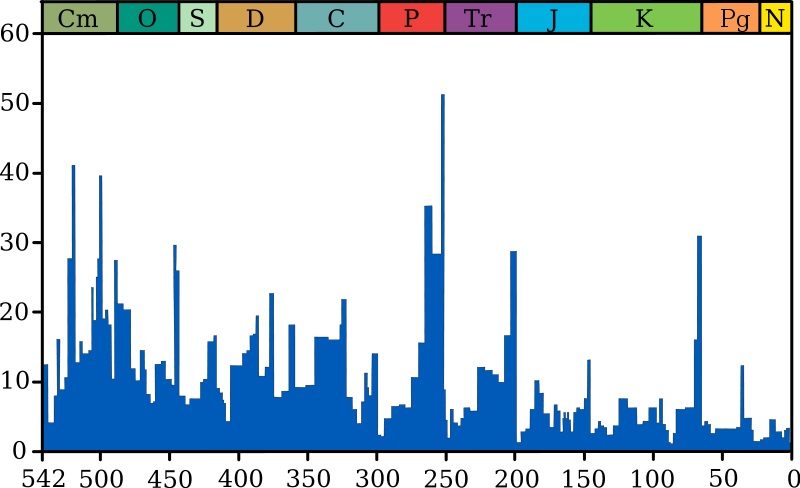
Extinction has many causes, some of which are caused directly by humans and others which are parts of natural cycles or apocalyptic events. An extinction event is when many species are driven to extinction by a particular species, natural disaster, or other phenomenon. While these mass extinctions sometimes wipe out a large majority of life, extinction itself is a continual part of evolution. Extinction happens on some scale all the time, as organisms adapt and outcompete others. It has been estimated that extinction has claimed at least 99 percent of all species that have ever lived. However, new species are also being generated through the process of speciation. As they spread, diversify, and recover the niches lost to extinction, the tree of life flourishes. However, it might flourish in a new direction.


Examples of Extinction


Thylacine


Welcome to Tasmania, mate! The year is 1800, and the island of Tasmania is overflowing with a variety of interesting marsupials. Among these is the thylacine, an apex predator resembling a mixture of a tiger and a wolf. Like other marsupials, the thylacine had an external pouch. Its newborn young, underdeveloped and tiny, would make their way to the pouch to continue developing in safety. Unfortunately for the thylacine, human expansion in Australia and Tasmania would lead to their extinction.


Bagged thylacine

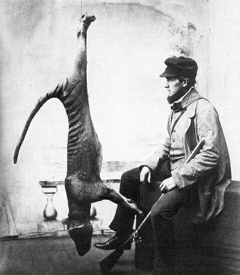
As seen above, the thylacine was often hunted. Thylacine were top predators, and the sheep and livestock of the new human population seemed no exception. As the human population spread on Tasmania, the competition grew fiercer, and bounties were put on the thylacine by the 1830s. Less than 100 years later, the thylacine would go extinct in the wild in 1930. While there were populations in zoos these too would die off by 1933. Thus, extinction for the thylacine was complete.


Passenger Pidgeon


Once a species that formed the vastest flocks known to man, the Passenger pigeon went extinct almost entirely at the hands of man. Before 1800, the Passenger pigeon enjoyed a range from New York to Denver, across most of the Continental United States. First described by Carl Linnaeus, the bird had been known to mankind for a long time. As the Europeans arrived in the New World, the saw the pigeon as useful and plentiful food source. At the time, hunting technology and population size would not allow for the mass harvesting of the birds, and they sustainably provided food.


Fast forward several hundred years, and man had multiplied across the North American continent. Where Native American populations were small, and more sustainable, the new colonizers needed vast resources to maintain their way of life. As such, the passenger pigeon started to see steady declines into the late 1800. By the end of the 1800s, there was a massive drop off. While bills were drafted and passed to protect the Passenger pigeon, it was too late. The biology of the Passenger pigeon made it an animal prone to gathering and flocking, driven by millions of years of evolving to escape solitary predators. This social feature of the bird which had protected it for so long, made it easy prey for human hunters. Extinction quickly ensued. By the early 1900s, the last Passenger pigeon had died in a zoo.


Megalodon


The largest known shark to ever live suffered extinction. Carcharocles megalodon, or simply Megalodon, has been identified from fossilized remains of its jaw and teeth. Possibly related to the Great White Shark, its teeth suggest it was much bigger. One of these teeth can be seen below, next to two Great White teeth.
Megalodon tooth with great white sharks teeth


Comparing measurements from these teeth and the jaw, scientists have estimated Megalodon to be somewhere around 60 feet long. The largest living shark currently, the whale shark, is only around 30 feet long, and even the Great White tops out at around 21 feet long. Scientific data suggests that extinction for Megalodon occurred around 2.6 million years ago. At this time, humans did not exist. It is suggested that extinction occurred because of a shift in the food supply for Megalodon as well as increased competition from other megapredators, such as early killer whales.


Interestingly, like other extinctions, there is always an air of doubt. Just because humans have not witnessed an animal thought to be extinct does not mean it is actually extinct. Extinction, in this regard, is simply a category used by the International Union for the Conservation of Nature (IUCN) and other agencies to categorize an animal thought to be extinct. For example, the black-footed ferret was thought to be extinct for several decades, until a population was found in Wyoming. Due to the unknown and vast nature of the ocean, even Megalodon is outliving its extinction. Often, claims of large sharks and attacks on boats are still attributed to Megalodon. However, no actual evidence has ever been found to refute that Megalodon suffered extinction.


Causes of Extinction


Ultimate Causes


Ultimately, every species has three “choices”. They can adapt to a situation, somehow evolving a novel or more efficient way to live. They can migrate, in the hopes that other areas will provide the resources they need with less competition. Or, as is the case for many animals, they can die. Extinction, as has been demonstrated in the fossil record, far surpasses survival for most species. While this may be seen as a negative thing, remember that extinction not only leaves new niches open to colonize, but can also be caused by a species becoming more successful. While one species may take over for a while, they usually undergo speciation into a variety of forms.


Proximate Causes


There are many more proximate causes of extinction. In mathematical terms, extinction happens any time the rate of reproduction is lower than the rate individuals are dying. This situation inevitably leads to extinction, but there are a number of factors which can drive these rates.


Predation, for example, is a major cause of extinction for many animals. Many species of fish in the Caribbean are currently threatened by the emergence of a new species, the Lionfish. Lionfish are not native to the Caribbean, and have no natural predators of their own. As such, they have pretty much free reign on the fish of the Caribbean. Many of these endemic species are being wiped out by the lionfish, and extinction is the likely result. In a similar story, extinction is plaguing many species of birds and lizards which have been exposed the brown tree snake. The snake, transported on cargo ships during WWII, has no natural predators on the islands to which is was transported. As such, the snake population has exploded and driven its prey items towards extinction, if not into it.


Other causes, which are directly the result of human action, involve habitat destruction and fragmentation. As we destroy the resources animals need to survive, we decrease the capacity an area can hold. As we further divide these areas with roads, fences, and other boundaries, we decrease the ability of species to migrate and successfully reproduce. This phenomena, as well as hunting and exploitation of animals for meat and game, has cause the extinction of a massive amount of animals. Scientists now speculate that, due to human interactions with the rest of nature, the world is entering another mass extinction event.


Quiz


1. How do we know an animal is really extinct?
A. We have no documented and confirmed sightings of the animal in recent times
B. We can never know
C. We find its fossils

Answer to Question #1
A is correct. While it may be believed by some that the thylacine and Megalodon are still out there, there would be so few individuals that they could not survive anyway. Due to the effects of genetic drift and bottlenecks in a small population, it is unlikely that a few individuals will survive an extinction. Remember that all animals leave fossil evidence, even animals which are still extant.

2. When considering extinct organisms which do not leave good fossils, how can scientists claim to pinpoint their extinctions?
A. Voodoo Magic
B. Only organisms with fossils can be determined
C. Chemical evidence points to many extinction events

Answer to Question #2
C is correct. While small microorganisms like bacteria and algae rarely leave reliable fossils, scientists have other ways of determining which organisms were most prominent. For example, by examining the composition of the air in modern times and comparing it to depositions in the soil, scientists can estimate the gas content of ancient times by looking at the composition of the soil and rocks.

3. Scientists want to revive the Woolly Mammoth. To do so, they supposed that they could use the DNA found in a frozen male mammoth to impregnate a female elephant. Would this “reverse” extinction?
A. Yes
B. No
C. Only if the baby comes out a Mammoth

Answer to Question #3
B is correct. First off, it is unlikely that the embryo would be viable, simply because the animals are separated by millions of years of evolution. Further, mixing two species is not actually recreating a mammoth, it is creating a hybrid. Lastly, a real way to bring the mammoth out of extinction would be to clone the mammoth DNA, and grow a new organism.

References


  • Feldhamer, G. A., Drickamer, L. C., Vessey, S. H., Merritt, J. F., & Krajewski, C. (2007). Mammology: Adaptation, Diversity, Ecology (3rd ed.). Baltimore: The Johns Hopkins University Press.

  • Pimiento, C., & Clements, C. (2014, October 22). When did Carcharocles megalodon become extinct? A new analysis of the fossil record. PLOS One. Retrieved from http://journals.plos.org/plosone/article?id=10.1371/journal.pone.0111086

  • Pough, F. H., Janis, C. M., & Heiser, J. B. (2009). Vertebrate Life. Boston: Pearson Benjamin Cummings.


Extinction

Extinction Event

An extinction event, or mass extinction, is a period of time in which a considerable portion of the world’s biodiversity is lost. An extinction event can have many causes, and can vary in intensity. There have been at least 5 major extinction events since the Cambrian explosion, each taking a large portion of the biodiversity with it. As seen in the graph below, these extinction events punctuate the fossil record. The following graph shows the intensity of extinction over time, which is a gradual and constant process. The spikes represent significant extinction events.
Extinction intensity


The highest bar represents the Permian-Triassic extinction event, which wiped out almost half the species on Earth in less than a million years. One ultimate reason that an extinction event may occur is the interdependent nature of food webs. If one species suffers and goes extinct, it often means changes for other species. As more and more species fall prey to the extinction event, the food web collapses and must be rebuilt from the bottom up. Oftentimes, a change in the Earth such as weather patterns will cause the extinction event. Other times, a species or group of species will change the environment and drive the extinction event.


Major Extinction Events


Ordovician-Silurian Extinction Event


One of the oldest mass extinctions, this extinction event occurred nearly 450 million years ago. At the time, many forms of multicellular life roamed the ocean. Just before this extinction event, many changes were happening. For instance, land plants had emerged, and were likely changing the composition of the atmosphere. In doing so, they shifted the balance from a carbon dioxide rich atmosphere to an oxygen rich atmosphere. Theoretically, this could have dramatically cooled the planet.


As much of the diversity of life was found within the oceans at this time, it suffered greatly as the planet cooled. As glaciers formed, sea levels fell. Many habitats on coastal areas were assumed to be destroyed as this happened. The change in atmosphere and global weather patterns ended up killing off up to 50 percent of the genera, and eliminating many marine species. Those species on land and in the sea which did not go extinct to glaciation expanded greatly once the glaciers melted and temperatures stabilized. It was after this mass extinction that major families of land plants and animals exploded.


Late Devonian Extinction Event


By the next great extinction event, the glaciers had melted and land was heavily colonized by plants and insects. These two groups had expanded rapidly in the newly available niche. The marine fauna had also rebounded, becoming greatly diversified and building huge coral reefs, which we can find evidence of today. The event may in fact be a series of events, so close in time that they are not well defined in the fossil record.


The causes of the Late Devonian event are not understood well, and many hypothesis abound. It is understood that marine, warm-water organisms and early jawed vertebrates were heavily affected. In fact, almost 97 percent of all vertebrate species disappeared. At least 75 percent of all species did not survive this era. One of the causes could have been an asteroid impact, which would change weather patterns and cause glaciation and sea levels to fall. Another theory presented involves the evolution of plants. It suggests that the new forms of plants, complete with roots and mechanisms to extract nutrients, had caused a massive influx of these nutrients into the ocean. As with fertilizer running into the ocean today, the increase in nutrients would cause massive growths of algae. As these blooms expanded, they would deplete the oxygen from large portions of ocean. Other fact supporting this is that many species of vertebrate got considerably smaller after the extinction event. This suggests that there was less oxygen and prey in the water. Other causes include volcanism, which may have added greenhouse gases to the atmosphere, changing its composition.


Permian-Triassic Extinction Event


The Permian-Triassic extinction event is the largest and most severe extinction event in the fossil record. The extinction event, also called the Great Dying, is supposed to have happened around 252 million years ago. Scientist have estimated that during this time 96 percent of all marine species went extinct. Further, terrestrial vertebrates, which had just expanded for the first time, lost nearly 70 percent of living species. Over 80 percent of all the known genera disappeared after this event. In today’s equivalent, it would be like wiping out all life on Earth, minus the insects and other invertebrates. That includes plants and fungi!


In the marine environment at one archeological site in China, for instance, nearly 87 percent of all the known invertebrate marine genera disappeared. It is though that ocean acidification, as a result of increasing carbon dioxide in the atmosphere, contributed greatly to this loss. On land, things were just as bad. At the end of the Permian, insects had grown and diversified on land. Some of the largest insects to walk or fly the Earth existed in this period. Nearly all of them would be extinct by the end of the extinction event. Plant communities, while they didn’t experience the same level of extinction, went through rapid periods of fluctuation. This likely cause the extinction of many of the terrestrial vertebrates alive at the time.


The causes of this extinction, like the ones before it, are heavily debated. While it has been shown that organisms sensitive to carbon dioxide were the most vulnerable, the source of that gas is debated. As with the extinction events before it asteroids, volcanoes, and greenhouse gases are the likely culprits. Several possible impact sites of asteroids have been identified, but it is hard to date them. Further, it is more likely that an asteroid would have hit the ocean, and evidence of it would be completely gone by now. Evidence of large-scale volcanic eruptions has been found. While it is clear that there were major chemical and physical changes happening the world at a time, it is difficult to pinpoint the exact cause of the extinction event.


Triassic-Jurassic Extinction Event


This mass extinction event, while much small than the one preceding it, allowed for many niches to be cleared for the rise of the dinosaurs. This extinction event took around somewhere around 30 percent of marine species. Interestingly, this extinction event coincides with the breakup of Pangea, a supercontinent that had formed as the continents drifted together. As the continent broke apart, there were massive changes to the flora and fauna.


Unlike the other extinction events, one cause of this extinction event might have been a decline in speciation as opposed to an increase in extinction. Theoretically, there is a level of background extinction, which is always taking place. If speciation slows, because organisms can’t adapt or all the niches are full, extinction wins out. While many species were lost over this time, the causes aren’t clear. Again, asteroids and climate change are presumed to be the culprits.


Cretaceous-Paleogene Extinction Event


Probably the most well-known extinction event, the Cretaceous-Paleogene is the one which wiped out the dinosaurs and cleared the way for mammals and humans. Unlike other mass extinction events, this extinction event happened relatively recently, only 66 million years ago. Also unlike the other extinction events, scientists have a fairly good idea of what caused the massive extinction.


An asteroid crater in the Gulf of Mexico was found that dated to the time of the extinction. At over 100 miles wide, the asteroid would have been able to completely shift the global atmosphere. One of the largest extinction events known, the impact is probably responsible for the die-off of around 75 percent of all living species. The main effect of the asteroid was to produce an impact winter. Dust and debris from the impact would float in the atmosphere for years, blocking the sun. As photosynthetic organisms died off, so too would the herbivores that feed on them and the carnivores which feed on them. As such, entire food webs in both the terrestrial and marine environments were lost.


Holocene Extinction Event


The most recent extinction event is the one we are currently living through. Since our recorded history, we have witnessed, if not caused, the extinction of many species. Changes in climate, whether man-made or natural, are driving the same conditions of greenhouse gas build up and ocean acidification which have driven other extinction events in the past. According to data from organizations which monitor threatened and endangered species, we are on the precipice of another extinction event. While some dismiss this theory, we should be careful. If the food webs we are dependent on collapse, we will not be able to survive.


Quiz


1. What is the difference between extinction and an extinction event?
A. Nothing
B. Extinction happens to a single species, an event happens to many
C. An extinction event is when an extinction happens

Answer to Question #1
B is correct. Extinction is a continual process. There is a certain level of extinction which is always happening. An extinction event occurs when a large global change causes a drastic increase in the level of extinction across many different species.

2. How can scientists determine we are entering an extinction event?
A. By comparing recent trends to historical records
B. By monitoring the extinctions we see today
C. All of the above

Answer to Question #2
C is correct. Based on the number of species going extinct, compared to the number we are away of, it seems that the extinction rate is much higher than normal. When we start to look at the types of species going extinct today, the link between historical causes and current human activity become clearer.

3. Your friend argues that a mass extinction would not affect humans, because we are so technologically advanced. Is he right?
A. Yes
B. No
C. It depends

Answer to Question #3
C is correct. It truly depends on the scale of the extinction and the extinction event itself. Already, humans have replace the majority of vertebrate life with themselves and their agricultural animals. If gas emissions and the greenhouse effect eventually lead to another ice age or global temperature spike, we are in trouble. If plants can’t survive, we can’t survive.

References


  • Feldhamer, G. A., Drickamer, L. C., Vessey, S. H., Merritt, J. F., & Krajewski, C. (2007). Mammology: Adaptation, Diversity, Ecology (3rd ed.). Baltimore: The Johns Hopkins University Press.

  • Pimiento, C., & Clements, C. (2014, October 22). When did Carcharocles megalodon become extinct? A new analysis of the fossil record. PLOS One. Retrieved from http://journals.plos.org/plosone/article?id=10.1371/journal.pone.0111086

  • Pough, F. H., Janis, C. M., & Heiser, J. B. (2009). Vertebrate Life. Boston: Pearson Benjamin Cummings.


Extinction Event

Stabilizing Selection

Stabilizing Selection Definition


Stabilizing selection is any selective force or forces which push a population toward the average, or median trait. Stabilizing selection is a descriptive term for what happens to an individual trait when the extremes of the trait are selected against. This increases the frequency of the trait in the population, and the alleles and genes which help form it. Many traits which are common across entire groups of species have been formed through the effects of stabilizing selection. Stabilizing selection can be seen in the image below, comparing the three types of selection.


Stabilizing Selection vs Directional Selection vs Diversifying Selection


Stabilizing Selection Examples


Robin eggs


In this case, the number of eggs in a robin nest has been selected for through a stabilizing selection. Robins are apparently not ably to raise more than 4 chicks with much success. This is probably because of the size of the birds and the amount of food that two adults can provide. By comparison, most penguins can only raise one chick at a time, due to the size of the chick and the amount of food it requires. While they stabilized on different numbers, both are forms of stabilizing selection which maximize the fitness of the species in their environment.


As opposed to the other forms of selection, you can clearly see in the stabilizing selection graph that the population of the median trait increases, while the other populations decrease. In this case, 5 eggs is too many and some would die. On the other hand, 3 is too few. Either the eggs are not viable enough to only rely on 3 eggs, or predation and other forces require more than 3 eggs to overcome them and carry on to another generation.


Hypothetical Lemurs


There is a population of multicolored lemurs on Crazy Island. This particular population of lemurs has been observed by scientists, and they have noticed the following changes in the lemur’s color.


Stabilizing Selection


As you can see, the lemurs have obviously undergone a stabilizing selection. The light and dark lemurs have almost disappeared, while the middle brown lemurs have increased. Without further information, it is not clear why this would be the case. It is the job of ecologists and evolutionary biologists to observe the population, noting peculiar aspects of the various forms to understand what may have caused the stabilizing selection. This is no easy question to answer and may have many more than one answer.


In the case if the lemurs, it could be that the darker and lighter lemurs were both easier to spot by predators. If the lemurs only have one predator, this is as easy hypothesis to test. A scientist would simply observe the predator, and see which lemurs it prefers. This would lend evidence to the hypothesis that the stabilizing selection is caused by predation. Further evidence could include the amount of lemurs the predators eat, and models showing how that level of predation could produce the coloration seen.


However, it is much more common that a species has multiple selective pressures, and that each pressure acts on various traits in different ways. For example, the lighter color could be suffering from predation, whereas the darker version could be overheating. (Dark colors absorb more solar heat.) Likewise, predation could be driving both traits, but not influencing them totally. Female lemurs might have a preference for brown lemurs, due to their increased survival. This would be a form of sexual selection, driving a trend of stabilizing selection.


Common Causes of Stabilizing Selection


Stabilizing selection, along with directional selection and disruptive selection, refer to the direction of individual traits. While stabilizing selection pushed the trait towards the average instead of one or both of the extremes, it can be driven by any form of selection. Some of the most common forms of selection are from predation, resource allocation, coloration of the environment, food type, and a wide variety of other forces.


Many traits we don’t talk about regularly have been driven by a variety of causes throughout history. Take the modest insect, for example. All insects have an exoskeleton, a miraculous structure made of chitin and other structural molecules which form a shield around their organs and allow them to maintain a water balance in the harshest of environments. This shield, while it has been modified in to an almost infinite number of forms, was first selected for out of stabilizing selection. The ancestors to insects did not have this adaptation, but once it evolved it was highly favored.


Simply stated, there is no common cause of stabilizing selection, besides the fact that the most average individual is selected for. In that way, like all forms of selection, the cause of stabilizing selection is the increased fitness and reproductive success that the median individuals have. The extreme versions or traits have a disadvantage, in one way or another. This disadvantage, in evolutionary terms, is decreased reproduction. The traits they carry are coded for in part by their DNA, which they can only pass on through reproduction. In stabilizing selection, the increase in the median traits represents their increased success. The other extreme traits are not as successful, possibly causing their owners to die. This increases the resources available to the median animals, further boosting their success. In this way, stabilizing selection is the cause of many traits that entire groups of animals share. These are known as synapomophies.


Quiz


1. Which of the following is NOT stabilizing selection?
A. A population of foxes shifts from mostly red to mostly grey
B. The most common color of rabbit increases after new predators are introduced
C. A population of purple sea urchins stays purple, as starfish eat other colors

Answer to Question #1
A is correct. In this case, the foxes are changing in majority from red to grey. This could indicate a number of driving factors, but is directional selection, not stabilizing selection. The other two cases represent situations in which the majority was selected for, and increased in frequency, due to the forces of predation.

2. Which of the following was not caused by forces driving stabilizing selection?
A. A species of moth, divided by selection, becomes two species
B. A species of rhino has 2 horns, instead of any other number
C. Most vertebrates with limbs have 4 or 5 toes

Answer to Question #2
A is correct. Again, anything which selects against the majority is not from stabilizing selection. Here, a group of moths becomes divided, a form of disruptive selection. In the other two answers, the number has been determined by stabilizing forces on both sides of the trait. Notice that questions one and two are very similar, but are asked in different ways. Don’t get tricked up by complex wording!

3. A trait has been selected for by stabilizing selection, to the extreme. There are no other forms left in the environment. How can variety be reintroduced into the population?
A. Genetic variation and recombination
B. Mutations
C. Both

Answer to Question #3
C is correct. Although there may be only one trait present within the population, remember that this is the phenotype. Many creatures are diploid or more, and carry multiple copies of the DNA. Some of the alleles present in the DNA are recessive and will not show up until it is the only allele present in an organism. When DNA is copied and divided during sexual reproduction, these genes get mixed up and the recessive alleles can come to the surface. If no more recessive alleles exist, mutations caused by toxins, sunlight, and various chemicals can induce a new allele to be present within a population. It may react differently to the selective pressures and change the direction of selection.

References


  • Brusca, R. C., & Brusca, G. J. (2003). Invertebrates. Sunderland, MA: Sinauer Associates, Inc.

  • Feldhamer, G. A., Drickamer, L. C., Vessey, S. H., Merritt, J. F., & Krajewski, C. (2007). Mammology: Adaptation, Diversity, Ecology (3rd ed.). Baltimore: The Johns Hopkins University Press.

  • Kaiser, M. J., Attrill, M. J., Jennings, S., Thomas, D. N., Barnes, D. K., Brierley, A. S., & Hiddink, J. G. (2011). Marine Ecology: Processes, Systems, and Impacts. New York: Oxford University Press.


Stabilizing Selection

Silent Mutation

Silent Mutation Definition


A silent mutation is a change in the sequence of nucleotide bases which constitutes DNA, without a subsequent change in the amino acid or the function of the overall protein. Sometimes a single amino acid will change, but if it has the same properties as the amino acid it replaced, little to no change will happen. A silent mutation can be caused many ways, but the key point is that it does not change the function of the amino acid or subsequent proteins. A silent mutation is just that: it does nothing significant, not making a sound in the orchestra of the cell.


Silent Mutation Examples


The Redundant Genome


The DNA is read in units of three nucleotides, called codons. Each codon specifies a certain amino acid, with a few reserved as stop and start signals. Sometimes, different codons specify the same amino acid. This redundancy allows a flexibility in the genetic code. This means that a silent mutation usually goes completely unnoticed. You can see a typical silent mutation in the graph below.


Different Types of Mutations


Here, a silent mutation is compared with both a nonsense mutation and a missense mutation. The silent mutation, which is an actual change at the DNA level from a thymine to a cytosine. This mutation could have been caused by a mistake in DNA replication, or from some sort of repair that happen after the DNA was damaged. Regardless, both of these three nucleotide codons tell the ribosome and machinery within to attach a lysine amino acid.


In this case, the entire structure of the protein will remain the same regardless of the silent mutation. With the exact same amino acid structure, the protein will function no differently unless it is placed in a different environment. A silent mutation can also happen at the protein level, with no functional effect on the protein.


Amino Acid Groups


The 4 nucleotides, in groups of three codons, can call for all 21 amino acids. Seen below, the amino acids are grouped by their structure and side chains. These features directly impact how they interact with other amino acids, and what effects they have on molecules in the environment.


Amino Acids


A silent mutation, which could easily include more than one nucleotide, could easily change an entire amino acid, or even series of amino acids. If a serine changed into a threonine, the effect might be minimal. The two amino acids are in the same category and are very similar shapes. This means that they will have a similar chemical reaction on the molecules around them. This will influence the shape and effect of the total protein. If the effect is negligible, the change is considered a silent mutation.


Place within Protein Structure


Several amino acids can be key to the entire structure or functionality of a protein. Many proteins have an active site, to which other molecules must bind. This site is constructed from a specific sequence of amino acids. When folded just right, certain amino acids and their side chains will have the exclusive ability to interact with another molecule. If these amino acids are changed by a mutation, the functionality of bonding may be seriously impaired. This can change the function or utility of a protein.


Other proteins, on the inside of the molecule, have complex structures which must be present to preform specific functions. Many proteins undergo a conformational change, which is a change in shape. This is driven by electrical stimulation or the binding of a molecule like a coenzyme or a substrate to the protein. The conformational change, literally changing the shape of the protein, can press molecules together or tear them apart. The energy supplied is dependent upon the internal structure and specific bonds within the protein. Sometimes, a single amino acid can be a key piece of this. If this amino acid is changed for a non-functional one, the mutation is not a silent mutation. The change on the inside of the protein can also affect the functionality of the protein. Regardless of what a silent mutation changes, it should not change the functionality of the resulting protein.


Within Non-coding DNA


Many portions of the DNA are used structurally, and their full purpose is not understood. There are many cases in which parts of the DNA are vastly different between individuals, yet their phenotypes seem the same. These changes, especially small structural changes in the DNA, do not become significant until they begin to change the interaction of the coding DNA with the environment. A silent mutation could easily happen in these areas without notice, yet over time many mutations may begin to change a population.


Bacteria, interestingly, usually have a single circle of DNA, which carries all the information they need. By contrast, the human genome is separated on multiple chromosomes, which are bundled and managed by specialized proteins so they can be wound up during cell division. One hypothesis as to how this much more complex DNA came about was that certain silent mutations began forming structures of DNA. In a more compact genome, more information can be stored, which may have led to the complexity of life from single celled organisms to more complex forms. The folding and protection of various parts of DNA is part of normal cell differentiation in eukaryotes. Supposedly, these could have arisen through silent mutation until they became useful and were selected for.


Quiz


1. One of the following examples includes a silent mutation. Which one?
A. A sunbather forgets to put sunscreen on. The DNA within many skin cells is damaged, and several mutations happen when it is repaired.
B. In the third spot of the codon, a C mutates to a G. Both codons call for threonine amino acid
C. Due to a mutation, a changed amino acid within an enzyme cause it to slow down.

Answer to Question #1
B is correct. If both of the codons call for the same amino acid, then there will be no noticeable change. The protein will function as normal. In answer A, we have no idea what the effect of the mutations will be, so we cannot say they are silent. In all likelihood, they may eventually lead to cancer. Answer C is the example of a regular mutation, which changes the function of a protein. This could be a good or bad trait, depending on the environment, as slower proteins might be preferred.

2. What is the difference between a nonsense mutation and a silent mutation?
A. Nothing, they both change protein function
B. A nonsense mutation does not create a change in the protein
C. A silent mutation does nothing, while a nonsense mutation can be significantly disruptive

Answer to Question #2
C is correct. These two terms are essentially the opposite. A silent mutation is unnoticeable, whereas a nonsense mutation will produce a protein which is much different than the original. This is because a nonsense mutation introduces a codon which is completely different than the one before, possibly stopping the protein synthesis short. This produces a drastic effect on protein function.

3. On the inside of a protein, a mutation causes the swap of one amino acid with another. This change does not affect the internal structure of the protein, and both proteins act similarly in their roles. Which of the following represents this situation?
A. Silent mutation
B. Point mutation
C. Missense mutation

Answer to Question #3
A is correct. In this case, nothing happened noticeably to the protein. In a missense mutation, a different amino acid causes slightly different function, even though it may be conservative and similar to the original. The different is that the change is noticeable. While this may be a point mutation, or a change of one nucleotide, a point mutation can be the source of any mutation.

References


  • Hartwell, L. H., Hood, L., Goldberg, M. L., Reynolds, A. E., & Silver, L. M. (2011). Genetics: From Genes to Genomes. Boston: McGraw Hill.

  • Lodish, H., Berk, A., Kaiser, C. A., Krieger, M., Scott, M. P., Bretscher, A., . . . Matsudaira, P. (2008). Molecular Cell Biology (6th ed.). New York: W.H. Freeman and Company.

  • Nelson, D. L., & Cox, M. M. (2008). Principles of Biochemistry. New York: W.H. Freeman and Company.


Silent Mutation

Channel Protein

Channel Protein Definition


A channel protein is a special arrangement of amino acids which embeds in the cell membrane, providing a hydrophilic passageway for water and small, polar ions. Like all transport proteins, each channel protein has a size and shape which excludes all but the most specific molecules. A generic channel protein is seen below, embedded within the membrane. Ions, the small green hexagons, travel through the channel protein. They move from an area of high concentration to an area with a lower concentration.


https://upload.wikimedia.org/wikipedia/commons/f/f4/0306_Facilitated_Diffusion_Channel_Protein_labeled.jpg


Types of Channel Protein


Non-gated


Like the image above, a channel protein may exist in a state which stays open all the time. This is called a non-gated channel protein. These proteins allow ions and water to flow through the cell membrane, which is normally hydrophobic and would resist the passage of these molecules. A non-gated channel protein is needed whenever the balance of water and ions must be assisted by the constant passage of water and ions into or out of the cell. However, it is often a disadvantage to leave a channel open all the time. The second type of channel protein addresses this problem.


Gated


A gated channel protein remains closed, until it receives a special chemical or electrical signal. These channel proteins are extremely important in many cellular functions. The ability to gate an ion channel allows electrical energy to be built up inside the cell. Nerve function is entirely based on this fact. Channel proteins on the surface of nerve cells react to electrical signals created by the flooding of ions through the membrane next to them. As they open, ions spill through and continue the electrical disturbance. This passes a signal very quickly through the body. A gated channel protein reacting to a signal molecule can be seen in the image below.


https://upload.wikimedia.org/wikipedia/commons/c/c7/Figure_09_01_04.jpg


Channel Protein Function


Depending on whether it is gated or non-gated, a channel protein has a slightly different function. A non-gated channel protein simple allows ions and water to flow freely from one side of a membrane to another. While these type of channels are not often found on the external cell membrane, they are more often found within organelles and places where ion gradients are not maintained.


When an ion gradient needs to be maintained, gated channel proteins serve the role of holding back the tide of ions until they are signaled to open. A closed channel acts as corked bottle. Water and ions move slowly through the plasma membrane, or not at all. If the channel protein is closed, they have little chance of obtaining an equilibrium. Cells use these proteins in many ways, everything from balancing their water content to actively building up charges.


Channel Protein Structure


Most channel proteins are made of several identical protein subunits which form a hydrophilic region in their center. Gated channels function by changing conformation upon receiving a signal, allowing access to the hydrophilic passageway. Non-gated channels are usually formed from identical subunits, which attach to each other in a circle. While the inside of the circle is hydrophilic, the amino acids exposed within the hydrophobic cell membrane are also non-polar. This helps to anchor the protein within the membrane. If the protein tried to slip out of the membrane, it would be pushed by polar forces back into place.


Channel Protein Example


When your muscles contract, this is the result of the action of gated channel proteins within your muscle cells. These cells respond to the neurotransmitter acetylcholine, which is present in high amount as the end of nerve cells. At the synapse or space where they release the neurotransmitter, the opposing nerve cell contains many channel proteins set to receive the signal. An electrical signal coming down the nerve (also driven by a type of channel protein) causes the acetylcholine to be released.


The neurotransmitter diffuses quickly across the synapse, and reaches channel proteins on the other side. Each channel protein opens, releasing sodium and potassium ions. The electrical disturbance travels down special channels within muscles, carrying the signal to each muscle cell. Here, another set of channel proteins is activated. These release sodium, causing the proteins actin and myosin to start their crawling motion against each other, contracting each cell. The full result is a full muscle contraction, moving a limb or operating a part of the body.


Channel Proteins and Carrier Proteins


There are four types of transport that occur within cells. Simple diffusion occurs with small gas molecules, such as oxygen and carbon dioxide, as well as many non-polar chemicals such as steroid hormones and medicinal drugs. These molecules have the right chemistry and size to pass right through the cell membrane.


More charged molecules, which are hydrophilic, have a hard time passing through the membrane. These include ions, water, and sugars such as glucose. Channel proteins carry out the majority of facilitated diffusion. While the chemicals are still moving in the direction of their concentration (from high to low), they are given a passageway through the cell membrane. This allows them to move at near diffusion speeds.


However, not all facilitated diffusion is carried out by channel proteins. Carrier proteins, proteins which bind and transport molecules across the membrane, are also involved in facilitated diffusion. Large molecules like glucose cannot pass through the narrow passageway created by channel proteins. Carrier proteins known as uniporters bind to glucose molecules one at a time. The binding action causes a conformational change in the protein, which causes it to deposit the molecule on the opposite side of the cell. These carrier proteins operate without energy, and move molecules down their concentration gradient.


When substances need to be moved against their concentration gradient, more complicated carrier proteins are needed. Active transport is the process of using a carrier protein and powering it with an interaction with ATP to move a molecule against the gradient. The energy is needed because molecules naturally want to diffuse, and spread out. It takes a lot of energy to move some ions and molecules, but is necessary for the way life has evolved. Other carrier proteins have evolved for cotransport. By transporting a molecule down its concentration gradient, another molecule can be moved against its gradient. This carrier protein type allows cells to transport materials using the ion gradient they build with other ATP carrier proteins.


The major difference between a channel protein and a carrier protein is stereospecificity. While channel proteins only allow certain sized molecules to pass, they do not bind the molecules. Carrier proteins have an active site, which the chemical to be transported must bind to. This site will bind specifically to only one molecule, and seeks to transport this molecule alone. The binding action is what allows the passage of the large molecule through the cell membrane.


Quiz


1. What is the difference between a channel protein and a carrier protein?
A. They move different types of molecules
B. A channel protein does not need energy
C. A channel protein does not bind the molecules it transports

Answer to Question #1
C is correct. Channel proteins are simply that: channels. Like a straw, or the drain on a tub, they simply allow water and ions to pass through them. While they can be gated or non-gated, they do not need energy to operate, but neither do uniporters nor other carrier proteins. Channel proteins and carrier proteins can move the same types of molecules.

2. A mutation in a person causes their ion channels to malfunction. Will this be a problem?
A. Yes
B. No
C. They can treat it

Answer to Question #2
A is correct. This is the cause of a terrible disease known as Cystic Fibrosis. Sufferers of this disease cannot regulate ions in their mucous membranes, causing suffocating phlegm buildups and organ failures at a young age. Channel proteins are very important for many other functions, and most of them are required to work for normal bodily functions.

3. In an experiment, a scientist separates two bodies of water with a thin phospholipid membrane, such as that found in a cell. He pours salt in one of the bodies of water. The membrane has channel proteins embedded. His control experiment is two bodies of water separated by the same membrane, but without the channel proteins. He adds salt to this control as well. Which of the following would you expect to happen?
A. In both experiments, the salt will quickly come to equilibrium between the bodies
B. In the control, equilibrium will come more slowly than the experimental membrane
C. The control will come to equilibrium faster

Answer to Question #3
B is correct. The control will come to equilibrium more slowly because it lacks a channel protein. These proteins will allow the ions in salt to distribute right through the membrane along with water. These two substances will work their way back and forth across the membrane until they are equal. Without the proteins, the membrane holds back water and ions, and the process will happen much more slowly, if it happens at all.

References


  • Lodish, H., Berk, A., Kaiser, C. A., Krieger, M., Scott, M. P., Bretscher, A., . . . Matsudaira, P. (2008). Molecular Cell Biology (6th ed.). New York: W.H. Freeman and Company.

  • Nelson, D. L., & Cox, M. M. (2008). Principles of Biochemistry. New York: W.H. Freeman and Company.

  • Widmaier, E. P., Raff, H., & Strang, K. T. (2008). Vander’s Human Physiology: The Mechanisms of Body Function (11th ed.). Boston: McGraw-Hill Higher Education.


Channel Protein

Cell Differentiation

Cell Differentiation Definition


Cellular differentiation, or simply cell differentiation, is the process through which a cell undergoes changes in gene expression to become a more specific type of cell. The process of cell differentiation allows multi-cellular organisms to create uniquely functional cell types and body plans. The process of cell differentiation is driven by genetics, and their interaction with the environment.


All organisms begin from a single cell. This single cell carries the DNA coding for all the proteins the adult organism will use. However, if this cell expressed all of these proteins at once it would not be functional. This cell must divide repeatedly, and the cells must begin the process of cell differentiation as they divide. The cell lines begin to emerge, and the cells get more and more specific. Eventually, an entire organism is formed with hundreds of different cell types from this process of cell differentiation.


The original mass of cells, which have not undergone differentiation, are known as stem cells. Unlike normal cell division, which creates two identical daughter cells, the division of stem cells is asymmetric cell division. In this case, one of the cells remains identical to the parent stem cell. In the other cell, chemical triggers activate the process of cell differentiation, and the cell will start to express the DNA of a specific cell type. Stem cells which can differentiate into entire organisms are known as embryonic stem cells and are said to be totipotent.


By contrast, the body also has many cells which are only pluripotent. These cells have already undergone some cell differentiation. These stem cells can only divide into a narrow range of cell types. Bone marrow, for instance, contains somatic stem cells which can only become red blood cells. These cells are necessary for the constant replenishment of blood cells, which are mostly inactive besides their oxygen-carrying ability.


Cell Differentiation Examples


In Animals


After the process of fertilization in animals, a single-celled organism called the zygote is formed. The zygote is totipotent, and will eventually become an entire organism. Even the largest animal on Earth, the blue whale, starts as a single cell. The complex tissues and organ systems, which are completely different in their form and function, all come from the zygote. The process of cell differentiation starts early within the organism. By the time the gastrula has formed, the cells have already started expressing various portions of the DNA.


These changes drive the first folding processes within the embryo. As the tissues continue to form, some cells begin releasing hormones, or chemical triggers which signal various cells to react. Hormone signals direct the expression of DNA in various body parts, which drives their cell differentiation further. In humans it only takes a little over a month for a rudimentary heart and circulatory system to form.


As the systems continue to form, many of the stem cells lose their totipotency, themselves undergoing cell differentiation. This allows for faster production of specialized cells, which the growing organism needs to sustain its growth and enter the world with success. Through cell differentiation, tissues as different as brain tissue and muscle are formed from the same single cell.


In Plants


While the plant lifecycle sometimes seems alien and complex, the process of cell differentiation is very similar. While there are different hormones involved, all plants also develop from a single cell. A seed is simply a protective housing for the zygote, which also provides a food supply. It is very similar to an egg in the animal world. The zygote inside undergoes cell division, and becomes a small embryo. Development is halted, as the seed is distributed into the world.


After winter, or anytime the environment is prime, the seed will soak up moisture and restart the process of development. The embryo will begin to form two meristems. A meristem is a unique portion of stem cells, which undergo cell differentiation as they grow outward. One will grow towards the surface, while the other will become the roots.


In the roots a layer of cells forms around the meristem, forming the root cap. This layer of cells sloughs off as the roots move through the soil, and are consistently replaced by the meristem. On the inside of the meristem, cell differentiation happens in a different direction. The hormones and environment here directs the cells to become vascular tissue and supporting cells. These will eventually carry water and nutrients to the top of the plant.


On the surface, the meristem acts in a similar way. As it divides upward, it creates both inward and outward cells. The inward cells undergo a differentiation similar to that of the roots, creating more vascular tissue. On the outside, the cells undergo cell differentiation into stems and leaves. These are equivalent to the different organs of animals, and are as different from the starting cells as animal cells. If you aren’t convinced, pick up an acorn and compare it to the massive tree it will become. Not only is it vastly smaller, it also contains completely different cell types. This can accounted for through the process of cell differentiation.


Cell Differentiation Process


One of the keys to the cell differentiation process is transcription factors. These hormones and chemicals direct the activities surrounding DNA, determining what gets transcribed and what is ignored. The factors present in cells from birth to death are determined by the body, and other cells in the vicinity.


For instance, the pancreas or thyroid may release a hormone calling for cellular growth. This transcription factor directly impacts the proteins which transcribe DNA, turning it eventually into functional proteins and more cells. However, when cells start to squeeze together, they will also signal to each other that there is no more room. Thus, the process of cell differentiation has a multitude of inputs and possible outcomes.


This complex process is still being studied. Scientist have made considerable advances in understanding cell differentiation, starting with the complete understanding of the nematode C. elegans. This tiny, worm-like creature has a total of 959 cells as an adult female. With such a small number, they are relatively easy to track from the zygote to the adult. Tracing their cell lineage, scientists have started to determine some of the complex and epigenetic forces working on cell differentiation. In other words, it matters not only what DNA a cell has, but where and how that DNA is expressed.


Quiz


1. Why is cell differentiation an important process?
A. It allows for multi-cellular life forms
B. It creates new species
C. We could do without it

Answer to Question #1
A is correct. Without the process of cell differentiation, multicellular organisms would not be possible. Some algae live in colonies, but this is nowhere near the level of complexity developed by insects or vertebrates. Cell differentiation allows the creation of tissues and organs, which can serve specific and useful functions for an organism.

2. What is the difference between cell differentiation and development?
A. Development does not include differentiation
B. Cell differentiation is part of development
C. There is no difference

Answer to Question #2
B is correct. Development is the entire process of creating a new organism from a single cell. It includes everything from forming the correct tissues to creating the neural connections to support a new body. Cell differentiation is simply the process through which cells begin to express only certain portions of DNA, thereby becoming specialized cell types.

3. If each stem cell divides into more specialized cells, where do you get more stem cells from?
A. You don’t
B. Stem cells divide asymmetrically
C. The zygote creates them

Answer to Question #3
B is correct. When most stem cells divide, one of them retains the original character of being a stem cell. The zygote divides without cell differentiation 3 times, creating 8 identical totipotent cells. These cells will continue to divide asymmetrically, and will eventually give rise to three general tissues, the ectoderm, mesoderm, and endoderm. These tissues will undergo further cell division to become specific tissues.

References


  • Lodish, H., Berk, A., Kaiser, C. A., Krieger, M., Scott, M. P., Bretscher, A., . . . Matsudaira, P. (2008). Molecular Cell Biology (6th ed.). New York: W.H. Freeman and Company.

  • McMahon, M. J., Kofranek, A. M., & Rubatzky, V. E. (2011). Plant Science: Growth, Development, and Utilization of Cultivated Plants (5th ed.). Boston: Prentince Hall.

  • National Institutes of Health. (2018, March 11). Stem Cell Basics III. Retrieved from Stemcells.nih.gov: https://stemcells.nih.gov/info/basics/3.htm


Cell Differentiation

Substitution Mutation

Substitution Mutation Definition


A substitution mutation is a type of replication error during DNA replication which places the wrong nucleotide or sequence of nucleotides in the wrong position. A type of substitution mutation, a point mutation, occurs which a single nucleotide is substituted. This can be seen in the image below.


https://upload.wikimedia.org/wikipedia/commons/3/37/Single_nucleotide_polymorphism_substitution_mutation_diagram_-_cytosine_to_thymine.png


Importantly, a substitution mutation results in DNA of the same length. It does not add or subtract from the number of nucleotides in the sequence. A single nucleotide substitution mutation is the most common, as most large-scale nucleotide swaps involve other mechanisms. For example, a reciprocal translocation involves the movement of entire portions of chromosomes, and swaps one portion for a portion of another chromosome.


As with all mutations, a substitution mutation can drastically change the proteins created by an organism. The proteins responsible for reading DNA process the molecule in units of three base pairs at a time. These codons each specify a different amino acid. If the sequence change even by one nucleotide, a different amino acid is placed within the protein. The function of each protein is dependent on the specific interaction between the amino acids they consist of. A substitution mutation can displace many more than one nucleotide. In this case, it may make the protein completely dysfunctional, or give it an entirely new function. New adaptations can arise this way, if they are transferred to the offspring and are beneficial. However, the large majority of mutations are deleterious, or cause negative effects.


What causes a Substitution Mutation?


A substitution mutation can be caused by a number of sources directly related to the reading and storage of DNA. For instance, every hour each cell in your body losses around 1,000 nucleotides from the DNA backbone. These nucleotides fall off due to the process of depurination. In the process of replacing them, the proteins that manage the DNA make a mistake approximately 75% of the time, because there are 4 nucleotides to choose from. Other proteins must come along after and check the DNA for errors. If they miss the substitution mutation, it may stay and be replicated.


Another factor which can drive a substitution mutation is deamination, the process by which amino groups degrade off of nucleotides. One of the only ways the protein machinery can differentiate between nucleotides is the amino groups attached to them. As these fall off, the protein machinery can misrecognize the nucleotide, and supply the wrong nucleotide pair. When the DNA replicates, the new nucleotide will become established in a new cell line.


On top of these internal drivers which can cause a substitution mutation, there are also external forces which can cause nucleotide swaps. Carcinogens and mutagens are a special classes of chemicals which drastically impede the protein machinery and cause lots of mutations. Even sunlight can degrade and impede with DNA function, driving a substitution mutation.


Substitution Mutation Examples


Sickle-Cell Anemia


The blood disease Sickle-cell anemia is caused by a simple substitution mutation. In the mutation, a single nucleotide is replaced in the portion of DNA which codes for a unit of hemoglobin. Hemoglobin is a multi-protein complex, responsible for carrying oxygen and supporting the shape of blood cells. The substitution mutation causes a glutamic acid in the protein to be changed to a valine amino acid.


While this might not seem like much of a change in a protein which contain over 140 amino acids, it makes all the difference. Valine, unlike glutamic acid, is hydrophobic. As such, it repels polar interactions where glutamic acid would attract them. This severely impacts the protein’s ability to function. Blood cells immediately reflect this change, becoming shriveled and sickle-shaped. With a lower ability to carry oxygen, these cells also are more prone to clot within the small capillaries of organs. This can lead to an increased risk of heart attack, stroke, and other cardiovascular diseases.


Interestingly, the substitution mutation has survived in the population for a surprising reason. The parasite which causes malaria depends on human blood cells for part of its life cycle. People with the sickle-cell substitution mutation are less susceptible to getting malaria. Apparently the different shape and function of the blood cells impedes their reproductive processes.


Color Blindness


In your eye, certain cells are responsible for picking up the colors red, green, and blue. These cells rely on different proteins, which react to the various colors. A substitution mutation in the DNA that codes for one of these proteins can lead to the condition of color blindness. People with this condition have a hard time distinguishing between the colors, while their vision is still clear otherwise. Oftentimes, only one color is knocked out. The various proteins are coded for on different places on the DNA, which makes a substitution unlikely to occur in all three genes.


Types of Substitution Mutations


Transition


There are two basic types which a substitution mutation can be. Within the four nucleotides, there are two types: the purines and pyrimidines. Adenine (A) and guanine (G) are both purines, while cytosine (C) and thymine (T) are pyrimidines. If a purine changes to a purine, the substitution mutation is considered a transition. Likewise, if a pyrimidine changes into a pyrimidine, the substitution mutation is also a transition. In the image below, transitions are labeled by the alpha lines.


https://upload.wikimedia.org/wikipedia/commons/b/b4/TsTvMutation.jpg


Transversion


The opposite of transition is transversion. In a substitution mutation involving a transversion, a purine is substituted for a pyrimidine, or vis versa. In the above image, a transversion is labeled by the beta lines. Transversions are much less likely than transitions. This is probably due to the fact that the machinery used to repair and proof-read the DNA are more specific for purine versus pyrimidine than specifying between individual nucleotides.


Quiz


1. What is the difference between a substitution mutation and a deletion mutation?
A. No difference
B. A deletion causes a frame-shift
C. A substitution causes a frame-shift

Answer to Question #1
B is correct. A substitution mutation may cause a difference in the protein, but a mutation can completely change the entire code. A frame-shift mutation happens whenever an insertion or deletion into the DNA causes the 3-codon frame to shift, which calls for entirely different amino acids.

2. Look at the following sequence of DNA


  • CTTGACTC

  • CCTGACTC

Which of the following happened?


A. Deletion Mutation

B. Insertion Mutation

C. Substitution Mutation
A. XXXX
B. XXXX
C. XXXX
D. XXXX

Answer to Question #2
C is correct. If a deletion or insertion had happened, the code would be a different length. A substitution occurred on the second nucleotide from the left, changing a T to a C. This would be considered a transition substitution mutation.

3. A substitution mutation occurred in an organism. It happened to change the sequence of amino acids just slightly, and the new amino acid is only slightly different than the old one. Will the mutation result in a functional change?
A. No
B. Yes
C. Maybe…

Answer to Question #3
C is correct. There are a lot of factors which affect the function of a protein. Even a single change in amino acid can drastically change the function. However, if the amino acid changed is very similar to the first one, it may not change the overall function.

References


  • Hartwell, L. H., Hood, L., Goldberg, M. L., Reynolds, A. E., & Silver, L. M. (2011). Genetics: From Genes to Genomes. Boston: McGraw Hill.

  • Lodish, H., Berk, A., Kaiser, C. A., Krieger, M., Scott, M. P., Bretscher, A., . . . Matsudaira, P. (2008). Molecular Cell Biology (6th ed.). New York: W.H. Freeman and Company.

  • Nelson, D. L., & Cox, M. M. (2008). Principles of Biochemistry. New York: W.H. Freeman and Company.


Substitution Mutation

Parenchyma Cells

Parenchyma Cells Definition


In plants, parenchyma is one of three types of ground tissue. Ground tissue is anything that is not vascular tissue or part of the dermis (skin) of the plant. In contrast to collenchyma and sclerenchyma cells, parenchyma cells primarily consists of all of the simple, thin walled, undifferentiated cells which form a large majority of many plant tissues.


Structure of Parenchyma Cells


Parenchyma cells are notable for their thin walls, and for being alive at maturity. Collenchyma cells tend to develop thicker secondary cell walls, to support structure. Sclerenchyma cells get both thicker walls and die off at maturity, producing tissues like bark and vascular tissue. The parenchyma cells have thinner walls and stay alive at maturity. While this makes them less useful in structural applications, the cells can move and store water and nutrients as well as divide quickly. This is important for the growth and repair functions of the parenchyma cells.


Each parenchyma cell may be a different shape, depending on its exact location and which tissue it is present in. However, it will always have a large central vacuole. This organelle is responsible for storing water and ions. This both creates a pressure between the parenchyma cells and their neighbors (called turgor pressure) and also allows the plant to store enormous amounts of water and nutrients. The thin walls of the parenchyma cells also allow the easy passage of sugars created in the leaves.


In fact, most photosynthesis takes place within specialized parenchyma cells found within leaves. These parenchyma cells, called chlorenchyma cells, contain chloroplasts. Chloroplasts are special organelles which carry out the process of photosynthesis, storing the energy of sunlight in the newly created bonds of sugar molecules. These sugars can then be converted into other sugars, fats, and oils, and stored in other parenchyma cells within the stems and roots. Potatoes, for example, are mostly parenchyma cells packed with stored starches. The plant would typically use the stores to survive the winter and get a boost the next spring.


Parenchyma Cells Functions


Healing and Repair


One of the most important functions of parenchyma cells is that of healing and repair. Parenchyma cells are unique in their meristematic nature. This means that the cells are pluripotent, having the ability to divide into a number of different cells. This plays an important role in how a plant can heal itself after a wound. While it may seem silly to think that a tree heals, the process is not much different to healing in a human body.


Parenchyma cells, once exposed to the outside when a wound occurs, are stimulated to start dividing. The cells divide towards the wound, differentiating into the different cell types which are needed, such as bark and epidermis. The parenchyma cells on the inside of the wound remain undifferentiated, and provide a source of meristematic cells in case the plant is attacked again. This process is responsible for healing in plants, from giant trees to a blade of grass.


Photosynthesis


Another important role parenchyma cells play is that of provider. While the other cell types provide much of the support and foundation on which the parenchyma cells operate, they produce a majority of the photosynthesis products. Simply through sheer numbers, parenchyma cells outnumber the other types. The chlorenchyma cells specifically do the majority of the photosynthesis.


However, photosynthesis would come to halt if the products had nowhere to go. Some parenchyma cells differentiate into part of the phloem, a special passageway for the sugars and products of photosynthesis to traverse the plant. These parenchyma cells allow the products to make it from the leaves, where they are created, all the way to the roots. The living cells have specialized proteins and channels which are used to help the sugars make their way efficiently to the roots and other tissues. These other parenchyma tissues need the sugars because they are internal and do not contain chloroplasts with which to create their own energy.


Nutrient and Food Storage


Humans rely on the storage ability of parenchyma cells as our main source of food. The entire food chain is based upon the storage of sugar within parenchyma cells. So, whether you eat meat or are a vegan, you need parenchyma cells. The large central vacuole within plant cells allows the storage of large amounts of soluble nutrients, which dissolve into the water. The plant can control the usage and distribution of the nutrients within cells via the activation of specific proteins and pathways. Parenchyma cells are a major storage place for ions, water, and all photosynthesis products. Many of the foods we know, like fruits and vegetables, are purposefully bred exaggerations of natural plant processes. Corn, potatoes, and wheat were all selected from less productive ancestors which stored higher amounts of nutrients in their parenchyma cells.


Quiz


1. What is the difference between a parenchyma and sclerenchyma cell?
A. Parenchyma cells typically don’t die at maturity
B. They are essentially the same
C. Parenchyma cells provide more structural support

Answer to Question #1
A is correct. Sclerenchyma cells typically have very thick walls, embedded with structural proteins like lignin. When these tissues die, they form rigid, tough structural support. Parenchyma cells stay alive, typically helping produce and store nutrients. They constitute the majority of most plants.

2. What is the difference between a parenchyma and chlorenchyma cell?
A. Chlorenchyma cells are internal, without chloroplasts
B. Parenchyma cells do not have chloroplasts
C. Chlorenchyma cells are a type of parenchyma cells, which contain chloroplasts

Answer to Question #2
C is correct. Chlorenchyma cells differentiate from parenchyma cells, and produce chloroplasts. To say that parenchyma cells do not have chloroplasts is false, because chlorenchyma cells are a type of parenchyma cell.

3. Could a plant survive without parenchyma cells?
A. No
B. Yes, if you water it
C. Yes under all circumstances

Answer to Question #3
A is correct. The parenchyma cells form a majority of the living cells in the plant. They carry out most of the metabolism reactions, and conduct most of the activities which constitute life, such as growth and photosynthesis. Without the parenchyma cells, a plant would be a hollow shell of mostly structural cells. Without chloroplasts or an ability to transport nutrients, they would be useless.

References


  • Feldhamer, G. A., Drickamer, L. C., Vessey, S. H., Merritt, J. F., & Krajewski, C. (2007). Mammology: Adaptation, Diversity, Ecology (3rd ed.). Baltimore: The Johns Hopkins University Press.

  • McMahon, M. J., Kofranek, A. M., & Rubatzky, V. E. (2011). Plant Science: Growth, Development, and Utilization of Cultivated Plants(5th ed.). Boston: Prentince Hall.

  • Nelson, D. L., & Cox, M. M. (2008). Principles of Biochemistry. New York: W.H. Freeman and Company.


Parenchyma Cells

Apical Meristem

Apical Meristem Definition


The apical meristem is the growth region in plants found within the root tips and the tips of the new shoots and leaves. Apical meristem is one of three types of meristem, or tissue which can differentiate into different cell types. Meristem is the tissue in which growth occurs in plants. Apical is a description of growth occurring at the tips of the plant, both top and bottom. Intercalary meristem is found between branches, while lateral meristem grow in girth, such as in woody plants.


Apical meristem is crucial in extending both access to nutrients and water via the roots and access to light energy via the leaves. Plants must expand in both of these directions in order to be successful. Some plants show apical dominance, in which only one main shoot apical meristem is the most prominent. In plants like this, there is a single main trunk which reaches to great heights. If the apical meristem is cut off, the branches below will start to assume the role of primary apical meristem, which will lead to a bushier plant. Horticulturalists use this phenomenon to increase the bushiness and yield of certain agricultural crops and ornamental plants.


Apical Meristem Function


The apical meristem, found just below the surface of the branches and roots furthest from the center of the plant, is continually dividing. Some cells divide into more meristematic cells, while other cells divide and differentiate into structural or vascular cells. There are two apical meristem locations in most plants. The shoot apical meristem is found in the tips of plants. This apical meristem is responsible for creating cells and growth to drive the plant into the light and air, where it can photosynthesize and exchange built up gases.


The root apical meristem is found at the tips of roots. Sensing the conditions of the soil around the root, signals are created within the apical meristem which direct the plant towards water and desired nutrients. It is for this reason that roots often invade pipes for water and drainage, which carry many of the nutrients they need. The apical meristem, protected by the root cap continues to produce cells even as the root cap is scraped away as it pushes through the dirt. The apical meristem must produce enough cells to not only extend into the soil, but also to replace the cells lost to abrasion.


Apical Meristem Structure


The apical meristem is located just below the root cap in the roots, as seen in the image below. The actual apical meristem is a cluster of densely packed and undifferentiated cells. From these cells will come all of the various cell structure the plant uses. An undifferentiated apical meristem cell will divide again and again, slowly becoming a specialized cell.


https://upload.wikimedia.org/wikipedia/commons/3/3f/Figure_25_01_03.jpg


In the root apical meristem, the cells are produced in two directions. In the shoot apical meristem, cells are only created in one direction. The shoot apical meristem may exist at the tips of plants, as in many dicots, or may start slightly below the soil and generate leaves which grow upward, like most monocots. However, in both groups the shoot apical meristem is the growth center of all above ground growth.


Interestingly, the shoot apical meristem in most plants is capable of producing an entire plant, whereas the root apical meristem cannot. Scientists have used the ability of the shoot apical meristem to clone many species of plant. By simply cutting off the apical meristem and transferring it to an appropriate growth medium, the apical meristem will develop roots and differentiate into a whole new plant. As an added benefit, more apical meristems form on the plant, and can be harvested for more clones. In this way, a desirable plant can be replicated almost indefinitely.


Regulation in the Apical Meristem


Diversification of cells in the apical meristem is a complex process controlled by a number of genes. In effect, these genes determine the shape and structure of a plant. As the apical meristem grows, it branches of smaller meristem locations, which will develop into branches of the stems and roots. The timing and number of these events are controlled by a series of genes within plants. The various expressions of these genes leads to different forms, some of which are more successful than others. The interaction between these genes and the growth of the apical meristem has led to the millions of different species of plants which exist today.


The variety of forms in plants is attributable almost solely to the differences in how their apical meristem functions. Some plants, like bushes, branch continuously and equally, while plants like pine trees have a single main branch. The root apical meristem is likewise responsible for root development. Roots can be deep, and focused on a single branch, such as tap-root, common to many weeds. Corn and bamboo, on the other hand, has much more dispersed and fibrous root system, which depends on lots of branching and lateral roots.


Quiz


1. What is the difference between an apical meristem and an intercalary meristem?
A. No difference
B. The apical meristem is at the tip
C. Intercalary meristems can be apical

Answer to Question #1
B is correct. The term apical simply means at the tip. A meristem is simply a portion of the organism with stem cells. Intercalary describes the space between apical meristems, in which smaller branches form.

2. How can the apical meristem be manipulated to increase the harvest of a crop?
A. They can be cut to create a bushy plant
B. More meristems means more fruit
C. They can’t be manipulated

Answer to Question #2
B is correct. While it might also create a bushy plant, most fruits and vegetables are the product of a fertilized flower. Flowers typically form at a meristem. Therefore, if clipping the apical meristem means more meristems, more flowers can be created.

3. How is the apical meristem similar to stem cells in a human fetus?
A. Both have the ability to differentiate
B. They are completely different
C. They divide in the same way

Answer to Question #3
A is correct. Both sets of cells are totipotent, in that they can differentiate into an entire organism. While the apical meristem may stay totipotent, the stem cells in humans typically reduce the stem cells to multipotent, able to only transform into a handful of related cell types. This is one reason it is much harder to clone a human.

References


  • Hartwell, L. H., Hood, L., Goldberg, M. L., Reynolds, A. E., & Silver, L. M. (2011). Genetics: From Genes to Genomes. Boston: McGraw Hill.

  • McMahon, M. J., Kofranek, A. M., & Rubatzky, V. E. (2011). Plant Science: Growth, Development, and Utilization of Cultivated Plants (5th ed.). Boston: Prentince Hall.

  • Reece, J. B., Urry, L. A., Cain, M. L., Wasserman, S. A., Minorsky, P. V., & Jackson, R. B. (2014). Campbell Biology, Tenth Edition (Vol. 1). Boston: Pearson Learning Solutions.


Apical Meristem

Adaptation

Adaptation Definition


An adaptation, or adaptive trait, is a feature produced by DNA or the interaction of the epigenome with the environment. While not all adaptations are totally positive, for an adaptation to persist in a population it must increase fitness or reproductive success. All offspring, whether formed sexually or asexually, inherit their traits from their parents. In asexual reproduction mostly identical clones are created.


Adaptation arises in asexual populations through mutations in the DNA, errors copying the DNA, or the interaction of the DNA with changes in the environment. In sexually reproducing populations, adaptation arises through similar mechanisms with the added effects of recombination during meiosis, and a more complex DNA molecule. An adaptation can become vestigial, or unused, when changes in the population or environment render it useless. An adaptation also has certain trade-offs, such as the energy it takes to create an adaptation or the increase in predation an adaptation may cause.


Types of Adaptation


Genetic Mutation and Recombination


Deoxyribonucleic acid, or DNA, is the molecule that carries the information necessary for creating and maintaining life. DNA is made from a series of nucleotides, 4 small chemicals which chain together. The sequence of these chemicals can be read by specialized enzymes and organelles within cells to produce new proteins. These proteins have various functions, and determine how the cell functions within its environment.


Since the first proteins and cellular constituents aggregated to form the first self-replicating cell, the interaction between DNA and the environment has driven adaptation. Single-celled organisms rely solely on molecular adaptation, since their basic structure prohibits the complex nature of developing new limbs other structures. Instead, an adaptation in a prokaryote comes from advantageous mutations within their DNA which create new proteins or alter the effects of current proteins. The chemical reactions enabled by these proteins allow the organisms to more efficiently collect nutrients, grow, and divide. The adaptation will persist in the population as long as it increases fitness and reproduction.


In eukaryotes and multi-cellular species, the process of mutation also drives adaptation. As in prokaryotes, the DNA is controlled by a system of proteins which interacts with the environment, known as the epigenome. In eukaryotes, the complexity of this system has increased. An adaptation can affect the organism on any level, from creating a different way to replicate DNA to developing entirely new organelles and structures of the body. Studies have shown that mutations are often deleterious, or do not adapt the organism to the environment. These mutations are not typically considered adaptations because they do not persist in the population at high levels. However, as the environment changes mal-adapted traits may become beneficial and persist as an adaptation to a new scenario.


Changes in Environment


Changes in the environment are second major category of adaptation. In many cases the epigenome is as or more important that the DNA itself. Large environmental changes, such as a change in ocean temperature or acidity, can affect a great number of species. As the environment changes, the proteins of the organisms start to function differently. Changes to the DNA or to how the epigenome interacts with the new environment can lead to a novel adaptation. For instance, life on Earth currently depends on a system of oxygen and carbon dioxide, which its organisms use for energy and respiration. Scientists have estimated that this environment was not present until photosynthetic organisms started creating oxygen and depositing it into the atmosphere. The new chemicals in the atmosphere started a wave of adaptation which has led to the current biome we have now.


As more and more species became differentiated, their interactions with each other started to drive adaptation as much as the simple composition of the atmosphere. Vast food webs developed and fell apart over the billions of years of life. These events were driven in part by the ability of organisms to quickly form an adaptation to a situation and continue reproducing. However, during many of these events, as many as 90 percent of species didn’t survive the abrupt change. While adaptation can make organisms more competitive in an environment, it can also make them less flexible to survive in a changing environment.


The complex interactions between animals have also led to diverse forms of selection which affect and form adaptation among the organisms involved. In sexual selection, for instance, differences and adaptation strategies between genders are not necessarily determined by the environment, but simply by the strange selection preferences of individuals trying to reproduce. Many birds show highly colored males, selected for by the dull colored females. The adaptation of color in the males is a characteristic used to attract more females. The females’ adaptation of dull color, on the other hand, is the result of a more directional selection of the predator prey relationship. Less colorful females are less likely to be spotted by predators. While these two adaptive traits contradict each other, they have persisted because they benefit the males and females in different ways.


Examples of Adaptation


Rhinocerous Beetle


If you’ve ever seen a Rhinoceros Beetle, you’ve probably wondered what it uses those huge horns for. Seen below is a male Rhino Beetle, with its distinctive headgear.


https://upload.wikimedia.org/wikipedia/commons/f/f3/Coleoptera_Scarabaedae_Dynastinae_Strategus_Aloeus_Julianus_–_Rhinoceros_Beetle_%2824274369714%29.jpg


Like all arthropods, the beetle is divided into segments. These various sections are very responsive to adaptation. In the Rhino Beetle, the head section has developed these large thorns. The male beetles use these large obtrusions to fight each other, in competition for females. It is presumed that ancestral beetles had little to no horns. As the beetles competed for mates over many generations, mutations which created a better way to peel the opponent off his feet were rewarded. Over time, this adaptation of large horns emerged. Horns with the greatest ability of defeating opponents allow those males to reproduce more and the adaptation will persist within the population.


Digestive Tract in Mammals


If you were to dissect various mammals, you would find something very peculiar in the size and composition of their digestive tract. Carnivores, like wolves and cats, have very short and simple digestive tracts. In fact, the more carnivorous an animal, the shorter and simpler the digestive tract is. Meat and animal products are easily digested. The adaptation of a short gut allows these animals to quickly process the energy out of their meaty meal, before it starts to rot in their gut.


Herbivores, on the other hand, have a long and complex digestive system. Some mammals, the ruminants, have multiple stomachs to process the energy out of grasses and other tough plants. Non-ruminant herbivores have complex twists and turns in their guts which increases the surface area and the amount of time food spends in the digestive tract. This adaptation allows the animals to process all of the energy out of the plant material. Interestingly, humans have a vastly complex gut, an adaptation for herbivores. Part of the complex story behind diet, nutrition, and health probably arises from the fact that the Western diet focuses on meat, rather than the foods our body has adapted to eat.


Quiz


1. A fox has a litter of 3 kits. 1 of the kits is randomly eaten by an eagle. Only 1 of the remaining kits learns how to successfully feed itself, the other starves to death. Which of the following could be considered an adaptation?
A. The learning that allowed the survivor to feed itself
B. Any genetic basis for the intelligence of the surviving fox
C. The luck of surviving the eagle

Answer to Question #1
B is correct. Learning itself is not an adaptation, because it cannot be passed on genetically. Behaviors which are inherited are known as innate behaviors, and can be considered adaptations. However, if the learning was enabled by some sort of change in the DNA or structure of the brain which is inheritable, it is an adaptation. Luck is an important part of evolution, but is not an adaptation.

2. There are somewhere around 80,000 species of animals with basis of a vertebral column, including everything from fish to elephants. Insects, on the other hand, represent somewhere around 5,000,000 species. What is one explanation for the difference in the number of species?
A. The adaptability of the insect body plan
B. Greater care for offspring
C. Global distribution

Answer to Question #2
A is correct. The insect body, made from a series of segments which connect together, presents a much more editable structure than the vertebrate endoskeleton. An exoskeleton can change and adapt without much restructuring of the muscles and internal organs. As such, insects can develop adaptations which would take mammals a much longer time to accomplish. That, plus their reproduction rate, allows them to diversify much faster.

3. A new technique known as CRISPR (Crisp-ur) is based on the immune system of certain bacteria. These bacteria, to protect against invasion from virus species, store information about the virus in their own DNA. Thus, when they replicate, their offspring have a defense to the virus. Which of the following accurately describes this process?
A. Adaptation
B. Learning
C. A little of both?

Answer to Question #3
C is correct. Although this form of learning is not the same as a child learning math, the bacteria is taking information from an attack and using it to protect itself in the future. Many scientists consider this a form of learning, as our immune system can do this as well. However, when the immunity is directly passed to the offspring, it becomes a case of adaptation. Scientist can use the same proteins and methods bacteria use to directly modify and edit DNA in living systems with this technique.

References


  • Brusca, R. C., & Brusca, G. J. (2003). Invertebrates. Sunderland, MA: Sinauer Associates, Inc.

  • Hartwell, L. H., Hood, L., Goldberg, M. L., Reynolds, A. E., & Silver, L. M. (2011). Genetics: From Genes to Genomes. Boston: McGraw Hill.

  • Pough, F. H., Andrews, R. M., Cadle, J. E., Crump, M. L., Savitzky, A. H., & Wells, K. D. (2004). Herpetology. Upper Saddle River, NJ: Pearson Prentice Hall.


Adaptation