Tuesday, December 26, 2017

Rhizome

Rhizome Definition


A rhizome (also known as rootstocks) is a type of plant stem situated either at the soil surface or underground that contains nodes from which roots and shoots originate (shown below). Rhizomes are unique in that they grow perpendicular, permitting new shoots to grow up out of the ground. When separated, each piece of a rhizome is capable of producing a new plant.


Zingiber officinale fresh rhizome


Rhizome Function


The primary function of the rhizome is the storage of nutrients, including carbohydrates and proteins, until the plant requires them for the growth of new shoots or to survive the winter in a process termed vegetative reproduction. Farmers use vegetative reproduction to laterally propagate plants such as hops, ginger and various grass species. Some rhizomes are also consumed or used as a seasoning, including ginger and turmeric.


Rhizome Examples


Underground Rhizomes


By far the most dominant type of rhizome is the underground rhizome (pictured below), which is situated underground and includes ginger, hops, poison oak, grass species, and bamboo. Many of these plants have rhizomes that are consumed by humans (e.g., ginger).


Jiaogulan Rhizome


Above-ground Rhizomes


While most rhizomes are situated underground, some plants have rhizomes that grow at the soil level or above (shown below). Examples of these plant species include ferns and irises.


Golden Chicken Fern


Multi-layered Rhizomes


The majority of rhizomes occur as a single layer from which shoots and roots originate. However, there are some plant species which form multiple layers in a complex network (e.g., Giant Horsetails [shown below]).


the Giant Horsetail


Quiz


1. True or False, rhizomes are always found underground.

Answer to Question #1

2. Which of the following statements is TRUE regarding the function of the rhizome:
A. The rhizome is an enzyme found in plants
B. The function of the rhizome is to store nutrients
C. The function of the rhizome is to provide a defense against pathogens
D. None of the above are true

Answer to Question #2

References


  • Holshouser D, Chandler J, and Wu H. (1996). Temperature-Dependent Model for Non-Dormant Seed Germination and Rhizome Bud Break of Johnsongrass (Sorghum halepense). Weed Science. 44(2): 257-265.

  • Kraemer G and Alberte R. (1993). Age-related patterns of metabolism and biomass in subterranean tissues of Zostera marina (eelgrass). Marine Ecology Progress Series. 95: 193-203.

  • Marba N and Duarte C. (1998). Rhizome elongation and seagrass clonal growth. Marine Ecology Progress Series. 174: 269-280.

  • McSteen P. (2009). Hormonal Regulation of Branching in Grasses. Plant Physiology. 149 149 (1) 46-55. DOI: 10.1104/pp.108.129056

  • Shaver G and Billings W. (1976). Carbohydrate Accumulation in Tundra Graminoid Plants as a Function of Season and Tissue Age. Flora. 165(3): 247 – 267.


Rhizome

Keystone Species

Keystone Species Definition


Keystone species are those which have an extremely high impact on a particular ecosystem relative to its population. Keystone species are also critical for the overall structure and function of an ecosystem, and influence which other types of plants and animals make up that ecosystem. Thus, in the absence of a keystone species, many ecosystems would fail to exist. A common example of keystone species in the context of conservation biology is the predator-prey relationship. Small predators that consume herbivorous species prevent such herbivores from decimating the plant species in the ecosystem, and are considered keystone species. In this scenario, despite the low number of predators required to maintain a low population of herbivorous species, without this keystone species, the herbivore population would continue to grow, and thus consume all of the dominant plant species in the ecosystem.


Keystone Species Examples


Sea Otter


The sea otter (shown below) is considered a keystone species as their consumption of sea urchins, preventing the destruction of kelp forests caused by the sea urchin population. Kelp forests are a critical habitat for many species in nearshore ecosystems. In the absence of sea otters, sea urchins feed on the nearshore kelp forests, thereby disrupting these nearshore ecosystems. However, when sea otters are present, their consumption of sea urchins restricts the sea urchin population to smaller organisms confined to protective crevices. Thus, the sea otter protects the kelp forests by reducing the local sea urchin population.


Sea Otter


Large Mammalian Predators


While small predators are important keystone species in many ecosystems, as mentioned above, large mammalian predators are also considered keystone species in larger ecosystems. For example, the lion, jaguar (shown below), and gray wolf are considered keystone species as they help balance large ecosystems (e.g., Central and South American rainforests) by consuming a wide variety of prey species.


Black jaguar


Sea Star


Sea stars (shown below) are another commonly recognized keystone species as they consume mussels in areas without natural predators. In many cases, when the sea star is removed from an ecosystem, the population of mussels proliferates uncontrollably, and negatively effects the resources available to other species within the ecosystem.


Sea Star


Quiz


1. Which of the following statements is TRUE regarding keystone species?
A. Keystone species are usually herbivores.
B. Keystone species are usually predators.
C. Keystone species are the most abundant species in an ecosystem.
D. Keystone species are non-essential species in an ecosystem.

Answer to Question #1

2. Which of the following statements is FALSE regarding keystone species?
A. Without a keystone species, many ecosystems would fail to exist.
B. Keystone species populations are typically small compared to other species in an ecosystem.
C. Kelp forests are considered a keystone species.
D. Sea otters, lions, and sea stars are also considered important keystone species.

Answer to Question #2

References


  • Bucci et al. (2017). Sea Star Wasting Disease in Asterias forbesi along the Atlantic Coast of North America. PLoS One. 12(12):e0188523.

  • Gooding, R and Harley, C. (2015). Quantifying the Effects of Predator and Prey Body Size on Sea Star Feeding Behaviors. Biol Bull. Jun;228(3):192-200.

  • Humphries et al. (2017). To Everything There Is a Season: Summer-to-Winter Food Webs and the Functional Traits of Keystone Species. Integr Comp Biol. 57(5):961-976. doi: 10.1093/icb/icx119.

  • Petes eta l. (2008). Effects of environmental stress on intertidal mussels and their sea star predators. Oecologia 156(3):671-80. doi: 10.1007/s00442-008-1018-x.


Keystone Species

Coevolution

Coevolution Definition


In the context of evolutionary biology, coevolution refers to the evolution of at least two species, which occurs in a mutually dependent manner. Coevolution was first described in the context of insects and flowering plants, and has since been applied to major evolutionary events, including sexual reproduction, infectious disease, and ecological communities. Coevolution functions by reciprocal selective pressures on two or more species, analogous to an arms race in an attempt to outcompete each other. Classic examples include predator-prey, host-parasite, and other competitive relationships between species. While the process of coevolution generally only involves two species, multiple species can be involved. Moreover, coevolution also results in adaptations for mutual benefit. An example is the coevolution of flowering plants and associated pollinators (e.g., bees, birds, and other insect species).


Coevolution Examples


Predator-Prey Coevolution


The predator-prey relationship is one of the most common examples of coevolution. In this respect, there is a selective pressure on the prey to avoid capture and thus, the predator must evolve to become more effective hunters. In this manner, predator-prey coevolution is analogous to an evolutionary arms race and the development of specific adaptations, especially in prey species, to avoid or discourage predation.


Herbivores and plants


Similar to the predator-prey relationship, another common example of coevolution is the relationship between herbivore species and the plants that they consume. One example is that of the lodgepole pine seeds, which both red squirrels and crossbills eat in various regions of the Rocky Mountains. Both herbivores have different tactics for extracting the seeds from the lodgepole pine cone; the squirrels will simply gnaw through the pine cone, whereas the crossbills have specialized mandibles for extracting the seeds. Thus, in regions where red squirrels are more prevalent, the lodgepole pine cones are denser, contain fewer seeds, and have thinner scales to prevent the squirrels from obtaining the seeds. However, in regions where crossbills are more prevalent, the cones are lighter and contain thick scales, so as to prevent the crossbills from accessing the seeds. Thus, the lodgepole pine is concurrently coevolving with both of these herbivore species.


Acacia ants and Acacias


An example of coevolution that is not characteristic of an arms race, but one which provides a mutual benefit to both a plant species and insect is that of the acacia ants and acacia plants. In this relationship, the plant and ants have coevolved to have a symbiotic relationship in which the ants provide the plant with protection against other potentially damaging insects, as well as other plants which may compete for nutrients and sunlight. In return, the plant provides the ants with shelter and essential nutrients for the ants and their growing larvae (shown below).


Ant - Pseudomyrmex species, on Bull Thorn Acacia


Flowering Plants and Pollinators


Another example of beneficial coevolution is the relationship between flowering plants and the respective insect and bird species that pollinate them. In this respect, flowering plants and pollinators have developed co-adaptations that allow flowers to attract pollinators, and insects and birds have developed specialized adaptations for extracting nectar and pollen from the plants (shown below).


Prima Vera


Research indicates that there are at least three traits that flowering plants have evolved to attract pollinators:


  • Distinct visual cues: flowering plants have evolved bright colors, stripes, patterns, and colors specific to the pollinator. For example, flowering plants seeking to attract insect pollinators are typically blue an ultraviolet, whereas red and orange are designed to attract birds.

  • Scent: flowering plants use scents as a means of instructing insects as to their location. Since scents become stronger closer to the plant, the insect is able to hone-in and land on that plant to extract its nectar.

  • Some flowers use chemical and tactile means to mimic female insect species to attract the male species. For example, orchids secrete a chemical that is the same as the pheromones of bee and wasp species. When the male insect lands on the flower and attempts to copulate, the pollen is transferred to him.


Hummingbirds are another type of pollinator that have coevolved for mutual benefit. The hummingbirds serve as pollinators and the flowers supply the birds with nutrient-rich nectar. The flowering plants attract the hummingbirds with certain colors, the shape of the flower accommodates the bird’s bill, and such flowers tend to bloom when hummingbirds are breeding. Coevolution of such flowering plants with various hummingbird species is evident by the distinct shape and length of the flower’s corolla tubes, which have adapted to the shape and length of the hummingbird bill that pollenates that plant. The shape of the flower has also adapted such that the pollen becomes attached to a particular region of the bird while it consumes the nectar from the flower (shown below).


Hummingbird seen on the Sonoran Desert


Quiz


1. Which of the following statements is TRUE regarding coevolution?
A. Coevolution can result in a symbiotic or mutually beneficial relationship between two species.
B. Coevolution can be the result of selective pressures between two species, resulting in an arms race between them.
C. Both A and B are correct
D. None of the above

Answer to Question #1

2. Which of the following is NOT an example of coevolution?
A. Acacia ants and lodgepole pines
B. Acacia ants and acacia plants
C. Crossbills and lodgepole pines
D. Red squirrels and lodgepole pines

Answer to Question #2

3. An example of coevolution for mutual benefit is:
A. Red Squirrels and lodgepole pines
B. Crossbills and lodgepole pines
C. Large mammalian predators (e.g., foxes, wolves) and hedgehogs or skunks
D. Acacia ants and acacia plants

Answer to Question #3

References


  • Benkman et al. (2003). Reciprocal Selection Causes a Coevolutionary Arms Race between Crossbills and Lodgepole Pine. The American Naturalist. June.

  • Endara et al. (2017). Coevolutionary arms race versus host defense chase in a tropical herbivore– plant system. PNAS. 114(36): E7499-E7505.

  • Eriksson, Ove. (2016). Evolution of angiosperm seed disperser mutualisms: the timing of origins and their consequences for coevolutionary interactions between angiosperms and frugivores. Biological Reviews. 91(1): 168-186.

  • Pauw, A. et al. (2017). Long-legged bees make adaptive leaps: linking adaptation to coevolution in a plant–pollinator network. Proceedings of the Royal Society B. September. DOI: 10.1098/rspb.2017.1707

  • Janzen, D. (1966). Coevolution of mutualism between ants and acacias in Central America. Evolution. 20(3): 249-275.

  • Smith, J and Benkman, C. (2006). A Coevolutionary Arms Race Causes Ecological Speciation in Crossbills. The American Naturalist. November.


Coevolution

Sunday, November 5, 2017

Difference between Spongy Bone and Compact Bone

Spongy bone and compact bone make up the long bones of the human skeleton. Long bones are longer than they are wide, like the tibia and the femur. In addition to long bones, the four other types of bones in the human skeleton are short bones (the tarsal bones of the wrists and feet), flat bones (skull, rib cage, sternum, scapula), sesamoid bones (knee cap) and irregular bones (vertebrae).


Spongy Bone


Spongy bone is also called cancellous or trabecular bone. It is found in the long bones and it is surrounded by compact bone. The term spongy comes from the fact that it is a highly vascularized and porous tissue. Trabeculae are spaces created in the tissue by thin areas of osteoblast cells. As a result, trabecular bone has about 10 times the surface area of compact bone. It also makes up about 20% of a human skeleton. Spongy bone is home to the bone marrow and hematopoietic stem cells that differentiate into red blood cells, white blood cells and platelets.


Compact Bone


Compact bone, also called cortical bone, surrounds spongy bone and makes up the other 80% of the bone in a human skeleton. It is smooth, hard and heavy compared to spongy bone and it is also white in appearance, in contrast to spongy bone which has a pink color. Compact bone is made up of units called lamellae which are sheets of collagen aligned in a parallel pattern that gives the bone strength. Blood vessels supply compact bone with oxygen and nutrients through structures called Haversian canals or osteons.


Bone cross-section

The image above shows the relationship between spongy bone and hard (compact) bone.


References


  • Bone (n.d.). In Wikipedia. Retrieved September 27, 2017 from https://en.wikipedia.org/wiki/Bone

  • Introduction to Bone. (n.d.). Retrieved September 27, 2017 from https://courses.lumenlearning.com/boundless-ap/chapter/introduction-to-bone/


Difference between Spongy Bone and Compact Bone

Tuesday, September 26, 2017

Difference between Mutualism and Commensalism

Commensalism and mutualism both describe a symbiotic relationship between two organisms. The main difference lies in whether one or both of the organisms benefits from the relationship. Mutualism is further subdivided into two categories that define how dependent the organisms are on each other for survival.


Mutualism


In mutualistic relationships, individuals of different species both benefit from their interaction. This is also called interspecies reciprocal altruism. These relationships can be obligate for both species, meaning they can’t live without each other, or facultative for both species, meaning they can live without each other.


Obligate Mutualism


An example of obligate mutualism is the relationship between termites and the protozoans that live in their digestive system. Termites cannot digest the cellulose they take in from eating wood to obtain the nutrients, but the protozoans in their gut can. In turn, the protozoans who cannot chew up wood, receive a reliable food supply from the termites.


Facultative Mutualism


Facultative mutualism exists between birds and the plants that produce the fruit they eat. The birds benefit from eating the fruit but they also have other food sources so they are not dependent on it. Likewise, the plant that bears the fruit benefits from the bird scattering its seeds in their droppings, but this seed dispersion also happens in other ways and with other species.


Commensalism


In commensalism, one of the organisms benefits in some way while the other is unaffected. An example of a commensal relationship is when an organism uses another organism (or part of a dead organism) for transportation or housing without having any effect on it. For example, hermit crabs use the abandoned shells of other creatures like sea snails to protect themselves. Other commensal relationships exist in nature such as when birds build a nest in a tree. The birds benefit from having a home, protection and a place to raise their young, but the tree is unaffected.


European honey bee extracts nectar

The image above shows the mutualistic relationship between bees and flowers. The bees benefit from the pollen and nectar they gather from the flowers and the flowers benefit by the bees transporting their pollen and pollinating other flowers.


Oceanic whitetip shark

The image above shows commensalism between some shark species and pilot fish. Pilot fish will feed on the leftovers in the water after the shark makes a kill, while the shark remains unaffected by this behavior.


References


  • OpenStax, Biology. OpenStax. May 20, 2013. http://cnx.org/content/col11448/latest/

  • Symbiosis. (n.d.). In Wikipedia. Retrieved September 19, 2017 from https://en.wikipedia.org/wiki/Symbiosis


Difference between Mutualism and Commensalism

Chemoheterotrophic Bacteria

There are two things that make chemoheterotrophic bacteria unique. They are unable to make their own food (like autotrophs do) so they get their energy from the oxidation of inorganic minerals in their environment. Also, these bacteria cannot make organic molecules from inorganic sources (they cannot “fix” carbon) so they eat other organisms to get the carbon they need.


An example of chemoheterotrophic bacteria is a sub-type called lithotrophic bacteria, also known as “rock eaters” or “stone eaters.” These bacteria are found in underground water sources and on the ocean floor where there are both mineral food sources and organic molecules available. The common food and energy sources for them are dead organic material and elemental sulfur and iron and gases having these elements such as hydrogen sulfide.


Troph flow chart

The image above is a flowchart that helps determine the trophic category an organism belongs to. It shows that chemoheterotrophs don’t make their own food (they don’t use photosynthesis to fix carbon) and they get their energy from the oxidation of inorganic molecules.


References


  • Lithotroph. (n.d.). In Wikipedia. Retrieved September 19, 2017 from https://en.wikipedia.org/wiki/Lithotroph


Chemoheterotrophic Bacteria

Chemoautotrophic Bacteria

Chemoautotrophic bacteria get their energy from oxidizing inorganic compounds. In other words, instead of using the energy of photons from the sun, they break the chemical bonds of substances that don’t contain carbon in order to get their energy. Some of the inorganic chemicals chemoautotrophic bacteria use are hydrogen sulfide, ammonia and iron. For example, the sulfur-eating bacteria Thiothrix oxidizes hydrogen sulfide to produce water and sulfur. The energy that is stored in the chemical bonds of the hydrogen sulfide molecule is released during the reaction. The bacteria use this energy along with carbon dioxide to make sugars and carbohydrates. Chemoautotrophic bacteria often live in extreme environments like deep sea vents in the ocean, hence their other name, extremophiles.


Giant tube worm

The image above shows giant tube worms, Riftia pachyptila, that live near hot sulfur vents in the ocean. Chemoautotrophic bacteria live in a symbiotic relationship with these worms which have no digestive tract, making organic molecules for the worms from hydrogen sulfide, carbon dioxide and oxygen. In return for this, the worms supply a special type of hemoglobin they make as food for the bacteria.


References


  • Chemoautotrophic and Chemolithotrophic Bacteria. World of Microbiology and Immunology. Retrieved September 18, 2017 from Encyclopedia.com: http://www.encyclopedia.com/science/encyclopedias-almanacs-transcripts-and-maps/chemoautotrophic-and-chemolithotrophic-bacteria


Chemoautotrophic Bacteria

Monday, September 25, 2017

Cell Theory Founder and Contributors

When it was first proposed in the early 1830s, the cell theory had two main components; the cell is the basic functional and structural unit of all living things and all living things are made of one or more cells. Credit for this theory is often given to the German scientists Theodor Schwann and Matthias Schleiden, although their fellow scientist and countryman Rudolf Virchow made significant contributions later.


There were several other people who helped lay the groundwork prior to the work of Schleiden and Schwann. Galileo Galilei’s historic invention of the microscope in 1625 was improved on by the work of Anton van Leeuwenhoek who made considerable improvements to the quality of the lenses in microscopes in 1670. But even before Leeuwenhoek’s lens improvements, the British scientist Robert Hooke had already coined the term “cell” in 1665 after looking at thin slices of cork under his microscope.


Further understanding of cells came from the work of J.H.F. Link and Karl Rudolphi who, in 1804 conducted experiments that proved cells had their own cell walls and were independent of each other. Then, in 1833 botanist Robert Brown discovered the nucleus of plant cells.


In 1855, Rudolf Virchow was recognized for his idea that became the third component of the cell theory at the time, Omnis cellula e cellula which is Latin for “cells only come from other cells.”


Theodor Schwann

The image above shows the German scientist Theodore Schwann who contributed to the first cell theory.


Matthias Jacob Schleiden

The image above shows the German scientist Matthias Schleiden who along with Theodore Schwann, developed the first cell theory.


Rudolf Virchow

The image above is that of Rudolf Virchow whose contributions to the cell theory are often overlooked in history.


References


  • Cell Theory. (n.d.). In Wikipedia. Retrieved September 18, 2017 from https://en.wikipedia.org/wiki/Cell_theory


Cell Theory Founder and Contributors

Cell Theory Timeline

The original cell theory states that the cell is the basic structural and functional unit of living organisms and all cells come from other cells. The scientists Matthias Schleiden and Theodor Schwann are credited with establishing the cell theory in 1839. However, there was a lot of work done over the previous centuries which paved the way.


1600s


The Italian scientist Galileo Galilei is credited with building the first microscope in 1625. It was a logical step for him to take from his groundbreaking work with telescopes and astronomy in 1609. In 1665, Robert Hooke, a British scientist, looked at a thin slice of cork under the microscope and saw a honeycomb structure made up of small compartments he called cells. The first person to see living cells under a microscope was Anton van Leeuwenhoek. In 1670, Leeuwenhoek significantly improved the quality of microscope lenses to the point that he could see the single-celled organisms that lived in a drop of pond water. He called these organisms “animalcules,” which means “miniature animals.”


1800s


Microscopes and science in general advanced throughout the 1700s, leading to several landmark discoveries by scientists at the beginning of the 1800s. In 1804, Karl Rudolphi and J.H.F. Link were the first to prove that cells were independent of each other and had their own cell walls. Prior to this work, it was thought that cells shared their walls and that was how fluids were transported between them. The next significant discovery occurred in 1833 when the British botanist Robert Brown first discovered the nucleus in plant cells.


From the years 1838-1839, the German scientist Matthias Schleiden proposed the first foundational belief about cells, that all plant tissues are composed of cells. His fellow scientist and countryman Theodor Schwann concluded that all animal tissues were made of cells as well. Schwann blended both statements into one theory which said 1) All living organisms consist of one or more cells and 2) The cell is the basic unit of structure for all living organisms. In 1845, the scientist Carl Heinrich Braun revised the cell theory with his interpretation that cells are the basic unit of life.


The third part of the original cell theory was put forth in 1855 by Rudolf Virchow who concluded that Omnis cellula e cellula which translates roughly from Latin to “cells only arise from other cells.”


The modern version of the cell theory includes several new ideas that reflect the knowledge that has been gained since the mid-1800s. These include the knowledge that energy flows within cells, hereditary information is passed from cell to cell, and cells are made of the same basic chemical components.


Hooke Microscope cork

The image above shows a drawing of the microscope set up used by Robert Hooke in 1665 in which he first saw cells in a thin slice of cork. The circular inset shows the drawing Hooke made of the honeycomb structure that he saw under the microscope.


References


  • Cell Theory. (n.d.). Retrieved September 14, 2017 from http://www.softschools.com/timelines/cell_theory_timeline/96/

  • Cell Theory. (n.d.). In Wikipedia. Retrieved September 14, 2017 from https://en.wikipedia.org/wiki/Cell_theory


Cell Theory Timeline

Purpose of Cell Division

Cell division has three main functions which are reproduction of unicellular organisms and the production of gametes and growth in eukaryotes. The process of meiosis in eukaryotes produces sex cells or gametes with half the chromosome compliment of somatic cells. Multicellular eukaryotic organisms use mitosis to grow and to repair their tissues. In contrast, prokaryotes (single-celled organisms) reproduce using a process similar to mitosis called binary fission. In binary fission, the organism divides to create an exact copy of itself, also called a clone.


Three cell growth types

The image above shows the three types of cell division. Binary fission is used for reproduction by single-celled organisms, mitosis is used for the growth and maintenance of eukaryotic organisms and the process of meiosis produces eggs and sperm in eukaryotes.


References


  • Cell Division. (n.d.). In Encyclopedia.com. Retrieved from http://www.encyclopedia.com/science-and-technology/biology-and-genetics/cell-biology/cell-division


Purpose of Cell Division

How Does Cell Division Solve the Problem of Increasing Size

When an organism grows, it’s because its cells are dividing not getting bigger. Cells divide for several reasons including to keep them from getting too big. As a cell gets bigger, it has a difficult time keeping up with taking in the extra nutrients it needs and expelling more waste. In other words, as the cell gets bigger, it has less surface area compared to its size—the surface area to volume ratio of the cell decreases as it gets bigger.


Cells grow during the three phases of interphase during which time the chromosomes are duplicated and more proteins and organelles are made. This is what increases the amount of cellular contents while the surface area of the cell membrane stays the same. Cell division solves the problem of increasing size by reducing the volume of cytoplasm in the two daughter cells and dividing up the duplicated DNA and organelles, thereby increasing surface to volume ratio of the cells.


Animal cell cycle

The image above shows the process of animal cell division. Notice the size of the cell starting to get bigger during interphase.


References


  • Mitosis. (n.d.) In Wikipedia. Retrieved September 13, 2017 from https://en.wikipedia.org/wiki/Mitosis


How Does Cell Division Solve the Problem of Increasing Size

What Role Do Centrioles Play in Cell Division

Centrioles are microscopic cylinders (microtubules) that are the building blocks of centrosomes. A single centriole consists of 9 microtubule triplets arranged in the shape of a cylinder with 2 centrioles making up each centrosome. Centrioles are responsible for organizing the spindle fibers in the mitotic spindle apparatus and are thought to participate in the completion of cytokinesis during the process of cell division.


It is interesting to note that a cell can still divide if the centrosome is removed, although mitosis takes a lot longer and there are more errors in chromosome division. In addition, plant cells do not have centrioles or centrosomes but are still capable of cell division. This information has led scientists to think that centrioles evolved as an improvement in cell division which made the process faster and less error-prone.


OSC Microbio 03 04 Centrosome

The image above shows (a) how centrioles are a component of centrosomes and (b) how centrosomes are involved in cell division.


References


  • Centriole. (n.d.). In Wikipedia. Retrieved September 13, 2017 from https://en.wikipedia.org/wiki/Centriole


What Role Do Centrioles Play in Cell Division

What Determines the Carrying Capacity of an Ecosystem

The carrying capacity of an ecosystem is the largest population that it can sustain indefinitely with the available resources, also called the “maximum load” by population biologists. Carrying capacity depends on many abiotic and biotic factors in the ecosystem and some are more obvious than others. For example, the availability of the basic needs of organisms such as food, water and shelter dictates how many individuals the ecosystem can sustain. This process is self-regulating to some extent because individuals will die when the carrying capacity is exceeded. Therefore, another way to look at carrying capacity is that it is the point at which the population growth reaches zero.


Other naturally-occurring factors that influence the carrying capacity of an ecosystem include disease, predator-prey interactions, the consumption rate of resources and the number of populations in the ecosystem. However, there are other factors that are hidden, less obvious and/or disregarded which have a significant impact on populations such as pollution, eradication of habitat and climate change.


Carrying Capacity

The image above shows a graph of the logistical growth of a population of individuals (N) over time (t). The K value is the carrying capacity. The line on the graph has the characteristic S-shape when the resources are limited. The line has more of a J-shape, indicating exponential growth, when there are unlimited resources available to the population.


References


  • Carrying Capacity. (n.d.). In Wikipedia. Retrieved September 12, 2017 from https://en.wikipedia.org/wiki/Carrying_capacity


What Determines the Carrying Capacity of an Ecosystem

Carbon Cycle Reservoirs

The carbon cycle reservoirs on Earth interact with each other through chemical, geological, physical and biological processes. The exchange of carbon between the reservoirs is balanced so that carbon levels remain stable, except when it comes to the influence of humans. The largest reservoir of carbon on Earth is the oceans. Below are all the major carbon reservoirs on Earth and the approximate amount of carbon they have sequestered in them.


  • Deep oceans = 38,400 gigatons

  • Fossil fuels = 4,130 gigatons

  • Terrestrial biosphere = 2,000 gigatons

  • Surface oceans = 1,020 gigatons

  • Atmosphere = 720 gigatons

  • Sediments = 150 gigatons


The main activities of humans that cause additional carbon to enter the carbon cycle beyond the natural processes are the burning of fossil fuels and deforestation. The burning of fossil fuels released almost 10 gigatons of carbon into the atmosphere in 2015.


Carbon cycle diagram

The image above shows Earth’s carbon cycle and the associated carbon reservoirs.


References


  • Carbon Cycle. (n.d.). In Wikipedia. Retrieved September 11, 2017 from https://en.wikipedia.org/wiki/Carbon_cycle


Carbon Cycle Reservoirs

What Role Do Producers Play in the Carbon Cycle

The Earths producer organisms are primarily its green terrestrial plants and the algae in the oceans. These plants use the carbon from carbon dioxide to create sugar molecules through the process of photosynthesis. Terrestrial plants get their carbon dioxide from the atmosphere while marine plants get it from carbonic acid, the dissolved form of carbon dioxide. Plants feed themselves (autotrophs) with the sugars they make and store excess sugar in the form of glucose, proteins, fats and polysaccharides.


When herbivores and omnivores eat plants (and when another animal eats them), the carbon-containing molecules are stored in their bodies or broken down and used to make energy through the process of cellular respiration. One of the products of respiration is carbon dioxide which is released and re-enters the atmosphere and the oceans. This constant back and forth exchange of carbon between plants and the animals that eat them is a major part of the carbon cycle on Earth.


Carbon cycle full

The image above shows the carbon cycle on earth including the important roles that animal/human respiration and photosynthesis play.


References


  • OpenStax, Biology. OpenStax. May 20, 2013. http://cnx.org/content/col11448/latest/


What Role Do Producers Play in the Carbon Cycle

How Are Oceans Involved in the Carbon Cycle

The largest amount of actively recycled carbon on Earth is located in its oceans. The surface layer of the oceans contains dissolved inorganic carbon that is rapidly and continually exchanged with the atmosphere in large amounts. The oceans take in carbon from carbon dioxide in the atmosphere by dissolving it and converting it to carbonate. Carbon also enters the oceans from rivers in the form of dissolved organic carbon that comes from the weathering of rocks.


Through the process of photosynthesis, green plants incorporate carbon into sugar molecules which are passed along through the food chain to animals in the sea, air and on land. Carbon is also deposited on the ocean floor through the settling and decomposition of dead organisms as well as the shells of some creatures which contain calcium carbonate. Carbon exists and circulates in the deep waters of the oceans as well, sometimes for many years, where it either settles into the sediment on the bottom or recirculates back to the surface via the process of thermohaline circulation.


The oceans are also an important storage (sequestering) location for carbon. Human activities such as the burning of fossil fuels introduce more carbon into the atmosphere which is taken up by the oceans and, in turn, makes the water more acidic. The disruption of this delicate balance in the carbon cycle will in time, reduce the amount of carbon the oceans are capable of sequestering.


Carbon cycle

The image above shows the carbon cycle on Earth. The numbers represent billions of tons of carbon. Numbers in red are carbon that comes from human activity, numbers in white are stored carbon and yellow number represent natural fluxes. Volcanic and tectonic activity also contribute to the carbon cycle, but are not shown.


References


Carbon Cycle. (n.d.). In Wikipedia. Retrieved from https://en.wikipedia.org/wiki/Carbon_cycle#Oceans


How Are Oceans Involved in the Carbon Cycle

Friday, September 8, 2017

Small Intestine

Small Intestine Definition


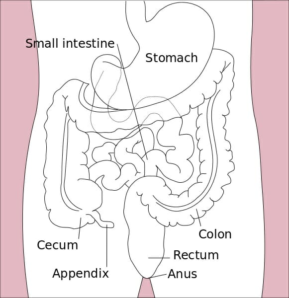
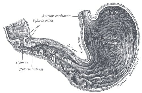
The small intestine is the part of our gastrointestinal tract where most of our nutrient absorption takes place. Everything we eat and drink throughout the course of our day will make its way to the small intestine. The small intestine is commonly known as the “small bowel;” but despite its naming, it still spans an impressive twenty feet long with a circular diameter of about an inch. It is amazing to think that such a long intestinal tract is all encased within the relatively small space inside our abdomen. In terms of location, the small intestine will span from the pylorus (or, the muscular opening that connects the stomach to the opening of the small intestine) to the cecum (or the fecal pouch).


Upon closer inspection, the small intestine appears narrow and coiled. Its coiled nature, of course, helps the intestine fit within its limited allotted space within our bodies. Inside, the small intestine is covered by a soft lining that is contains many villi and microvilli. These little ridges or projections give the small intestine even more surface area from which to absorb nutrients. It is no surprise, then, that the small intestine is the principal site of our food’s molecular digestion. The small intestine is able to absorb these nutrients and shuttle them to the rest of the body via the bloodstream. Let us discuss the small intestine’s many functions in more detail.


Stomach colon rectum diagram

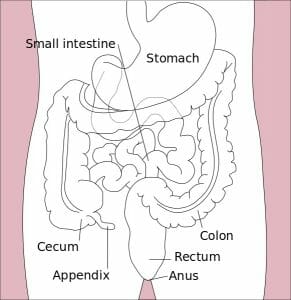
The image depicts an illustration of the organs of the alimentary canal.


Small Intestine Function


The small intestine is the site where up to ninety percent of our total nutrient and mineral absorption takes place. The remainder of the absorption is left to the stomach and the large intestine.


While the small intestine’s main function lies in absorbing nutrients from broken down food particles, it is important to note that the actual digestion of food undergoes two phases. Digestion begins with mechanical digestion in our mouths. By chewing and churning food with our teeth, the bonds that hold food particles together are physically broken down. This process is only carried forward by the starch-breaking action of amylase enzyme in our saliva. This digestion continues on in the stomach with the help of acids. This brings us to a second digestive phase, which is chemical digestion. Chemical digestion differs from mechanical digestion, in part because there are actual enzyme reactions taking place to break apart the molecular bonds that bind our food. This is made possibly with the help of bile acids that are released from the liver and the gall bladder. Likewise, chemical digestion relies on bile acids and enzymes that break the food down, and then give way to the release of minerals into our bloodstream and our body’s many tissues. Chemical digestion is a process that really only occurs in the small intestine, which is another fact that separates it from standard mechanical digestion that takes place at several points along the alimentary canal.


It is worth mentioning that the small intestine is a site that is very rich in enzyme activity. While some chemical activity does occur in the stomach with the help of acidic enzyme pepsin, chemical digestion continues, and thrives, in the small intestine. Even more distinctions are elucidated when we are tasked with investigating the digestion of the different macromolecules in our diets. In general, the main molecules that are absorbed by the small intestine include amino acids derived from proteins, fatty acids from lipids, and simple sugars derived from starches or complex carbohydrates, which we will discuss in more detail below. Furthermore, up to eighty percent of the water in our bodies is absorbed by the small intestine, as well as electrolytes like chloride, iron, potassium, and sodium ion. Ion channels will be crucial in replenishing and driving this life-sustaining process. Likewise, the small intestine has the important role of absorbing vitamins and minerals from our diet. Fat soluble vitamins K, A, D, and E are absorbed by simple diffusion along with dietary fats. Meanwhile, water soluble vitamins B and C will be absorbed by facilitated diffusion, as their hydrophilic nature precludes their simple entry into our cells. Vitamin B12, likewise, will be absorbed at the small intestine’s Ileum via active transport.


Digestion of Proteins


Proteolytic enzymes are those that target and break down peptide bonds within the proteins in our food. We, as a society, are quite familiar with what constitutes as protein in our diets; and in truth, a lot of protein is consumed throughout the world in the form of chicken, beef, tofu, and legumes. These enzymes will include trypsin and chymotrypsin, which are first released by the pancreas and will make their way to the small intestine to cleave proteins. Carboxypeptidase is an even more refined intestinal enzyme that is also released by the pancreas, but will split amino acids into singular amino acids. Protein digestion starts at the mouth and will continue, to a lesser extent, in the large intestine. Notably, amino acids are hydrophilic, or “water loving,” and will therefore require some help passing through the lipid barrier of our cells. They will generally follow primary active transport where an ATP molecule will be expended.


Digestion of Lipids


Lipases are likewise secreted by the pancreas and act on the fats in our diets. Lipases will break triglycerides into free fatty acids that can circulate within our bodies. But their action is further helped by bile salts that our liver and gallbladder secrete. Fatty triglycerides are very averse to the watery environments of our tissues. The bile salts act by enclosing the triglycerides within their structures until lipases can come and break them down. Transport of lipids and short-chained fatty acids will follow the rules of passive or simple diffusion through the hydrophobic lipid bilayers of our cells.


Digestion of Carbohydrates


The carbohydrates in our food will often consist of complex sugars. Their digestion into simpler sugars such as glucose is absolutely essential. Pancreatic amylase will help break down some of these carbohydrates, while the more tenacious fibers will experience bacterial breakdown in the large intestine. While fructose can be structurally absorbed by cells via facilitated diffusion, glucose will require secondary active transport.


Small Intestine Parts


The small intestine is further divided into three sections: the duodenum, the jejunum, and the ileum. The duodenum is the first and shortest section of the small intestine, which measures about fifteen inches long. It will receive chyme (or, a mix of partially digested food particles that is mixed with bile) from our stomachs. The duodenum’s intestinal cells will also secrete amylase, sucrase, and lipase enzymes that break down fats and sugars. The jejunum will follow suit, and is located near our belly buttons. The jejunum marks the end of our digestion of fats and carbohydrates. It is covered in villi and microvilli that make it the principal site of digestion. It is also a coiled structure that is thicker and has more blood vessels than the third and final section, the ileum. The ileum lies in our pelvic area, more or less, and is thinner and less vascular than the jejunum. The ileum’s main role is in absorption and it will absorb amino acids, lipids, fat soluble vitamins, and vitamin B12.


Quiz


1. Which method of transport do amino acids follow?
A. Passive diffusion
B. Simple diffusion
C. Primary active transport
D. Secondary active transport

Answer to Question #1

2. Correctly label the first, middle, and third sections of the small intestine:
A. Ileum, duodenum, jejunum
B. Duodenum, ileum, jejunum
C. Jejunum, duodenum, ileum
D. Duodenum, jejunum, ileum

Answer to Question #2

3. Correctly identify the main site of digestion, and the site of vitamin B12 absorption:
A. Duodenum; jejunum
B. Jejunum; ileum
C. Ileum; jejunum
D. Ileum: duodenum

Answer to Question #3

References


  • Hoffman, Matthew (2017). “Picture of the Intestines.” Web MD: Human Anatomy. Retrieved on 2017-08-30 from http://www.webmd.com/digestive-disorders/picture-of-the-intestines#1

  • Mandal, Ananya MD (2017). “What does the small intestine do?” News Medical Life Sciences. Retrieved on 2017-09-01 from https://www.news-medical.net/health/What-Does-the-Small-Intestine-Do.aspx

  • Schmidler, Cindy (2017). “Anatomy and Function of the Digestive System.” Health Pages. Retrieved on 2017-09-02 from https://www.healthpages.org/anatomy-function/anatomy-function-digestive-system/#Jejunum_Function

  • Coleman, Ruth (2017). “What are the digestive enzymes that occur in each section of the small intestine.” Livestrong. Retrived on 2017-09-03 from http://www.livestrong.com/article/420133-what-are-the-digestive-enzymes-that-occur-in-each-section-of-the-small-intestine/


Small Intestine

Stomach

Stomach Definition


The stomach is a muscular organ that is found in our upper abdomen. If we were to locate it on our bodies, it can be found on our left side just below the ribs. In simple terms, the stomach is a kind of digestive sac. It is a continuation of the esophagus and receives our churned food from it. Therefore, the stomach serves as a kind of connection between the esophagus and the small intestine, and is a definite pit stop along our alimentary canal. Muscular sphincters, which are similar to valves, allow some separation between these organs.


The stomach’s functions benefit from several morphological attributes. The stomach is able to secrete enzymes and acid from its cells, which enables it to perform its digestive functions. With its muscular lining, the stomach is able to engage in peristalsis (in other words, to form the ripples that propel the digested food forward) and in the general “churning” of food. Likewise, the abundant muscular tissue of the stomach has ridges in its linings called rugae. These increase the surface area of the stomach and facilitate its functions, which we will describe in more detail below.


Stomach diagram

The image illustrates the esophagus, stomach, and intestinal regions of the human body


Functions of the Stomach


As mentioned before, the stomach is first and foremost a principal site of digestion. In fact, it is the first site of actual protein digestion. While sugars can begin to be lightly digested by salivary enzymes in the mouth, protein degradation will not occur until the food bolus reaches the stomach. This breakdown is carried out by the stomach’s pepsin enzyme. The stomach’s roles can essentially be distilled down to three functions.


Much like an elastic bag, the stomach will provide a place for varied amounts of swallowed food to rest and digest in. Hence, the stomach is a storage site. The stomach will also introduce our swallowed food to essential acids. The cells in the stomach’s lining will excrete a strong acidic mixture of hydrochloric acid, sodium chloride, and potassium chloride. This gastric acid, or colloquially known as gastric “juice,” will work to break down the bonds within the food particles at the molecular level. Pepsin enzyme will have the unique role of breaking the strong peptide bonds that hold the proteins in our food together, further preparing the food for the nutrient absorption that takes place in the small (mainly) and large intestines. This brings us to the third task the stomach has, which is to send off the churned watery mixture to the small intestine for further digestion and absorption. It takes about three hours for this to occur once the food is a liquid mix.


The stomach’s main roles:


  1. Food storage

  2. Acidic breakdown of swallowed food

  3. Sends mixture on to the next phase in the small intestine

Structure of the Stomach


Stomach

The archaic illustration depicts the different regions of the stomach


Although we have briefly discussed the location and physical traits of the stomach, it is important to detail the structure of the stomach, as well. The stomach begins at the lower esophageal sphincter that discerns the cut-off point of the esophagus. The stomach itself is very muscular. When the muscularis externa layers are dissected, one can visualize three distinct layers coined the longitudinal, circular, and oblique layers. The first region of the stomach is called the cardia. It is the layer closest to the esophagus and it contains cardiac glands that secrete mucus. Mucus protects the delicate epithelial lining of many tissues in the human body. This region is followed by the fundus, which is the superior arch of the stomach. Importantly, the fundus has the special function of containing gastric glands that release a cocktail of gastric juices. This region is followed by the body of the stomach, which is coated with rugae and is the largest region. Rugae, in turn, help facilitate digestion by increasing the site’s surface area. Finally, this section is followed by the pylorus region, which is closest to the exit into the duodenum of the small intestine and is pinched off by the pyloric sphincter.


Four regions of the stomach:


  • Cardiac

  • Fundus

  • Body

  • Pylorus


Common Stomach Issues


Almost every person has experienced a stomach related issue at one point in their lives. Perhaps the most common ones are indigestion and heartburn. These issues can be resolved quite easily with over-the-counter tablets (i.e. tums), but there is no denying that they are unpleasant experiences. However, there are more chronic illnesses that afflict many people. One of these is the gastroesophageal reflux disease, commonly known as GERD or Acid Reflux. GERD afflicts up to 3 million Americans each year, and is a physiological result of when the lower esophageal sphincter will not close properly. The principal function of this sphincter is to prevent food and stomach acids from regurgitating up the esophageal canal. While a healthy stomach has tons of mucus and barriers strong enough to prevent stomach acids from wreaking havoc on the epithelium, the esophagus is not quite so lucky. This, of course, has the long-term implications of damaging those delicate epithelial cells. When a patient does not have the sufficient barriers to prevent damage within the stomach, a medical issue that arises are peptic ulcers. Ulceration refers to the sores that pierce through an organ. When the stomach is not sufficiently protected from contact with these highly acidic acids, we do run into the issue of perforating the tissue and potentially having the stomach juices leak – which by all means requires urgent medical attention. These sores are very painful and recurrent in patients with peptic ulcer disease.


There are other red flag symptoms that present in the urgent or emergency care setting that indicate a stomach issue. These include a burning sensation in the chest (heartburn), piercing or diffuse abdominal pain, blood in the stool, and vomiting or diarrhea.


Quiz


1. How many hours does it take for the stomach to release the food to the small intestine?
A. 1
B. 2
C. 3
D. 4

Answer to Question #1

2. Which of the following is the largest region of the stomach?
A. Cardia
B. Fundus
C. Body
D. Pylorus

Answer to Question #2

3. Which region of the stomach releases gastric juice?
A. Cardia
B. Fundus
C. Body
D. Pylorus

Answer to Question #3

References


  • Hoffman, Matthew MD (2017). “Picture of the Stomach: Human Anatomy.” Web MD. Retrieved on 2017-08-26 from http://www.webmd.com/digestive-disorders/picture-of-the-stomach#1

  • MedicineNet (2017). “Medical Definiton of Stomach.” MedicineNet. Retrieved on 2017-08-27 from http://www.medicinenet.com/script/main/art.asp?articlekey=5560

  • Medline Plus (2017). “Stomach Disorders.” Medline Plus. Retrieved on 2017-08-28 from https://medlineplus.gov/stomachdisorders.html


Stomach

Uterus

Uterus Definition


The uterus, otherwise known as the womb, is the female sex organ that carries a huge significance in many species’ survival – ours included. The uterus itself is a hollow organ that is shaped in the form of a pear, and interestingly enough measures about that size. It is neatly tucked into the pelvic area of most mammals and, of course, in humans. It is important to dissect the anatomy of the human uterus. In the female body, the upper end of the uterus, called the fundus, will join the fallopian tubes at either side while the lower end will open into the vagina. The wide portion at the top of the uterus is called the fundus, and will be the superior-most region that will host a fertilized embryo as it grows into a baby. A little below the fundus lies the muscular corpus region.


The corpus, in turn, is composed of three tissue layers. Post-pubescent women will have an innermost endometrium, which is the layer of muscle that is shed when the menstrual cycle commences in non-pregnant women. The endometrial tissue will thicken as the month’s cycle goes by in preparation for a fertilized egg to implant itself there. But in the absence of a fertilized egg, this layer will simply be shed away in what we know as menstruation. The middle muscle layer is called the myometrium, and is the layer that will expand during pregnancy and contract during childbirth. The outermost layer, the parametrium, will likewise expand and contract at these stages. Expanding will allow the uterus to house a growing baby, while the contractions will facilitate the newborn’s exit from the womb.


Uterus diagram

The image above depicts a diagram of the Uterus, with labeled tissue and arterial landmarks.


Function of the Uterus


Perhaps the principal, albeit lofty function of the uterus is to preserve life. It is the site of nourishment for the growing baby, making it one of the most important reproductive organs in the female body. This all begins when an egg, or ovum, is fertilized by a sperm and will make its downward trek in search of a better home. The tight fallopian tubes will not provide enough space to house the growing embryo! This is where the uterus meets all of these requirements, and more! The uterus’s thick, muscular nature will allow it to contract and expand to make room for the developing baby. The uterus is also rich in vasculature. There are many blood vessels supplying the muscle layers at any given time. This especially applies to the endometrium which is highly vascular and will come to nourish the embryo. In fact, many of the endometrial vessels that will come to supply the embryo will form just for this purpose. All of this explains why the fertilized ovum will choose to implant itself in the uterine lining – coined, the “site of implantation.” Thus, the uterus is the site that allows ours, and many species, to continue reproducing!


Location of the Uterus


The uterus measures about three inches long and two inches wide, and has a thick muscular lining within its walls. The lowest tip of the uterus will dip into the vagina in the area of the cervix, while the top most part will connect with the fallopian tubes through which the eggs travel. But a better way to pin its location is by describing its region as the area that lies between the belly button and the hip bones.


Abnormalities of the Uterus in Pregnancy


Nothing quite demonstrates the reproductive role of the uterus as the difficulties that arise from having an abnormal uterus. While the normal uterus will roughly measure three by two by one inches, some women will have uteruses that differ in shape and size. Many species’ evolution, including our own, has depended on having these precise dimensions to best support the growing embryo and to bring it to full term. But women with uterine abnormalities may realize they have this only once they have attempted and failed to conceive. These complications will surface either while trying to become pregnant, or after experiencing miscarriage. Examples of physical deformations of the uterus may include having a uterus with two inner cavities or vaginas (affecting roughly one in 350 women), having only one fallopian tube that will connect to the uterus, or having a heart shaped uterus instead of a pear-shaped one that is evolutionarily optimized to bear a child. Moreover, some afflicted women will have a septum that parts the uterus, or a slight indentation at the top of the uterus that will likewise compromise the ability for these women to have a baby. These abnormalities, however, are not all an absolute guarantee of infertility or miscarriage, but instead may lessen the probability of carrying a child to full-term.


Quiz


1. Which of the following is the outermost muscle layer of the corpus?
A. Endometrium
B. Parametrium
C. Myometrium
D. None of the above

Answer to Question #1

2. Which uterine layer nourishes the growing embryo?
A. Endometrium
B. Parametrium
C. Myometrium
D. None of the above

Answer to Question #2

3. Which of the following provides the best explanation for why the uterus is the main female reproductive organ?
A. It is the site of fertilization of the ovum
B. It is muscular and able to contract and expand
C. Its myometrial layer supplies the growing embryo
D. Both A and B

Answer to Question #3

References


  • MedicineNet (2017). “Uterus.” Medicine Net. Retrieved on 2017-08-26 from http://www.medicinenet.com/script/main/art.asp?articlekey=5918

  • BabyCentre Medical Advisory Board (2016). “Abnormalities of the uterus in pregnancy.” Retrieved on 2017-08-26 https://www.babycentre.co.uk/a551934/abnormalities-of-the-uterus-in-pregnancy

  • Danielsson, K. “Abnormal Uterus Shapes and iscarriage Risk.” Very Well. Retrieved on 2017-08-27 from https://www.verywell.com/abnormal-uterus-and-miscarriage-risk-2371694


Uterus

Extracellular Matrix

Extracellular Matrix Definition


The extracellular matrix can be thought of as a suspension of macromolecules that supports everything from local tissue growth to the maintenance of an entire organ. These molecules are all secretions made by neighboring cells. Upon being secreted, the proteins will undergo scaffolding. Scaffolding, in turn, is a term used to describe the ephemeral structures that form between individual proteins to make more elaborate protein polymers. These rigid, albeit temporary protein structures will lend the matrix a viscous consistency. One can think of the extracellular matrix as essentially a cellular soup, or gel mixture of water, polysaccharides (or linked sugars), and fibrous protein. This leads us to another category of molecule found within the extracellular matrix called the proteoglycan. The proteoglycan is a hybrid cross of a protein and a sugar, with a protein core and several long chain sugar groups surrounding it. All of the molecular groups that make up these macromolecules will lend them special properties that will dictate the kind of hydrophobic or hydrophilic interactions they can participate in.


Much like the ephemeral interactions they form in this aqueous solution, the actual structures of the proteins themselves are notably dynamic. The molecular components found within their structures are always changing. The remodeling they undergo is certainly aided by protease enzymes found in the matrix and can be modified by post-translational changes. The extracellular matrix has a functional value in buffering the effects of local stressors in the area. But we will discuss many more of the functions the matrix serves in detail below.


Extracellular Matrix Function


Living tissue can be thought of as a dynamic meshwork of cells and liquid. Despite their close proximity to each other, the cells of a tissue are not simply tightly wound together. Instead, they are spaced out with the help of the extracellular meshwork. The matrix will act as a kind of filler that lies between the otherwise tightly packed cells in a tissue. Furthermore, not only is the matrix filling the gaps in between these cells but it is also retaining a level of water and homeostatic balance. Perhaps the most important role of the extracellular matrix, however, can be distilled down to the level of support it provides for each organ and tissue.


The extracellular matrix directs the morphology of a tissue by interacting with cell-surface receptors and by binding to the surrounding growth factors that then incite signaling pathways. In fact, the extracellular matrix actually stores some cellular growth factors, which are then released locally based on the physiological needs of the local tissue. On the other hand, a tissue’s morphology is another way to describe the “look” or appearance of the organ or tissue. The physical presence of proteins and sugars in the matrix also have the benefit of cushioning any forces that may be placed upon the surrounding area. This prevents the cellular structures from collapsing or the delicate cells from going into shock. Since the extracellular matrix is thick and mineralized despite its water rich content, it has the additional function of keeping the cells in a tissue separate and physically distinct.


More direct applications of the extracellular matrix include its role in supporting growth and wound healing. For instance, bone growth relies on the extracellular matrix since it contains the minerals needed to harden the bone tissue. Bone tissue will need to become opaque and inflexible. The extracellular matrix will allow this by letting these growth processes take ample opportunity to recruit extracellular proteins and minerals to build and fortify the growing skeleton. Likewise, forming scar tissue after an injury will benefit from the extracellular matrix and its rich meshwork of water insoluble proteins.


Extracellular Matrix Components


The extracellular matrix is mostly made up of a few key ingredients: water, fibrous proteins, and proteoglycans. The main fibrous proteins that build the extracellular matrix are collagens, elastins, and laminins. These are all relatively sturdy protein macromolecules. Their sturdiness lends the extracellular matrix its buffering and force-resisting properties that can withstand environmental pressures without collapsing. Collagen is actually a main structural component of not only the matrix, but also of multicellular animals. Collagen is the most abundant fibrous protein made by fibroblasts, making up roughly one third of the total protein mass in animals. In the matrix, collagen will give the cell tensile strength and facilitate cell-to-cell adhesion and migration. Elastin is another fiber that will lend tissues an ability to recoil and stretch without breaking. In fact, it is because elastin and collagen bind and physically crosslink that this stretching is limited to a certain degree by collagen. Fibronectin is first secreted by fibroblast cells in water soluble form, but this quickly changes once they assemble into an un-dissolvable meshwork. Fibronectin regulates division and specialization in many tissue types, but it also has a special embryonic role worth mentioning where it will aid in the positioning of cells within the matrix. Laminin is a particularly important protein. It is particularly good at assembling itself into sheet-like protein networks that will essentially be the ‘glue’ that associates dissimilar tissue types. It will be present at the junctions where connective tissue meet muscle, nerve, or epithelial lining tissue.


Collagen triple helix

The image depicts a computerized illustration of the three-dimensional structure of collagen protein


Roles of fibrous protein:


  • Collagen – stretch resistance and tensile strength (i.e. scar formation during wound healing)

  • Elastin – stretch and resilience

  • Fibronectin – cell migration and positioning within the ECM, and cell division and specialization in various tissues

  • Laminin – sheet-like networks that will ‘glue’ together dissimilar types of tissue


On the contrary to fibrous proteins that resist against stretching, proteoglycans will resist against compression. This refers to the forces pushing down on the tissue that would otherwise “squash” or collapse it. This ability stems from the glycosaminoglycan group in the proteoglycan. Glycosaminoglycan, or GAGs, are chains of sugar that will vary and thus lend the molecules different chemical properties. Moreover, GAGs are the most highly negatively charged molecule animal cells produce. This charge will attract GAGs to positively charged sodium ions. In living tissue, water follows the movement of sodium. This will bring us to a situation where water and GAGs will attract as well, which will lend water within the extracellular matrix a characteristic resistance to compression.


Quiz


1. Which of the following is not a fibrous protein type mentioned?
A. Elastin
B. Proteoglycan
C. Collagen
D. Laminin

Answer to Question #1

2. Identify the distinction between fibrous protein and proteoglycans, per the article:
A. Fibrous protein is more capable of handling aqueous environments
B. Proteoglycans serve more of a filler role in the spaces between the cells in a tissue
C. Fibrous proteins resist against compressive forces
D. Proteoglycans resist against compressive forces

Answer to Question #2

References


  • Franz et al (2010). “The extracellular matrix at a glance.” J Cell Sci. 123(24): 4195-4200. https://www.ncbi.nlm.nih.gov/pmc/articles/PMC2995612/

  • Alberts, B et al (2002). “The Extracellular Matrix of Animals.” Molecular Biology of the Cell: 4th edition. https://www.ncbi.nlm.nih.gov/books/NBK26810/

  • Study (2017). “Extracellular Matrix.” Study.com. Retrieved on 2017-08-15 from http://study.com/academy/lesson/extracellular-matrix-function-components-definition.html


Extracellular Matrix