Tuesday, September 26, 2017

Difference between Mutualism and Commensalism

Commensalism and mutualism both describe a symbiotic relationship between two organisms. The main difference lies in whether one or both of the organisms benefits from the relationship. Mutualism is further subdivided into two categories that define how dependent the organisms are on each other for survival.


Mutualism


In mutualistic relationships, individuals of different species both benefit from their interaction. This is also called interspecies reciprocal altruism. These relationships can be obligate for both species, meaning they can’t live without each other, or facultative for both species, meaning they can live without each other.


Obligate Mutualism


An example of obligate mutualism is the relationship between termites and the protozoans that live in their digestive system. Termites cannot digest the cellulose they take in from eating wood to obtain the nutrients, but the protozoans in their gut can. In turn, the protozoans who cannot chew up wood, receive a reliable food supply from the termites.


Facultative Mutualism


Facultative mutualism exists between birds and the plants that produce the fruit they eat. The birds benefit from eating the fruit but they also have other food sources so they are not dependent on it. Likewise, the plant that bears the fruit benefits from the bird scattering its seeds in their droppings, but this seed dispersion also happens in other ways and with other species.


Commensalism


In commensalism, one of the organisms benefits in some way while the other is unaffected. An example of a commensal relationship is when an organism uses another organism (or part of a dead organism) for transportation or housing without having any effect on it. For example, hermit crabs use the abandoned shells of other creatures like sea snails to protect themselves. Other commensal relationships exist in nature such as when birds build a nest in a tree. The birds benefit from having a home, protection and a place to raise their young, but the tree is unaffected.


European honey bee extracts nectar

The image above shows the mutualistic relationship between bees and flowers. The bees benefit from the pollen and nectar they gather from the flowers and the flowers benefit by the bees transporting their pollen and pollinating other flowers.


Oceanic whitetip shark

The image above shows commensalism between some shark species and pilot fish. Pilot fish will feed on the leftovers in the water after the shark makes a kill, while the shark remains unaffected by this behavior.


References


  • OpenStax, Biology. OpenStax. May 20, 2013. http://cnx.org/content/col11448/latest/

  • Symbiosis. (n.d.). In Wikipedia. Retrieved September 19, 2017 from https://en.wikipedia.org/wiki/Symbiosis


Difference between Mutualism and Commensalism

Chemoheterotrophic Bacteria

There are two things that make chemoheterotrophic bacteria unique. They are unable to make their own food (like autotrophs do) so they get their energy from the oxidation of inorganic minerals in their environment. Also, these bacteria cannot make organic molecules from inorganic sources (they cannot “fix” carbon) so they eat other organisms to get the carbon they need.


An example of chemoheterotrophic bacteria is a sub-type called lithotrophic bacteria, also known as “rock eaters” or “stone eaters.” These bacteria are found in underground water sources and on the ocean floor where there are both mineral food sources and organic molecules available. The common food and energy sources for them are dead organic material and elemental sulfur and iron and gases having these elements such as hydrogen sulfide.


Troph flow chart

The image above is a flowchart that helps determine the trophic category an organism belongs to. It shows that chemoheterotrophs don’t make their own food (they don’t use photosynthesis to fix carbon) and they get their energy from the oxidation of inorganic molecules.


References


  • Lithotroph. (n.d.). In Wikipedia. Retrieved September 19, 2017 from https://en.wikipedia.org/wiki/Lithotroph


Chemoheterotrophic Bacteria

Chemoautotrophic Bacteria

Chemoautotrophic bacteria get their energy from oxidizing inorganic compounds. In other words, instead of using the energy of photons from the sun, they break the chemical bonds of substances that don’t contain carbon in order to get their energy. Some of the inorganic chemicals chemoautotrophic bacteria use are hydrogen sulfide, ammonia and iron. For example, the sulfur-eating bacteria Thiothrix oxidizes hydrogen sulfide to produce water and sulfur. The energy that is stored in the chemical bonds of the hydrogen sulfide molecule is released during the reaction. The bacteria use this energy along with carbon dioxide to make sugars and carbohydrates. Chemoautotrophic bacteria often live in extreme environments like deep sea vents in the ocean, hence their other name, extremophiles.


Giant tube worm

The image above shows giant tube worms, Riftia pachyptila, that live near hot sulfur vents in the ocean. Chemoautotrophic bacteria live in a symbiotic relationship with these worms which have no digestive tract, making organic molecules for the worms from hydrogen sulfide, carbon dioxide and oxygen. In return for this, the worms supply a special type of hemoglobin they make as food for the bacteria.


References


  • Chemoautotrophic and Chemolithotrophic Bacteria. World of Microbiology and Immunology. Retrieved September 18, 2017 from Encyclopedia.com: http://www.encyclopedia.com/science/encyclopedias-almanacs-transcripts-and-maps/chemoautotrophic-and-chemolithotrophic-bacteria


Chemoautotrophic Bacteria

Monday, September 25, 2017

Cell Theory Founder and Contributors

When it was first proposed in the early 1830s, the cell theory had two main components; the cell is the basic functional and structural unit of all living things and all living things are made of one or more cells. Credit for this theory is often given to the German scientists Theodor Schwann and Matthias Schleiden, although their fellow scientist and countryman Rudolf Virchow made significant contributions later.


There were several other people who helped lay the groundwork prior to the work of Schleiden and Schwann. Galileo Galilei’s historic invention of the microscope in 1625 was improved on by the work of Anton van Leeuwenhoek who made considerable improvements to the quality of the lenses in microscopes in 1670. But even before Leeuwenhoek’s lens improvements, the British scientist Robert Hooke had already coined the term “cell” in 1665 after looking at thin slices of cork under his microscope.


Further understanding of cells came from the work of J.H.F. Link and Karl Rudolphi who, in 1804 conducted experiments that proved cells had their own cell walls and were independent of each other. Then, in 1833 botanist Robert Brown discovered the nucleus of plant cells.


In 1855, Rudolf Virchow was recognized for his idea that became the third component of the cell theory at the time, Omnis cellula e cellula which is Latin for “cells only come from other cells.”


Theodor Schwann

The image above shows the German scientist Theodore Schwann who contributed to the first cell theory.


Matthias Jacob Schleiden

The image above shows the German scientist Matthias Schleiden who along with Theodore Schwann, developed the first cell theory.


Rudolf Virchow

The image above is that of Rudolf Virchow whose contributions to the cell theory are often overlooked in history.


References


  • Cell Theory. (n.d.). In Wikipedia. Retrieved September 18, 2017 from https://en.wikipedia.org/wiki/Cell_theory


Cell Theory Founder and Contributors

Cell Theory Timeline

The original cell theory states that the cell is the basic structural and functional unit of living organisms and all cells come from other cells. The scientists Matthias Schleiden and Theodor Schwann are credited with establishing the cell theory in 1839. However, there was a lot of work done over the previous centuries which paved the way.


1600s


The Italian scientist Galileo Galilei is credited with building the first microscope in 1625. It was a logical step for him to take from his groundbreaking work with telescopes and astronomy in 1609. In 1665, Robert Hooke, a British scientist, looked at a thin slice of cork under the microscope and saw a honeycomb structure made up of small compartments he called cells. The first person to see living cells under a microscope was Anton van Leeuwenhoek. In 1670, Leeuwenhoek significantly improved the quality of microscope lenses to the point that he could see the single-celled organisms that lived in a drop of pond water. He called these organisms “animalcules,” which means “miniature animals.”


1800s


Microscopes and science in general advanced throughout the 1700s, leading to several landmark discoveries by scientists at the beginning of the 1800s. In 1804, Karl Rudolphi and J.H.F. Link were the first to prove that cells were independent of each other and had their own cell walls. Prior to this work, it was thought that cells shared their walls and that was how fluids were transported between them. The next significant discovery occurred in 1833 when the British botanist Robert Brown first discovered the nucleus in plant cells.


From the years 1838-1839, the German scientist Matthias Schleiden proposed the first foundational belief about cells, that all plant tissues are composed of cells. His fellow scientist and countryman Theodor Schwann concluded that all animal tissues were made of cells as well. Schwann blended both statements into one theory which said 1) All living organisms consist of one or more cells and 2) The cell is the basic unit of structure for all living organisms. In 1845, the scientist Carl Heinrich Braun revised the cell theory with his interpretation that cells are the basic unit of life.


The third part of the original cell theory was put forth in 1855 by Rudolf Virchow who concluded that Omnis cellula e cellula which translates roughly from Latin to “cells only arise from other cells.”


The modern version of the cell theory includes several new ideas that reflect the knowledge that has been gained since the mid-1800s. These include the knowledge that energy flows within cells, hereditary information is passed from cell to cell, and cells are made of the same basic chemical components.


Hooke Microscope cork

The image above shows a drawing of the microscope set up used by Robert Hooke in 1665 in which he first saw cells in a thin slice of cork. The circular inset shows the drawing Hooke made of the honeycomb structure that he saw under the microscope.


References


  • Cell Theory. (n.d.). Retrieved September 14, 2017 from http://www.softschools.com/timelines/cell_theory_timeline/96/

  • Cell Theory. (n.d.). In Wikipedia. Retrieved September 14, 2017 from https://en.wikipedia.org/wiki/Cell_theory


Cell Theory Timeline

Purpose of Cell Division

Cell division has three main functions which are reproduction of unicellular organisms and the production of gametes and growth in eukaryotes. The process of meiosis in eukaryotes produces sex cells or gametes with half the chromosome compliment of somatic cells. Multicellular eukaryotic organisms use mitosis to grow and to repair their tissues. In contrast, prokaryotes (single-celled organisms) reproduce using a process similar to mitosis called binary fission. In binary fission, the organism divides to create an exact copy of itself, also called a clone.


Three cell growth types

The image above shows the three types of cell division. Binary fission is used for reproduction by single-celled organisms, mitosis is used for the growth and maintenance of eukaryotic organisms and the process of meiosis produces eggs and sperm in eukaryotes.


References


  • Cell Division. (n.d.). In Encyclopedia.com. Retrieved from http://www.encyclopedia.com/science-and-technology/biology-and-genetics/cell-biology/cell-division


Purpose of Cell Division

How Does Cell Division Solve the Problem of Increasing Size

When an organism grows, it’s because its cells are dividing not getting bigger. Cells divide for several reasons including to keep them from getting too big. As a cell gets bigger, it has a difficult time keeping up with taking in the extra nutrients it needs and expelling more waste. In other words, as the cell gets bigger, it has less surface area compared to its size—the surface area to volume ratio of the cell decreases as it gets bigger.


Cells grow during the three phases of interphase during which time the chromosomes are duplicated and more proteins and organelles are made. This is what increases the amount of cellular contents while the surface area of the cell membrane stays the same. Cell division solves the problem of increasing size by reducing the volume of cytoplasm in the two daughter cells and dividing up the duplicated DNA and organelles, thereby increasing surface to volume ratio of the cells.


Animal cell cycle

The image above shows the process of animal cell division. Notice the size of the cell starting to get bigger during interphase.


References


  • Mitosis. (n.d.) In Wikipedia. Retrieved September 13, 2017 from https://en.wikipedia.org/wiki/Mitosis


How Does Cell Division Solve the Problem of Increasing Size

What Role Do Centrioles Play in Cell Division

Centrioles are microscopic cylinders (microtubules) that are the building blocks of centrosomes. A single centriole consists of 9 microtubule triplets arranged in the shape of a cylinder with 2 centrioles making up each centrosome. Centrioles are responsible for organizing the spindle fibers in the mitotic spindle apparatus and are thought to participate in the completion of cytokinesis during the process of cell division.


It is interesting to note that a cell can still divide if the centrosome is removed, although mitosis takes a lot longer and there are more errors in chromosome division. In addition, plant cells do not have centrioles or centrosomes but are still capable of cell division. This information has led scientists to think that centrioles evolved as an improvement in cell division which made the process faster and less error-prone.


OSC Microbio 03 04 Centrosome

The image above shows (a) how centrioles are a component of centrosomes and (b) how centrosomes are involved in cell division.


References


  • Centriole. (n.d.). In Wikipedia. Retrieved September 13, 2017 from https://en.wikipedia.org/wiki/Centriole


What Role Do Centrioles Play in Cell Division

What Determines the Carrying Capacity of an Ecosystem

The carrying capacity of an ecosystem is the largest population that it can sustain indefinitely with the available resources, also called the “maximum load” by population biologists. Carrying capacity depends on many abiotic and biotic factors in the ecosystem and some are more obvious than others. For example, the availability of the basic needs of organisms such as food, water and shelter dictates how many individuals the ecosystem can sustain. This process is self-regulating to some extent because individuals will die when the carrying capacity is exceeded. Therefore, another way to look at carrying capacity is that it is the point at which the population growth reaches zero.


Other naturally-occurring factors that influence the carrying capacity of an ecosystem include disease, predator-prey interactions, the consumption rate of resources and the number of populations in the ecosystem. However, there are other factors that are hidden, less obvious and/or disregarded which have a significant impact on populations such as pollution, eradication of habitat and climate change.


Carrying Capacity

The image above shows a graph of the logistical growth of a population of individuals (N) over time (t). The K value is the carrying capacity. The line on the graph has the characteristic S-shape when the resources are limited. The line has more of a J-shape, indicating exponential growth, when there are unlimited resources available to the population.


References


  • Carrying Capacity. (n.d.). In Wikipedia. Retrieved September 12, 2017 from https://en.wikipedia.org/wiki/Carrying_capacity


What Determines the Carrying Capacity of an Ecosystem

Carbon Cycle Reservoirs

The carbon cycle reservoirs on Earth interact with each other through chemical, geological, physical and biological processes. The exchange of carbon between the reservoirs is balanced so that carbon levels remain stable, except when it comes to the influence of humans. The largest reservoir of carbon on Earth is the oceans. Below are all the major carbon reservoirs on Earth and the approximate amount of carbon they have sequestered in them.


  • Deep oceans = 38,400 gigatons

  • Fossil fuels = 4,130 gigatons

  • Terrestrial biosphere = 2,000 gigatons

  • Surface oceans = 1,020 gigatons

  • Atmosphere = 720 gigatons

  • Sediments = 150 gigatons


The main activities of humans that cause additional carbon to enter the carbon cycle beyond the natural processes are the burning of fossil fuels and deforestation. The burning of fossil fuels released almost 10 gigatons of carbon into the atmosphere in 2015.


Carbon cycle diagram

The image above shows Earth’s carbon cycle and the associated carbon reservoirs.


References


  • Carbon Cycle. (n.d.). In Wikipedia. Retrieved September 11, 2017 from https://en.wikipedia.org/wiki/Carbon_cycle


Carbon Cycle Reservoirs

What Role Do Producers Play in the Carbon Cycle

The Earths producer organisms are primarily its green terrestrial plants and the algae in the oceans. These plants use the carbon from carbon dioxide to create sugar molecules through the process of photosynthesis. Terrestrial plants get their carbon dioxide from the atmosphere while marine plants get it from carbonic acid, the dissolved form of carbon dioxide. Plants feed themselves (autotrophs) with the sugars they make and store excess sugar in the form of glucose, proteins, fats and polysaccharides.


When herbivores and omnivores eat plants (and when another animal eats them), the carbon-containing molecules are stored in their bodies or broken down and used to make energy through the process of cellular respiration. One of the products of respiration is carbon dioxide which is released and re-enters the atmosphere and the oceans. This constant back and forth exchange of carbon between plants and the animals that eat them is a major part of the carbon cycle on Earth.


Carbon cycle full

The image above shows the carbon cycle on earth including the important roles that animal/human respiration and photosynthesis play.


References


  • OpenStax, Biology. OpenStax. May 20, 2013. http://cnx.org/content/col11448/latest/


What Role Do Producers Play in the Carbon Cycle

How Are Oceans Involved in the Carbon Cycle

The largest amount of actively recycled carbon on Earth is located in its oceans. The surface layer of the oceans contains dissolved inorganic carbon that is rapidly and continually exchanged with the atmosphere in large amounts. The oceans take in carbon from carbon dioxide in the atmosphere by dissolving it and converting it to carbonate. Carbon also enters the oceans from rivers in the form of dissolved organic carbon that comes from the weathering of rocks.


Through the process of photosynthesis, green plants incorporate carbon into sugar molecules which are passed along through the food chain to animals in the sea, air and on land. Carbon is also deposited on the ocean floor through the settling and decomposition of dead organisms as well as the shells of some creatures which contain calcium carbonate. Carbon exists and circulates in the deep waters of the oceans as well, sometimes for many years, where it either settles into the sediment on the bottom or recirculates back to the surface via the process of thermohaline circulation.


The oceans are also an important storage (sequestering) location for carbon. Human activities such as the burning of fossil fuels introduce more carbon into the atmosphere which is taken up by the oceans and, in turn, makes the water more acidic. The disruption of this delicate balance in the carbon cycle will in time, reduce the amount of carbon the oceans are capable of sequestering.


Carbon cycle

The image above shows the carbon cycle on Earth. The numbers represent billions of tons of carbon. Numbers in red are carbon that comes from human activity, numbers in white are stored carbon and yellow number represent natural fluxes. Volcanic and tectonic activity also contribute to the carbon cycle, but are not shown.


References


Carbon Cycle. (n.d.). In Wikipedia. Retrieved from https://en.wikipedia.org/wiki/Carbon_cycle#Oceans


How Are Oceans Involved in the Carbon Cycle

Friday, September 8, 2017

Small Intestine

Small Intestine Definition


The small intestine is the part of our gastrointestinal tract where most of our nutrient absorption takes place. Everything we eat and drink throughout the course of our day will make its way to the small intestine. The small intestine is commonly known as the “small bowel;” but despite its naming, it still spans an impressive twenty feet long with a circular diameter of about an inch. It is amazing to think that such a long intestinal tract is all encased within the relatively small space inside our abdomen. In terms of location, the small intestine will span from the pylorus (or, the muscular opening that connects the stomach to the opening of the small intestine) to the cecum (or the fecal pouch).


Upon closer inspection, the small intestine appears narrow and coiled. Its coiled nature, of course, helps the intestine fit within its limited allotted space within our bodies. Inside, the small intestine is covered by a soft lining that is contains many villi and microvilli. These little ridges or projections give the small intestine even more surface area from which to absorb nutrients. It is no surprise, then, that the small intestine is the principal site of our food’s molecular digestion. The small intestine is able to absorb these nutrients and shuttle them to the rest of the body via the bloodstream. Let us discuss the small intestine’s many functions in more detail.


Stomach colon rectum diagram

The image depicts an illustration of the organs of the alimentary canal.


Small Intestine Function


The small intestine is the site where up to ninety percent of our total nutrient and mineral absorption takes place. The remainder of the absorption is left to the stomach and the large intestine.


While the small intestine’s main function lies in absorbing nutrients from broken down food particles, it is important to note that the actual digestion of food undergoes two phases. Digestion begins with mechanical digestion in our mouths. By chewing and churning food with our teeth, the bonds that hold food particles together are physically broken down. This process is only carried forward by the starch-breaking action of amylase enzyme in our saliva. This digestion continues on in the stomach with the help of acids. This brings us to a second digestive phase, which is chemical digestion. Chemical digestion differs from mechanical digestion, in part because there are actual enzyme reactions taking place to break apart the molecular bonds that bind our food. This is made possibly with the help of bile acids that are released from the liver and the gall bladder. Likewise, chemical digestion relies on bile acids and enzymes that break the food down, and then give way to the release of minerals into our bloodstream and our body’s many tissues. Chemical digestion is a process that really only occurs in the small intestine, which is another fact that separates it from standard mechanical digestion that takes place at several points along the alimentary canal.


It is worth mentioning that the small intestine is a site that is very rich in enzyme activity. While some chemical activity does occur in the stomach with the help of acidic enzyme pepsin, chemical digestion continues, and thrives, in the small intestine. Even more distinctions are elucidated when we are tasked with investigating the digestion of the different macromolecules in our diets. In general, the main molecules that are absorbed by the small intestine include amino acids derived from proteins, fatty acids from lipids, and simple sugars derived from starches or complex carbohydrates, which we will discuss in more detail below. Furthermore, up to eighty percent of the water in our bodies is absorbed by the small intestine, as well as electrolytes like chloride, iron, potassium, and sodium ion. Ion channels will be crucial in replenishing and driving this life-sustaining process. Likewise, the small intestine has the important role of absorbing vitamins and minerals from our diet. Fat soluble vitamins K, A, D, and E are absorbed by simple diffusion along with dietary fats. Meanwhile, water soluble vitamins B and C will be absorbed by facilitated diffusion, as their hydrophilic nature precludes their simple entry into our cells. Vitamin B12, likewise, will be absorbed at the small intestine’s Ileum via active transport.


Digestion of Proteins


Proteolytic enzymes are those that target and break down peptide bonds within the proteins in our food. We, as a society, are quite familiar with what constitutes as protein in our diets; and in truth, a lot of protein is consumed throughout the world in the form of chicken, beef, tofu, and legumes. These enzymes will include trypsin and chymotrypsin, which are first released by the pancreas and will make their way to the small intestine to cleave proteins. Carboxypeptidase is an even more refined intestinal enzyme that is also released by the pancreas, but will split amino acids into singular amino acids. Protein digestion starts at the mouth and will continue, to a lesser extent, in the large intestine. Notably, amino acids are hydrophilic, or “water loving,” and will therefore require some help passing through the lipid barrier of our cells. They will generally follow primary active transport where an ATP molecule will be expended.


Digestion of Lipids


Lipases are likewise secreted by the pancreas and act on the fats in our diets. Lipases will break triglycerides into free fatty acids that can circulate within our bodies. But their action is further helped by bile salts that our liver and gallbladder secrete. Fatty triglycerides are very averse to the watery environments of our tissues. The bile salts act by enclosing the triglycerides within their structures until lipases can come and break them down. Transport of lipids and short-chained fatty acids will follow the rules of passive or simple diffusion through the hydrophobic lipid bilayers of our cells.


Digestion of Carbohydrates


The carbohydrates in our food will often consist of complex sugars. Their digestion into simpler sugars such as glucose is absolutely essential. Pancreatic amylase will help break down some of these carbohydrates, while the more tenacious fibers will experience bacterial breakdown in the large intestine. While fructose can be structurally absorbed by cells via facilitated diffusion, glucose will require secondary active transport.


Small Intestine Parts


The small intestine is further divided into three sections: the duodenum, the jejunum, and the ileum. The duodenum is the first and shortest section of the small intestine, which measures about fifteen inches long. It will receive chyme (or, a mix of partially digested food particles that is mixed with bile) from our stomachs. The duodenum’s intestinal cells will also secrete amylase, sucrase, and lipase enzymes that break down fats and sugars. The jejunum will follow suit, and is located near our belly buttons. The jejunum marks the end of our digestion of fats and carbohydrates. It is covered in villi and microvilli that make it the principal site of digestion. It is also a coiled structure that is thicker and has more blood vessels than the third and final section, the ileum. The ileum lies in our pelvic area, more or less, and is thinner and less vascular than the jejunum. The ileum’s main role is in absorption and it will absorb amino acids, lipids, fat soluble vitamins, and vitamin B12.


Quiz


1. Which method of transport do amino acids follow?
A. Passive diffusion
B. Simple diffusion
C. Primary active transport
D. Secondary active transport

Answer to Question #1

2. Correctly label the first, middle, and third sections of the small intestine:
A. Ileum, duodenum, jejunum
B. Duodenum, ileum, jejunum
C. Jejunum, duodenum, ileum
D. Duodenum, jejunum, ileum

Answer to Question #2

3. Correctly identify the main site of digestion, and the site of vitamin B12 absorption:
A. Duodenum; jejunum
B. Jejunum; ileum
C. Ileum; jejunum
D. Ileum: duodenum

Answer to Question #3

References


  • Hoffman, Matthew (2017). “Picture of the Intestines.” Web MD: Human Anatomy. Retrieved on 2017-08-30 from http://www.webmd.com/digestive-disorders/picture-of-the-intestines#1

  • Mandal, Ananya MD (2017). “What does the small intestine do?” News Medical Life Sciences. Retrieved on 2017-09-01 from https://www.news-medical.net/health/What-Does-the-Small-Intestine-Do.aspx

  • Schmidler, Cindy (2017). “Anatomy and Function of the Digestive System.” Health Pages. Retrieved on 2017-09-02 from https://www.healthpages.org/anatomy-function/anatomy-function-digestive-system/#Jejunum_Function

  • Coleman, Ruth (2017). “What are the digestive enzymes that occur in each section of the small intestine.” Livestrong. Retrived on 2017-09-03 from http://www.livestrong.com/article/420133-what-are-the-digestive-enzymes-that-occur-in-each-section-of-the-small-intestine/


Small Intestine

Stomach

Stomach Definition


The stomach is a muscular organ that is found in our upper abdomen. If we were to locate it on our bodies, it can be found on our left side just below the ribs. In simple terms, the stomach is a kind of digestive sac. It is a continuation of the esophagus and receives our churned food from it. Therefore, the stomach serves as a kind of connection between the esophagus and the small intestine, and is a definite pit stop along our alimentary canal. Muscular sphincters, which are similar to valves, allow some separation between these organs.


The stomach’s functions benefit from several morphological attributes. The stomach is able to secrete enzymes and acid from its cells, which enables it to perform its digestive functions. With its muscular lining, the stomach is able to engage in peristalsis (in other words, to form the ripples that propel the digested food forward) and in the general “churning” of food. Likewise, the abundant muscular tissue of the stomach has ridges in its linings called rugae. These increase the surface area of the stomach and facilitate its functions, which we will describe in more detail below.


Stomach diagram

The image illustrates the esophagus, stomach, and intestinal regions of the human body


Functions of the Stomach


As mentioned before, the stomach is first and foremost a principal site of digestion. In fact, it is the first site of actual protein digestion. While sugars can begin to be lightly digested by salivary enzymes in the mouth, protein degradation will not occur until the food bolus reaches the stomach. This breakdown is carried out by the stomach’s pepsin enzyme. The stomach’s roles can essentially be distilled down to three functions.


Much like an elastic bag, the stomach will provide a place for varied amounts of swallowed food to rest and digest in. Hence, the stomach is a storage site. The stomach will also introduce our swallowed food to essential acids. The cells in the stomach’s lining will excrete a strong acidic mixture of hydrochloric acid, sodium chloride, and potassium chloride. This gastric acid, or colloquially known as gastric “juice,” will work to break down the bonds within the food particles at the molecular level. Pepsin enzyme will have the unique role of breaking the strong peptide bonds that hold the proteins in our food together, further preparing the food for the nutrient absorption that takes place in the small (mainly) and large intestines. This brings us to the third task the stomach has, which is to send off the churned watery mixture to the small intestine for further digestion and absorption. It takes about three hours for this to occur once the food is a liquid mix.


The stomach’s main roles:


  1. Food storage

  2. Acidic breakdown of swallowed food

  3. Sends mixture on to the next phase in the small intestine

Structure of the Stomach


Stomach

The archaic illustration depicts the different regions of the stomach


Although we have briefly discussed the location and physical traits of the stomach, it is important to detail the structure of the stomach, as well. The stomach begins at the lower esophageal sphincter that discerns the cut-off point of the esophagus. The stomach itself is very muscular. When the muscularis externa layers are dissected, one can visualize three distinct layers coined the longitudinal, circular, and oblique layers. The first region of the stomach is called the cardia. It is the layer closest to the esophagus and it contains cardiac glands that secrete mucus. Mucus protects the delicate epithelial lining of many tissues in the human body. This region is followed by the fundus, which is the superior arch of the stomach. Importantly, the fundus has the special function of containing gastric glands that release a cocktail of gastric juices. This region is followed by the body of the stomach, which is coated with rugae and is the largest region. Rugae, in turn, help facilitate digestion by increasing the site’s surface area. Finally, this section is followed by the pylorus region, which is closest to the exit into the duodenum of the small intestine and is pinched off by the pyloric sphincter.


Four regions of the stomach:


  • Cardiac

  • Fundus

  • Body

  • Pylorus


Common Stomach Issues


Almost every person has experienced a stomach related issue at one point in their lives. Perhaps the most common ones are indigestion and heartburn. These issues can be resolved quite easily with over-the-counter tablets (i.e. tums), but there is no denying that they are unpleasant experiences. However, there are more chronic illnesses that afflict many people. One of these is the gastroesophageal reflux disease, commonly known as GERD or Acid Reflux. GERD afflicts up to 3 million Americans each year, and is a physiological result of when the lower esophageal sphincter will not close properly. The principal function of this sphincter is to prevent food and stomach acids from regurgitating up the esophageal canal. While a healthy stomach has tons of mucus and barriers strong enough to prevent stomach acids from wreaking havoc on the epithelium, the esophagus is not quite so lucky. This, of course, has the long-term implications of damaging those delicate epithelial cells. When a patient does not have the sufficient barriers to prevent damage within the stomach, a medical issue that arises are peptic ulcers. Ulceration refers to the sores that pierce through an organ. When the stomach is not sufficiently protected from contact with these highly acidic acids, we do run into the issue of perforating the tissue and potentially having the stomach juices leak – which by all means requires urgent medical attention. These sores are very painful and recurrent in patients with peptic ulcer disease.


There are other red flag symptoms that present in the urgent or emergency care setting that indicate a stomach issue. These include a burning sensation in the chest (heartburn), piercing or diffuse abdominal pain, blood in the stool, and vomiting or diarrhea.


Quiz


1. How many hours does it take for the stomach to release the food to the small intestine?
A. 1
B. 2
C. 3
D. 4

Answer to Question #1

2. Which of the following is the largest region of the stomach?
A. Cardia
B. Fundus
C. Body
D. Pylorus

Answer to Question #2

3. Which region of the stomach releases gastric juice?
A. Cardia
B. Fundus
C. Body
D. Pylorus

Answer to Question #3

References


  • Hoffman, Matthew MD (2017). “Picture of the Stomach: Human Anatomy.” Web MD. Retrieved on 2017-08-26 from http://www.webmd.com/digestive-disorders/picture-of-the-stomach#1

  • MedicineNet (2017). “Medical Definiton of Stomach.” MedicineNet. Retrieved on 2017-08-27 from http://www.medicinenet.com/script/main/art.asp?articlekey=5560

  • Medline Plus (2017). “Stomach Disorders.” Medline Plus. Retrieved on 2017-08-28 from https://medlineplus.gov/stomachdisorders.html


Stomach

Uterus

Uterus Definition


The uterus, otherwise known as the womb, is the female sex organ that carries a huge significance in many species’ survival – ours included. The uterus itself is a hollow organ that is shaped in the form of a pear, and interestingly enough measures about that size. It is neatly tucked into the pelvic area of most mammals and, of course, in humans. It is important to dissect the anatomy of the human uterus. In the female body, the upper end of the uterus, called the fundus, will join the fallopian tubes at either side while the lower end will open into the vagina. The wide portion at the top of the uterus is called the fundus, and will be the superior-most region that will host a fertilized embryo as it grows into a baby. A little below the fundus lies the muscular corpus region.


The corpus, in turn, is composed of three tissue layers. Post-pubescent women will have an innermost endometrium, which is the layer of muscle that is shed when the menstrual cycle commences in non-pregnant women. The endometrial tissue will thicken as the month’s cycle goes by in preparation for a fertilized egg to implant itself there. But in the absence of a fertilized egg, this layer will simply be shed away in what we know as menstruation. The middle muscle layer is called the myometrium, and is the layer that will expand during pregnancy and contract during childbirth. The outermost layer, the parametrium, will likewise expand and contract at these stages. Expanding will allow the uterus to house a growing baby, while the contractions will facilitate the newborn’s exit from the womb.


Uterus diagram

The image above depicts a diagram of the Uterus, with labeled tissue and arterial landmarks.


Function of the Uterus


Perhaps the principal, albeit lofty function of the uterus is to preserve life. It is the site of nourishment for the growing baby, making it one of the most important reproductive organs in the female body. This all begins when an egg, or ovum, is fertilized by a sperm and will make its downward trek in search of a better home. The tight fallopian tubes will not provide enough space to house the growing embryo! This is where the uterus meets all of these requirements, and more! The uterus’s thick, muscular nature will allow it to contract and expand to make room for the developing baby. The uterus is also rich in vasculature. There are many blood vessels supplying the muscle layers at any given time. This especially applies to the endometrium which is highly vascular and will come to nourish the embryo. In fact, many of the endometrial vessels that will come to supply the embryo will form just for this purpose. All of this explains why the fertilized ovum will choose to implant itself in the uterine lining – coined, the “site of implantation.” Thus, the uterus is the site that allows ours, and many species, to continue reproducing!


Location of the Uterus


The uterus measures about three inches long and two inches wide, and has a thick muscular lining within its walls. The lowest tip of the uterus will dip into the vagina in the area of the cervix, while the top most part will connect with the fallopian tubes through which the eggs travel. But a better way to pin its location is by describing its region as the area that lies between the belly button and the hip bones.


Abnormalities of the Uterus in Pregnancy


Nothing quite demonstrates the reproductive role of the uterus as the difficulties that arise from having an abnormal uterus. While the normal uterus will roughly measure three by two by one inches, some women will have uteruses that differ in shape and size. Many species’ evolution, including our own, has depended on having these precise dimensions to best support the growing embryo and to bring it to full term. But women with uterine abnormalities may realize they have this only once they have attempted and failed to conceive. These complications will surface either while trying to become pregnant, or after experiencing miscarriage. Examples of physical deformations of the uterus may include having a uterus with two inner cavities or vaginas (affecting roughly one in 350 women), having only one fallopian tube that will connect to the uterus, or having a heart shaped uterus instead of a pear-shaped one that is evolutionarily optimized to bear a child. Moreover, some afflicted women will have a septum that parts the uterus, or a slight indentation at the top of the uterus that will likewise compromise the ability for these women to have a baby. These abnormalities, however, are not all an absolute guarantee of infertility or miscarriage, but instead may lessen the probability of carrying a child to full-term.


Quiz


1. Which of the following is the outermost muscle layer of the corpus?
A. Endometrium
B. Parametrium
C. Myometrium
D. None of the above

Answer to Question #1

2. Which uterine layer nourishes the growing embryo?
A. Endometrium
B. Parametrium
C. Myometrium
D. None of the above

Answer to Question #2

3. Which of the following provides the best explanation for why the uterus is the main female reproductive organ?
A. It is the site of fertilization of the ovum
B. It is muscular and able to contract and expand
C. Its myometrial layer supplies the growing embryo
D. Both A and B

Answer to Question #3

References


  • MedicineNet (2017). “Uterus.” Medicine Net. Retrieved on 2017-08-26 from http://www.medicinenet.com/script/main/art.asp?articlekey=5918

  • BabyCentre Medical Advisory Board (2016). “Abnormalities of the uterus in pregnancy.” Retrieved on 2017-08-26 https://www.babycentre.co.uk/a551934/abnormalities-of-the-uterus-in-pregnancy

  • Danielsson, K. “Abnormal Uterus Shapes and iscarriage Risk.” Very Well. Retrieved on 2017-08-27 from https://www.verywell.com/abnormal-uterus-and-miscarriage-risk-2371694


Uterus

Extracellular Matrix

Extracellular Matrix Definition


The extracellular matrix can be thought of as a suspension of macromolecules that supports everything from local tissue growth to the maintenance of an entire organ. These molecules are all secretions made by neighboring cells. Upon being secreted, the proteins will undergo scaffolding. Scaffolding, in turn, is a term used to describe the ephemeral structures that form between individual proteins to make more elaborate protein polymers. These rigid, albeit temporary protein structures will lend the matrix a viscous consistency. One can think of the extracellular matrix as essentially a cellular soup, or gel mixture of water, polysaccharides (or linked sugars), and fibrous protein. This leads us to another category of molecule found within the extracellular matrix called the proteoglycan. The proteoglycan is a hybrid cross of a protein and a sugar, with a protein core and several long chain sugar groups surrounding it. All of the molecular groups that make up these macromolecules will lend them special properties that will dictate the kind of hydrophobic or hydrophilic interactions they can participate in.


Much like the ephemeral interactions they form in this aqueous solution, the actual structures of the proteins themselves are notably dynamic. The molecular components found within their structures are always changing. The remodeling they undergo is certainly aided by protease enzymes found in the matrix and can be modified by post-translational changes. The extracellular matrix has a functional value in buffering the effects of local stressors in the area. But we will discuss many more of the functions the matrix serves in detail below.


Extracellular Matrix Function


Living tissue can be thought of as a dynamic meshwork of cells and liquid. Despite their close proximity to each other, the cells of a tissue are not simply tightly wound together. Instead, they are spaced out with the help of the extracellular meshwork. The matrix will act as a kind of filler that lies between the otherwise tightly packed cells in a tissue. Furthermore, not only is the matrix filling the gaps in between these cells but it is also retaining a level of water and homeostatic balance. Perhaps the most important role of the extracellular matrix, however, can be distilled down to the level of support it provides for each organ and tissue.


The extracellular matrix directs the morphology of a tissue by interacting with cell-surface receptors and by binding to the surrounding growth factors that then incite signaling pathways. In fact, the extracellular matrix actually stores some cellular growth factors, which are then released locally based on the physiological needs of the local tissue. On the other hand, a tissue’s morphology is another way to describe the “look” or appearance of the organ or tissue. The physical presence of proteins and sugars in the matrix also have the benefit of cushioning any forces that may be placed upon the surrounding area. This prevents the cellular structures from collapsing or the delicate cells from going into shock. Since the extracellular matrix is thick and mineralized despite its water rich content, it has the additional function of keeping the cells in a tissue separate and physically distinct.


More direct applications of the extracellular matrix include its role in supporting growth and wound healing. For instance, bone growth relies on the extracellular matrix since it contains the minerals needed to harden the bone tissue. Bone tissue will need to become opaque and inflexible. The extracellular matrix will allow this by letting these growth processes take ample opportunity to recruit extracellular proteins and minerals to build and fortify the growing skeleton. Likewise, forming scar tissue after an injury will benefit from the extracellular matrix and its rich meshwork of water insoluble proteins.


Extracellular Matrix Components


The extracellular matrix is mostly made up of a few key ingredients: water, fibrous proteins, and proteoglycans. The main fibrous proteins that build the extracellular matrix are collagens, elastins, and laminins. These are all relatively sturdy protein macromolecules. Their sturdiness lends the extracellular matrix its buffering and force-resisting properties that can withstand environmental pressures without collapsing. Collagen is actually a main structural component of not only the matrix, but also of multicellular animals. Collagen is the most abundant fibrous protein made by fibroblasts, making up roughly one third of the total protein mass in animals. In the matrix, collagen will give the cell tensile strength and facilitate cell-to-cell adhesion and migration. Elastin is another fiber that will lend tissues an ability to recoil and stretch without breaking. In fact, it is because elastin and collagen bind and physically crosslink that this stretching is limited to a certain degree by collagen. Fibronectin is first secreted by fibroblast cells in water soluble form, but this quickly changes once they assemble into an un-dissolvable meshwork. Fibronectin regulates division and specialization in many tissue types, but it also has a special embryonic role worth mentioning where it will aid in the positioning of cells within the matrix. Laminin is a particularly important protein. It is particularly good at assembling itself into sheet-like protein networks that will essentially be the ‘glue’ that associates dissimilar tissue types. It will be present at the junctions where connective tissue meet muscle, nerve, or epithelial lining tissue.


Collagen triple helix

The image depicts a computerized illustration of the three-dimensional structure of collagen protein


Roles of fibrous protein:


  • Collagen – stretch resistance and tensile strength (i.e. scar formation during wound healing)

  • Elastin – stretch and resilience

  • Fibronectin – cell migration and positioning within the ECM, and cell division and specialization in various tissues

  • Laminin – sheet-like networks that will ‘glue’ together dissimilar types of tissue


On the contrary to fibrous proteins that resist against stretching, proteoglycans will resist against compression. This refers to the forces pushing down on the tissue that would otherwise “squash” or collapse it. This ability stems from the glycosaminoglycan group in the proteoglycan. Glycosaminoglycan, or GAGs, are chains of sugar that will vary and thus lend the molecules different chemical properties. Moreover, GAGs are the most highly negatively charged molecule animal cells produce. This charge will attract GAGs to positively charged sodium ions. In living tissue, water follows the movement of sodium. This will bring us to a situation where water and GAGs will attract as well, which will lend water within the extracellular matrix a characteristic resistance to compression.


Quiz


1. Which of the following is not a fibrous protein type mentioned?
A. Elastin
B. Proteoglycan
C. Collagen
D. Laminin

Answer to Question #1

2. Identify the distinction between fibrous protein and proteoglycans, per the article:
A. Fibrous protein is more capable of handling aqueous environments
B. Proteoglycans serve more of a filler role in the spaces between the cells in a tissue
C. Fibrous proteins resist against compressive forces
D. Proteoglycans resist against compressive forces

Answer to Question #2

References


  • Franz et al (2010). “The extracellular matrix at a glance.” J Cell Sci. 123(24): 4195-4200. https://www.ncbi.nlm.nih.gov/pmc/articles/PMC2995612/

  • Alberts, B et al (2002). “The Extracellular Matrix of Animals.” Molecular Biology of the Cell: 4th edition. https://www.ncbi.nlm.nih.gov/books/NBK26810/

  • Study (2017). “Extracellular Matrix.” Study.com. Retrieved on 2017-08-15 from http://study.com/academy/lesson/extracellular-matrix-function-components-definition.html


Extracellular Matrix

Autopsy

Autopsy Definition


An autopsy is a surgical procedure performed on a corpse after death (a period called, “post-mortem.”). It is typically conducted in an attempt to understand the person’s cause of death. The autopsy will be conducted by a trained physician who has specialized in pathology, as determining the cause of death will require a vast understanding of disease and injury.


A brief history of autopsies will tell us that humans have been performing autopsies since the dawn of time, it seems. There is historical data to support that autopsies were performed in ancient Egyptian times. Ancient Egypt was notably known for performing elaborate death rituals and valuing the afterlife, so it makes sense that an autopsy would be part of that ritual. However, a distinction between those early times and now is that whereas ancient Egyptian examiners were removing organs for preservation, autopsies today are done with the intention to explain disease and death.


Autopsy of a Japanese victim killed in the Jinan Incident

The image depicts an old photograph of a Japanese victim of the Jinan Incident who is undergoing an autopsy


Autopsy Method


An autopsy, at its base, is a surgical dissection. There are different correct ways to perform it, but the Letulle method has become the principle protocol for training pathologists. This particular method finds the pathologist or medical student commencing the dissection at the abdomen. After piercing the abdominal area, the abdominal organs will be removed in one block per this method – understandably called the “en bloc” method. The direction the Letulle method will take is called a retroperitoneal approach. This essentially means that the organs will be removed starting with the organs situated directly behind the peritoneum, and moving backward. The peritoneum, in turn, is a serous lining located in the abdominal cavity. It appears like a light colored sheet that will cover and thus protect our abdominal organs. The retroperitoneal organs will include the adrenal glands, the pancreas, the lower segments of the small intestine’s duodenum, and the ascending and descending parts of the colon. The examiner will take out organs by layer, all while using the vasculature (or veins and arteries) as reference points to orient themselves. This method is considered to be best for the pathologist in –training, as it follows anatomical relationships learned in class. However, there are faster ways to perform an autopsy, as well.


The Virchow technique will see that the organs are removed one by one, starting at the cranium and moving down to the thoracic, abdominal, and cervical organs. In other words, it may follow a top-down approach, if you will.


The Rokitansky technique is an in situ (or local) dissection that starts at the neck and trails down, and the organ is removed as a bloc as well. The first cut pierces the larynx to separate the esophagus and pharynx, then the larynx and trachea, followed by the chest organs that are cut to expose those in the abdomen.


Finally, the Ghon technique is similar to the Rokitansky in that the thoracic, cervical, and abdominal organs are removed using the bloc method, but the Ghon will not employ an in situ dissection instead opting for “en bloc” removal.


Types of Autopsy


An autopsy is performed for three main reasons that we will discuss shortly. A clinical autopsy will be done on a patient that has died while under the care of a hospital or clinical staff and in cases where the physicians have failed to identify the cause of a sudden death. This type of autopsy will be useful for attaining the time and cause of death, as well as for giving doctors a cause of death to appropriately file a death certificate.


A forensic autopsy, on the other hand, will be a type of autopsy performed when a corpse has been retrieved from a crime or murder site. This autopsy will reveal any trace of bullets, blows or injuries, and poison in the system. A medical examiner must be present, and will decide if the cause of death was an accident, murder, or suicide. This autopsy will guide police through their investigation.


Lastly, an academic autopsy is one that is performed by medical students to teach them about human anatomy. Likewise, some may be used for research purposes, as well. The source of the bodies will be patients who have willingly donated their bodies to science, or unclaimed bodies after filing the needed legal paperwork.


Importance of Autopsies


While any family, or next of kin, can request an autopsy of their deceased loved one, autopsies are most certainly the golden standard when the cause of death is uncertain. This may take place if a person was found deceased from a possibly accidental circumstance, or if a person was murdered. This clearly has crucial significance in the realm of criminal law, but also within the human experience of grieving and finding closure. For this reason, the autopsy has been performed for legal and medical reasons for a long time.


Quiz


1. Which of the following characterizes the Rokitansky method of autopsy?
A. Start at the abdominal cavity and start removing organs by layer
B. Remove organs employing in situ removal
C. Begin at the cranium and move downward
D. Remove organs employing en bloc removal

Answer to Question #1

2. Which type of autopsy is one that requires the presence of a medical examiner?
A. Clinical
B. Forensic
C. Academic
D. None of the above

Answer to Question #2

References


  • Newsmax Health (2017). “What happens during an Autopsy?” Newsmax. Retrieved on 2017-8-19 from http://www.newsmax.com/Health/Health-Wire/autopsy-death-organs-forensic/2014/04/29/id/568274/

  • Culora GA, Roche WR (1996). “Simple method for necropsy dissection of the abdominal organs after abdominal surgery.” J Clini Pathol 49(9):776-9.

  • Forensic Pathology (2017). “Methods/Technique.” NCSSM Forensic Pathology. Retrieved on 2017-08-19 from http://ncssmforensicpathology.weebly.com/methodstechniques.html


Autopsy

Endocrine Glands

Endocrine Glands Definition


Endocrine glands are tissue or entire organs that excrete chemical substances (or hormones) directly into the blood rather than through a system of ducts. These two methods of transport mark the difference between exocrine and endocrine glands. While in the bloodstream, the hormones will be able travel through the body’s circulatory system to reach distant targets. Hormones, in turn, will carry out varied functions in the body depending on the receptors they bind and the quantity of hormone that is present. These voluble changes will reflect the balance of secretion and excretion of hormones in the body. Their duration will depend on the hormone’s inherent half-life and activity levels.


The actual release of hormones will be tempered by our nervous system. Hormone release will be directly tied to the body’s response to certain neural or hormonal stimuli. Hormones come in various forms; some may present as fatty steroids or long chained amino acids. These substances will travel through our blood stream to reach specific tissues or organs. The endocrine system will regulate our metabolic processes, our appetite, our growth, and even our sleeping patterns. Our endocrine glands will essentially help regulate our body’s energy distribution in order to wire all of these varied processes. Many tissues in our bodies have the ability to release chemical substances into our blood, but we will discuss the most major endocrine glands in more detail.


List of Endocrine Glands


Endocrine system

The figure depicts the major endocrine glands of the human body.


Among the most important endocrine glands in the human body is the hypothalamus. In spite of its small size, this part of the brain releases crucial chemicals that influence the body’s internal homeostasis as well as the pituitary gland. Its hormones include oxytocin and growth hormone, among many others. The pituitary gland, in turn, is another endocrine tissue that releases hormones related to growth, mental development, and sexual reproduction. Moving on the pineal gland in the brain, the pineal body will create and release various hormones, including melatonin, which regulates our sleep and waking cycles and eventual sexual maturation. The thyroid is an endocrine gland in the neck that releases thyroid hormones that help maintain our body’s metabolic and energetic processes. The parathyroid gland, on the other hand, lies behind the thyroid gland and secretes chemicals that allow for normal bone development. The thymus has much more important roles in immune health during our childhood (via T cell production), as it is eventually phased out by fat in post pubescent children. The pancreas is another endocrine gland that will release insulin in the body, which importantly allows for sugar in the blood to be metabolized. Moving southward to the kidneys, the adrenal glands that lie above each will secrete adrenaline hormone during strenuous fight or flight situations. This modulation will likewise influence the way our bodies uses energy. Lastly, our sex organs are considered a major type of endocrine gland. Ovaries in women will create estrogen and progesterone derivatives that help with our sexual development and will aid in the release of eggs for future fertilization. Thus, all of these glands orchestrate large processes that keep our species alive and thriving. Hence, the evolutionary importance of having endocrine tissue!


Major Endocrine Glands:


  • Hypothalamus

  • Thyroid

  • Parathyroid

  • Pituitary gland

  • Adrenal glands

  • Pineal gland

  • Pancreas


Function of Endocrine Glands


The endocrine system derives its power from coordinating the interactions that take place between the hormones that are released by this network of glands. Endocrine glands themselves will inherently be able to make, secrete, and store hormones for future use. This ability to store hormones for later release is useful for modulating a response to a certain stimulus. Depending on our developmental needs at whichever stage in life we are in, our endocrine system will ensure that a proper hormonal balance is in place so that we release more or less of certain hormone based on these needs. Many factors can compromise this balance, however, resulting in endocrine disease.


One such instance is when too much or too little hormone is released from a given endocrine gland. Another problematic scenario is if an afflicted patient’s blood supply is not strong enough to carry the hormones the distance they need to be carried to reach their target organs. Therefore, a vital process mediated by the endocrine system is compromised, if not many. Furthermore, once the hormones reach their target site, the tissue must have an adequate number of hormone receptors to maintain this intricate balance. The targets that receive the hormone must also be able to respond as they should to the signal. For instance, when TSH made by the pituitary gland travels through the blood to get to the thyroid, the thyroid must be able to respond by making enough thyroid hormone.


Quiz


1. Which of the following is not an endocrine gland property?
A. Production of hormones
B. Regulate wake and sleep processes
C. Expel primarily to local receptors
D. Secrete to blood

Answer to Question #1

2. Which of the following is not an endocrine gland?
A. Adrenal glands
B. Salivary glands
C. Pineal glands
D. Pancreas

Answer to Question #2

3. Which is not a complication of the endocrine system, as posed by the article?
A. Too little hormone is expelled
B. The patient does not have adequate blood flow to allow adequate passage of substances
C. The incorrect hormone is transmitted through a target tissue
D. Not enough hormone receptors are present

Answer to Question #3

References


  • Hormone Health Network (2017). “The Endocrine System.” Hormone. Retrieved on 2017-08-11 from http://www.hormone.org/hormones-and-health/the-endocrine-system

  • Teens Health (2017). “Endocrine System: Body Basics.” Kids Health Org. Retrieved on 2017-08-10 from http://kidshealth.org/en/teens/endocrine.html

  • Sargis, Robert MD. “About the Endocrine System.” Endocrine Web. Retrieved on 2017-08-13 from https://www.endocrineweb.com/endocrinology/about-endocrine-system


Endocrine Glands

Pituitary Gland

Definition of Pituitary Gland


The pituitary gland, also known as hypophysis, is a diminutive, pea sized gland located at the base of our brains. It is commonly referred to as the “master gland” of the human body, as it releases a ton of hormones that circulate our system and aid in maintaining our internal homeostasis. Moreover, the pituitary gland is also the “master” or dominant gland controlling the activity of other glands, as well. The pituitary gland is both responsible for producing and storing an assortment of important hormones that we will discuss in more detail.


Pituitary gland in brain

The image is an illustration of the pituitary gland as it is situated in the human brain. The depiction shows its relative size.


Pituitary Gland Location


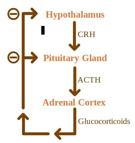
The pituitary gland lies roughly in the center of the human skull. It rests below the hypothalamus of the brain and behind the bridge of our noses. Its setting actually makes sense, in light of the hypothalamus’s role in fine tuning the activity of the pituitary gland. This is made possible by the nerve fibers that span these two structures and allow for easy communication. Likewise, a thin vascular connection that is forged within the pituitary stalk, or infundibulum, facilitates the hypothalamus’s control. Further, the pituitary gland itself is supplied by branches off of the internal carotid artery. Its regulation is fine-tuned by a negative feedback relationship between the pituitary and hypothalamus.


ACTH Negative Feedback

The concept map illustrates the complex regulatory relationship between the superseding hypothalamus and the pituitary gland. The relationship follows a negative feedback loop.


Structurally speaking, the pituitary gland is notably parsed into three sections: the anterior (front), intermediate, and posterior (back) lobes. Each can be described according to their unique functions. The anterior lobe has primary roles in the development of the human body. This involves secreting hormones that orchestrate our reproduction and sexual maturation. These hormones will control growth as well as activate the adrenal and thyroid glands and sexual organs. The intermediate lobe will secrete hormones that stimulate the cells in our body that produce pigment, called melanocytes. These melanocytes are the reason there is such a variation in our skin color. Lastly, the posterior lobe makes ADH, which is the hormone that allows our kidneys to reabsorb water into the bloodstream to prevent dehydration. Oxytocin is also made in the posterior lobe and will induce contractions during childbirth. While these hormones are supremely important to our species’ survival, they are few among the many hormones made by the pituitary gland.


Pituitary Gland Function


The main function of the pituitary gland lies in its ability to make hormones that retain many of our bodily functions. The front and back lobes are the primary secretory glands. As discussed before, the posterior lobe secretes oxytocin and ADH. Oxytocin not only stimulates uterine contractions to facilitate birth but also causes breast tissue to make milk, in preparation of caring for a child. The anterior pituitary gland has a bigger roster of hormones. It produces prolactin, which like the posterior’s oxytocin will trigger milk production post-partum. Follicle-stimulating hormone (or FSH) is released to stimulate sperm production and egg maturation in women that are able to produce estrogen. Likewise, luteinizing Hormone (LH) will stimulate testosterone release in men and egg release in ovulating women. One of the most important products of the anterior lobe is thyroid-stimulating hormone (TSH). The thyroid helps coordinate metabolic activity, and likewise, the TSH will stimulate thyroid activity. Therefore, TSH indirectly allow the thyroid to assume all of its roles. Adrenocorticotropic (ACTH) is released as well and will stimulate the creation of stress hormone, cortisol. Cortisol is essential to our survival and will keep our blood pressure and sugar levels at a healthy normal – of course, in healthy amounts. Any over or under expression has negative consequences. Lastly, the anterior pituitary lobe also releases growth hormone (GH), which is responsible for the muscle and bone mass growth that occurs during development. When growth is unaccounted for, as with a dysregulation of GH production, it can lead to serious illness, if not cancer as we will discuss below.


Posterior Pituitary Lobe:


  • ADH

  • Oxytocin


Anterior Pituitary Lobe:


  • Prolactin

  • Follicle Stimulating Hormone (FSH)

  • Luteinizing Hormone (LH)

  • Thyroid-Stimulating Hormone (TSH)

  • Adrenocorticotropic (ACTH)

  • Growth Hormone (GH)


Pituitary Gland Disorders


Deficiencies in any of the hormones mentioned above can cause illness, which vary in gravity. Starting with the posterior lobe, a deficiency of ADH will increase our thirst and urination. A lack of prolactin will quite predictably lead to an inability to lactate, which to this day cannot be treated. TSH deficiency has symptoms similar to those from a compromised thyroid gland, which includes fatigue, memory loss, and bodily weakness. A lack of LH or FSH will result in a decrease in libido, irregular menses, erectile dysfunction, and mood changes. ACTH deficiency will cause nausea, body aches, poor appetite, and even low blood sugar and pressure. Lastly, deficiency growth hormone will lower muscle mass and bone density, which has long term ramifications on the quality of our lives.


An overproduction of hormone has its own consequences. Too much growth hormone can lead to gigantism and acromegaly, or too much growth of bones and soft tissues leading to heart issues and sleep apnea. Too much TSH will result in shakiness, irritability, and high blood pressure. Too much prolactin will cause inappropriate expression of breast milk that can occur in women or men, and a weakening of bones. Excess ACTH will cause weight gain among brittle bones and mood instability. Lastly, excess FSH and LH are linked to infertility and irregular menstruation.


The most common type of pituitary gland disorder, however, are tumors. The grand majority of pituitary tumors are benign, or just a noncancerous swelling in the gland that may not cause any symptoms and may never be symptomatic. Unlike many types of tumors, most people afflicted with pituitary tumors have no prior family history of issues with the pituitary gland and is not usually genetically inherited. One of these exceptions is multiple endocrine neoplasia (or MEN) which is a set of inherited disorders that lead the body’s endocrine glands, including the pituitary gland, to overexpress hormones. But pituitary tumors, specifically, still remain by and large benign.


Tumors on the Pituitary Gland


Various types of pituitary tumors exist. In general, people with a pituitary gland tumor will experience a series of telltale symptoms. Most will have vision problems, headaches, menstrual changes, infertility, mood changes, fatigue, and even Cushing’s syndrome – which has its own set of symptoms including, but not limited to, high blood pressure and weight gain secondary to too much ACTH release.


The most common type of pituitary tumor is called a “non-functioning” tumor. The name derives from its inability to make hormones. These patients will have issues with their vision and headaches. Furthermore, pituitary tumors can be divided into three groups according to their problematic actions.


Hypersecretion refers to the making of too much hormone, and this is an issue afflicted by a secretory pituitary tumor. Hyposecretion, in contrast, is too little hormone production and is normally caused by a large pituitary tumor that will physically block the pituitary gland from making hormone. It can also result from surgical resection of a tumor. Lastly, tumor mass effects are the issues that arise from a growing pituitary tumor that is pressing against the pituitary gland and may result in compromised vision and headaches, as well.


Other pituitary conditions worth noting include craniopharyngioma. This is a type of cyst or tumor that is congenital, meaning it is present at birth. It can swell and fill with fluid, and may cause headaches and vision issues as well as sleep issues. ESS, or empty sella syndrome is a disorder that arises from an affliction in the bony structure that encases the brain and surrounds the pituitary. A primary ESS will be a small defect that give rise to high pressure in the bony base that causes the gland to flatten. This is linked to high blood pressure and obesity in females. On the other hand, secondary ESS will result from surgery or an injury that has caused the pituitary gland to regress. The symptoms will be related to pituitary function loss, such as infertility and fatigue.


Quiz


1. Which of the following is released by the posterior pituitary?
A. Prolactin
B. ACTH
C. Oxytocin
D. Growth Hormone

Answer to Question #1

2. Match the correct symptom with a deficiency in ACTH, per the article:
A. Erectile dysfunction
B. Nausea
C. Thirst
D. Reduced muscle mass

Answer to Question #2

3. Match the correct symptom with an overexpression of LH, per the article:
A. Infertility
B. Weakened bones
C. Weight gain
D. Acromegaly

Answer to Question #3

References


  • Hormone Health Network. “Pituitary Disorders.” Hormone Health Network. Retrieved on 2017-07-28 from http://www.hormone.org/diseases-and-conditions/pituitary

  • Cancer Editorial Board (2016). “Pituitary Gland Tumor: Syptoms and Signs.” Cancer.Net. Retrieved on 2017-07-29 from http://www.cancer.net/cancer-types/pituitary-gland-tumor/symptoms-and-signs

  • Health Line Medical Team (2017). “Pituitary Gland.” Health Line. Retrieved on 2017-07-28 from http://www.healthline.com/human-body-maps/pituitary-gland

  • Pituitary Foundation (2017). “What is the pituitary gland?” Pituitary. Retrieved on 2017-07-29 from https://www.pituitary.org.uk/information/what-is-the-pituitary-gland/


Pituitary Gland