Friday, April 21, 2017

Top 10 Money Administration Tips For College Students

For many people, choosing a dog for family is as simple since finding a cute puppy. The truth is however , this method is looking for problems and its the number one reason canines end up in a shelter. Choosing the right puppy dog for your family is an organized procedure and by following a couple of simple steps, you can get a puppy gowns perfect for your lifestyle.


Let’s explore that a little more. Think about most professionals. I am just talking about an established professional having a business built around his/her name. The name of the business might be something OTHER than their real name, but none-the-less these are still the name and encounter of their company.


A list of target viewers is what you really need. If you possibly can get a list of subscribers that are really interested in your business, you may make large money from them. E-mail marketing campaign should started having a target. A massive database, unless of course grown organically, will almost certainly become scraped together with random emails of people with different interests, demographics, requirements etc . The best way to boost the convert rate is to supply useful and interesting info for audiences.


It is still probable to be a top earner, best producer, top affiliate, best salesman, and most importantly a highly regarded leader. It will take a lot of excitement to climb the step ladder at Nu Skin. It might take some help from your close friends, family, and other like-minded business owners.


I cannot stress this enough, becoming different and making you aren’t letter connect with the reader ought to be the first goal. In my opinion, the assistance I got from a resume plus cover letter program I got produced the difference. My results were far better when I followed the tip on the program and I was known as in to more interviews. I actually didn’t expect to walk into any interview and property the job I wanted to create a lot more opportunities for face to face conversation. Your letter is your marketing and advertising, make it a good piece that will sells you and sets a person apart.


Alex: Ah, that’s the crux from it, isn’t it? What do you say that people save the nuts plus bolts of the exact way of next time Cole. That way we are able to get back together in the not as well distant future.


Included in the educational experience, I get my kids on a lot of industry trips. We particularly such as the Museum of Science plus Nature. We usually carry on weekdays. The weekends are very crowded. It’s totally “legal” to spend the day at the Technology Museum and call it college. Ditto for the zoo and also a bunch of other locations.


Top 10 Money Administration Tips For College Students

Friday, April 14, 2017

Spirochete

Spirochete Definition


A spirochete is a type of bacteria within the phylum Spirochetes. The spirochetes are so called because they are very long, thin shape and helically coiled, hence they have a spiraling corkscrew shape. The helical shape is one of three morphological categories of the prokaryotes.


The spirochetes are a diverse phylum, occupying a wide range of ecological niches; some are free-living in aquatic environments, while some can only survive by parasitizing the cells of other organisms. Most are anaerobic (can sustain themselves without the presence of oxygen), although certain species are aerobic. They are very difficult to culture and are gram-negative, meaning they are not easily visible.


Although some play an important role as symbionts within the stomach of other animals, many members of the spirochete class are responsible for common diseases such as Lyme disease and syphilis. Some spirochetes have also been found present within marine bivalves although with no apparent positive or negative effects; this neutral relationship is called a commensalism.


Spirochetes are distinguishable from other bacteria in that they move with unique endoflagella. The flagella are tightly wound around the corkscrew shape of the bacteria, between the outer membrane and the cell wall, within the periplasm. Together, the endoflagella make up a structure called the axial filament. The axial filament is rotated by the rotation of the flagella, causing the spirochete to move with a twisting motion. This method of motility is unique to the spirochetes and—much like the way a corkscrew can penetrate the cork of a wine bottle—allows them to move through viscous materials such as mucus, blood, mud and host connective tissues including cartilage and dental plaque; external flagella do not allow effective movement through these mediums.


Additionally, the presence of endoflagella allows the spirochetes to move backward and forward with equal ease, this allows them to move freely in their environment and target the best site of host attachment or resources.


Spirochaete Schemamobility

The image shows the spiral structure of the spirochete form (top) and a cross section (bottom) with the following labeled: 1-Bacterial Cell Envelope; 2-Cytoplasm; 3-Flagella; 4-The Attachment Point of the Flagella.


Spirochetes as Parasites


Spirochetes are best known by their prevalence as causative agents of disease. Three genera contain pathogenic species:


  • Treponema: The Treponema palidum species causes the sexually transmitted disease, syphilis. Closely related to this is are three other species which cause yaws, pinta and bejel; these are diseases transmitted either sexually or through skin contact and result in symptoms of mouth and skin sores, lumps in the bone, growths and thick discolored patches of skin. These bacteria can be killed with a penicillin injection.

  • Leptospira: Certain species within this genus cause a disease called Leptospirosis (also known as Weil’s disease). The symptoms of this are flu-like; including fever, headaches, and muscle pains, although can become as severe as meningitis and can cause bleeding into the lungs.

  • Borrelia: There are 52 species of the Borrelia genus, which transmit through the bites of ticks and lice. Of the 52 species, 21 are responsible for causing borreliosis (also known as Lyme disease) while 29 cause Relapsing Fever. Relapsing fever victims experience fever, chills, headaches, nausea and rashes. If left untreated, the symptoms often subside and then return several weeks later.


Lyme Disease


Lyme disease is a common disease, transmitted by tick bites after they have been attached for at least 24 hours. Usually a small rash appears on the body, which expands over several days. As the disease progresses, symptoms can include severe headaches, neck stiffness, loss of muscle use in the face, pains and numbing of the hands and feet, palpitations and irregular heartbeat, arthritis, and inflammation of the brain and spiral chord. In some cases Lyme disease is fatal.


Lyme disease is hard to cure unless it is caught immediately after infection and symptoms often do not occur for several days or even weeks after transmission. This is because of evolved features in the Lyme-causing spirochetes, which provide a high chance of transmission and the ability to evade the immune system of their host. Firstly, when the tick feeds, a surface protein of the Borrelia binds to a protein within the saliva of the tick. This protein coats the spirochete and makes it undetectable to the immune system. Because antibodies are not produced to fight the intruder, the spirochete is free to establish itself before it is recognized. In the following weeks the bacterium spreads throughout the body—it is able to do so effectively because of the motility provided by the endoflagella system.


The movement of the flagella stimulates the immune system, however the spirochete is able to alter the expression of proteins of its outer cell wall in order to ‘disguise’ it. This makes it extremely difficult for the immune system to produce the correct antibodies to attack the bacteria. The immune system responds by producing multiple types of antibody, which fail to kill the bacteria due to the changing protein structure but have a toxic effect on the host, causing inflammation and damage to tissues throughout the body


The spirochetes are also able to ‘cloak’ themselves within the extracellular matrix of the host cells, making it easier still to avoid detection. They have also been found to enter the lymph nodes, where antibodies are produced, and alter the normal function of antibody production.


Finally, the Borrelia have been found to protect themselves using a biofilm—a slimy film substance, which they generate themselves, containing the bacteria and other microorganisms. The biofilm protects the bacteria from the harsh conditions created by antibiotics and allows the invader to lie dormant for long periods of time until the conditions are more favorable and they can begin the attack again.


Spirochetes as Symbionts


Not all spirochetes are parasitic, in fact many of them hold symbiotic relationships or mutualisms with their host; in this case, both organisms depend on the other for their survival.


Symbiosis with Termites


Within the species of termite Mastotermes darwiniensis is a wood eating flagellate called “Mixotricha“. The surface of the mixotricha is covered by Treponema spirochetes, giving the appearance of cilia. The spirochetes use coordinated undulation to propel the symbionts through the intestinal fluid of the termite, while the anterior flagella of the mixotricha is used for steering.


The mixotricha helps to break down the cellulose within the wood that the termite has eaten into sugars, hydrogen, carbon dioxide and acetate. It is thought that the spirochetes oxidize the acetate, and use it to support the respiration requirements of the termite, whilst deriving nutrition from the other products produced by the mixotricha.


Symbiosis with Ruminants


Ruminants such as cows, deer, sheep, giraffes and elk have four chambers within their stomachs in which to break down hard-to-digest cellulose. The first chamber of the stomach is the ’rumen’. This chamber contains a diverse range of fungi, bacteria and protozoans, which secrete enzymes that the ruminant cannot produce independently, in order to digest the cellulose in the plant matter. Treponema spirochetes make up a small percentage of the organisms found within the rumen and, in a similar way to those found within termite intestines, aid and enhance the breakdown of cellulose, while using the by-products produced by the other bacteria for their growth.


The three other chambers of the stomach, the ‘reticulum’, the ‘obassum’ and the ‘abomasum’ perform functions such as absorption of liquids into the body and true digestion with gastric acids.


Related Biology Terms


  • Bacteria – A group of single-celled organisms, which have cell walls although lack organelles such as nuclei or mitochondria.

  • Parasite – An organism that interacts in some way with another ‘host’ organism in order to benefit itself, while (intentionally or not) causing damage to host.

  • Flagellum – A thread-like appendage, which enables microorganisms such as bacteria and protozoa to swim or move.

  • Symbiosis – The relationship between two organisms in both are dependent on the other either in part or the whole of their life cycles.

Quiz


1. In which way are the spirochetes unique among bacteria?
A. They are parasitic
B. They are smaller than other bacteria
C. The presence of endoflagella
D. They can survive in different environments

Answer to Question #1

2. Which of the following is NOT a method utilized by Treponema spirochetes to evade the immune system of their host?
A. Changing the protein expression of the outer membrane
B. Hiding within the extra cellular matrix of the host cells
C. Use protein coating from the tick saliva to disguise it
D. Fighting against the antibodies produced by the host

Answer to Question #2


Spirochete

Deuterostome

Deuterostome Definition


The Deuterostomes are a clade of animals that undergo deuterostomy during their embryonic development. They are a sister-clade of the Protostomes, and the two together with the Xenacoelomorpha form the major group of animals called the Bilateria—a major group animals which display bilateral symmetry and are mostly triploblastic.


Deuterostomy


During embryonic development, the fused gametes from the male and female—the sperm and the egg—form the zygote.


In order to develop, the zygote undergoes a process called cleavage. Cleavage involves splitting into multiple cells called blastomeres, and results in a dense ball of these cells called a morula. In deuterostomy, radial cleavage occurs, whereby the blastomeres are arranged along a central axis and is characterized by several tiers of cells stacked on top of each other. Radial cleavage is one of the defining features of the deuterostome development, contrasting the spiral cleavage that is typical of the protostomes. Additionally, most of the deuterostomes display indeterminate cleavage, in which the developmental fate of each cell is not predetermined in the embryo and therefore each cell has the ability to develop into a complete embryo if isolated.


Protovsdeuterostomes

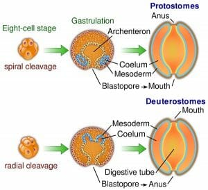
The image shows the defining differences between deuterostome and protostome embryonic development.


The blastula is the resulting structure, consisting of at least 128 cells surrounding a cavity of mainly empty space, called the blastocoel. Through a process called gastrulation, the cells of the blastula are reorganized to form the three primary germ layers of the gastrula that are present in all triploblastic organisms. Gastrulation begins with a small indentation in the blastula called the blastopore, the cells of which migrate to the opposite end of the embryonic structure, establishing the endoderm layer; the endoderm eventually gives rise to the digestive system.


In deuterostomes, the first cavity formed by the blastopore ends up as the organism’s anus, while the mouth is formed secondarily on the opposite side. This is the next major distinction between deuterostomes and protostomes; the protostomes form the mouth from the primary cavity and the anus second.


It is useful to note that the two names are derived from the Greek proto- “first” and deutero- “second”, and stoma meaning “mouth”. The deuterostomes develop a “second-mouth”.


In many egg-laying deuterostomes the peripheral layer of cells in the gastrula forms the ectoderm, which ultimately gives rise to the epidermis (the skin and hair) and the nervous system. In between the endoderm and the ectoderm is the mesoderm, which ends up as connective tissues, skeletal system, blood, the heart and kidneys and muscle.


In mammal development the outer layer of the blastula equivalent—the blastocyst—becomes the placenta and the inner cells give rise to the three primary germ layers.


Types of Deuterostome


The Deuterostomes can be taxonomically grouped into three clades.


Echinodermata


The echinoderms are a group of marine animals, which although are radially symmetrical in adult life, display bilateral symmetry in their larval stage and are thus classed within the Bilateria.


The echinoderms have anendoskeleton just below the skin made from calcium carbonate which provides rigidity and protection. Additionally, they have a hydrostatic skeleton—a fluid filled cavity present in many developed animals called the coelom, supported by hydrostatic pressure to allow movement.


Many echinoderms have structures called ‘tube feet’, which they use to grasp substrate in order to move, as well as for feeding and respiration. Predatory species use the tube feet to pry open bivalves and then feed by extruding the stomach out of the mouth to digest the prey. Non-predatory species use the tube feet for suspension feeding, whereby they flick food to their cilia, which then pass the food into the mouth.


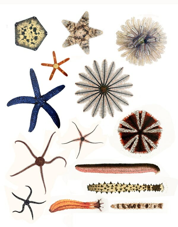
The Echinoderms are separated into six taxonomic classes.


  • Crinoidea—Feather stars and sea lilies

  • Asteroidea—Sea stars

  • Ophiuroidea—Brittle stars

  • Echinoidea—Sea urchins and sand dollars

  • Holothuroidea & Concentricycloidea—Sea cucumbers & Sea Daisies


Echinodermata

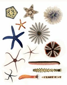
The image above shows an illustrative example of echinoderms from each taxonomic class.


Chordates


The chordates are a phylum of animals within the deuterostomes, which have the following common similarities:


  • A notochord. A flexible, supportive rod, made from material similar to cartilage. In the vertebrates this is replaced by the vertebral column during development.

  • A hollow dorsal nerve chord—This is formed from the ectoderm and runs the length of the body. In vertebrates, this makes up the central nervous system.

  • A post-anal tail. A tail that extends beyond the anus in at least some point of their development.

  • Pharyngeal gill slits in at least some point of their development. These are openings within the throat that allow the animal to breathe underwater. In marine organisms these become functioning gills, and in terrestrial animals they are modified for alternative functions.


Note that: All vertebrates are chordates—not all chordates are vertebrates.


The chordates can be separated into 3 subphyla:


The Cephalochordata


These cephalochordates are small invertebrate marine animals known as lancelets. They are simple fish-like organisms, which live with their tails buried in the sand and employ a filter feeding system. They are most likely the closest link between the chordates and other simple organisms.


The Urochordata


The urochordata includes the tunicates—also known as ‘sea squirts’. These invertebrate organisms are usually sessile and possess a U-shapes gut and two siphons, which allows them to take in food. The gills slits are modified in the adult form to allow filter feeding.


Sea Squirts

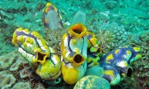
The image shows sea squirts, members of the urochordata which intake food and through their siphons (the visible holes).


The Vertebrata


The vertebrata is the largest subphylum within the chordates and the most morphologically complex. In addition to the typical characteristics of chordates, the vertebrates all posses a skull or cranium, which encases the brain and a backbone or vertebral column, which protects the dorsal nerve chord and internal organs as well as providing support.


They are also notable for the following evolutionary developments:


  • A hinged jaw, which allows the animal to capture food in a highly effective way.

  • The amniotic egg, containing a protective inner membrane through which gases and nutrients can be transferred to the embryo during development.

  • Limbs, either as fins or evolved into legs for improved movement ability.

  • The evolution of the pharyngeal gills into lungs.


The vertebrates are separated into seven extant taxonomic classes:


  • Agnatha—The hagfish and lampreys. Although they have a skull and basic vertebrae, these lack a jaw and vertebral column.

  • Condrichthyes—The sharks, skates, rays and sawfish. These have gills and an endoskeleton made from cartilage.

  • Osteichthyes—The bony fish. These have gills, an endoskeleton made from bone, and a ‘swim bladder’, which helps with depth control.


The ‘tetrapods’ are four-limbed vertebrates within the chordates:


  • Amphibia—Frogs, toads and salamanders. These are both marine and terrestrial organisms. Although in the adult form most of them have lungs, they can also breathe through their skin.

  • Reptilia—Turtles, snakes, crocodiles, lizards. These are ectothermic animals with scales and lungs.

  • Aves—Birds. These are endothermic animals with feathers and beaks.

  • Mammalia—These are endothermic amniotes with the defining characteristics of: 1)hair, which aids in insulation 2)mammary glands for producing milk 3)a neocortex, which allows complex brain function 4)three middle ear bones for enhanced hearing sensitivity 5)internal fertilization


Within the mammalia are the Eutherians or ‘placental mammals’; a group which includes the primates, such as monkeys and humans, cetaceans (whales and dolphins), rodents, cats, dogs and most other animals that are familiar to us.


The class Mammalia also includes the marsupials such as kangaroos, wombats and opossums, in which the offspring are born under-developed and complete development within a maternal pouch on the mother’s stomach.


Further included are the Monotremes, of which only the duck billed platypus and echidna species are extant. The monotremes are mammals that lay hard shelled eggs, additionally they lack nipples, so secrete milk through specialized hair follicles.


Hemichordata


The hemichordates are marine deuterostomes, which are characterized by a body that is comprised of three distinct sections: The anterior (front) prosome, the middle mesosome and the posterior (back end) metasome.


These are generally worm-like filter feeders, deposit feeders and detritivores and are considered to be the closest existing relatives to the vertebrates. Like other chordate deuterostomes, the hemichordates have pharyngeal gill slits and most have a dorsal nerve chord, although they lack the notochord.


They are divided into two classes:


  • Enteropneusta — The acorn worms. These can reproduce both through sexual and a-sexual reproduction.

  • Pterobranchia


Related Biology Terms


  • Protostomes – A clade of animals in which spiral cleavage occurs during embryonic development and the blastopore develops into the mouth.

  • Coelom – The fluid filled cavity present in most animals, which surrounds the digestive tracts and other organs.

  • Phylogenetic Tree – A diagram representing the evolutionary relationships between living organisms.

  • Bilateral Symmetry – A characteristic of the Bilateria Clade, in which the two sides of the body are mirror images of each other.

Quiz


1. Which form of cleavage is characteristic of all deuterostomes?
A. Radial cleavage
B. Spiral cleavage
C. Indeterminate cleavage
D. Rotational cleavage

Answer to Question #1

2. Which of the following is a feature not associated with the chordates?
A. Dorsal nerve chord
B. Hydrostatic skeleton
C. Pharyngeal gill slits
D. Amniotic egg

Answer to Question #2

3. In the deuterostomes, what is the fate of the blastopore?
A. The mesoderm
B. The mouth
C. The anus
D. The placenta

Answer to Question #3


Deuterostome

Gap Junction

Gap Junction Definition


Gap junctions are a type of cell junction in which adjacent cells are connected through protein channels. These channels connect the cytoplasm of each cell and allow molecules, ions, and electrical signals to pass between them. Gap junctions are found in between the vast majority of cells within the body because they are found between all cells that are directly touching other cells. Exceptions include cells that move around and do not usually come into close contact with other cells, such as sperm cells and red blood cells. Gap junctions are only found in animal cells; plant cells are connected by channels called plasmodesmata instead.


Function of Gap Junctions


The main function of gap junctions is to connect cells together so that molecules may pass from one cell to the other. This allows for cell-to-cell communication, and makes it so that molecules can directly enter neighboring cells without having to go through the extracellular fluid surrounding the cells. Gap junctions are especially important during embryonic development, a time when neighboring cells must communicate with each other in order for them to develop in the right place at the right time. If gap junctions are blocked, embryos cannot develop normally.


Gap junctions make cells chemically or electrically coupled. This means that the cells are linked together and can transfer molecules to each other for use in reactions. Electrical coupling occurs in the heart, where cells receive the signal to contract the heart muscle at the same time through gap junctions. It also occurs in neurons, which can be connected to each other by electrical synapses in addition to the well-known chemical synapses that neurotransmitters are released from.


When a cell starts to die from disease or injury, it sends out signals through its gap junctions. These signals can cause nearby cells to die even if they are not diseased or injured. This is called the “bystander effect”, since the nearby cells are innocent bystanders that become victims. However, sometimes groups of adjacent cells need to die during development, so gap junctions facilitate this process. In addition, cells can also send therapeutic compounds to each other through gap junctions, and gap junctions are being researched as a method of therapeutic drug delivery.


Gap Junction Structure


In vertebrate cells, gap junctions are made up of connexin proteins. (The cells of invertebrates have gap junctions that are composed of innexin proteins, which are not related to connexin proteins but perform a similar function.) Groups of six connexins form a connexon, and two connexons are put together to form a channel that molecules can pass through. Other channels in gap junctions are made up of pannexin proteins. Relatively less is known about pannexins; they were originally thought only to form channels within a cell, not between cells. Hundreds of channels are found together at the site of a gap junction in what is known as a gap junction plaque. A plaque is a mass of proteins.


Gap cell junction

This figure depicts channels at a gap junction.


Other Cell Junctions


The two other types of cell junctions in vertebrates are anchoring junctions and tight junctions. Anchoring junctions adhere cells through proteins that are connected to the cell’s cytoskeleton. Tight junctions are areas where cells are bound very closely together to form a barrier, and they are often found in epithelial cells, which are cells found on the surface of the body and lining organs.


Plant cells do not have gap junctions, but they do have plasmodesmata, which are channels that connect the cytoplasm of two adjacent plant cells. Plasmodesmata are structured differently than gap junctions due to plant cells having thick cell walls, their function is essentially the same. Plant cells can regulate the passage of small molecules and communicate with each other through their plasmodesmata.


Related Biology Terms


  • Anchoring junction – A type of cell junction in which cells are connected by a mass of proteins.

  • Tight junction – A type of cell junction where cells are tightly bonded to form a barrier.

  • Plasmodesmata – Channels that connect the cytoplasm of adjacent plant cells.

  • Connexin – A family of proteins that makes up gap junctions.

Quiz


1. How many connexins are found in one gap junction channel?
A. 6
B. 4
C. 12
D. 2

Answer to Question #1

2. What is the “bystander effect” in relation to gap junctions?
A. Molecules can enter neighboring cells without passing through extracellular fluid.
B. Cells next to a cell that is undergoing cell death can also die.
C. Cells can transmit therapeutic compounds to one another.
D. Gap junctions are only found in cells that are located next to other cells.

Answer to Question #2

3. Which is NOT a function of gap junctions?
A. Forming a barrier
B. Allowing molecules to pass between cells
C. Electrically coupling cells
D. Ensuring correct embryonic development

Answer to Question #3

References


  • Alberts, Bruce, et al. (1994). Molecular Biology of the Cell, 3rd. ed. Ch. 1. Garland Science: New York. ISBN: 978-0815316206.

  • Davidson, Michael W. (2015-11-13). “Plasmodesmata”. Molecular Expressions Cell Biology. Retrieved 2017-04-13 from https://micro.magnet.fsu.edu/.

  • Evans, W.H. and Martin, P.E. (2002). “Gap junctions: structure and function (Review)”. Mol. Membr. Biol. 19 (2): 121–36.

  • Kimball, John W. (2015-03-02). “Junctions Between Cells”. Kimball’s Biology Pages. Retrieved 2017-04-12 from http://www.biology-pages.info/.

  • Russell, Peter. J., and Hertz, Paul E. (2011). Biology: The Dynamic Science, 2nd ed. Ch. 5. Brooks/Cole: Boston. ISBN: 978-0538494182.

  • Wei, C.J., Xu, X., and Lo, C.W. (2004). “Connexins and cell signaling in development and disease”. Annu. Rev. Cell Dev. Biol. 20: 811–38.


Gap Junction

Tight Junctions

Tight Junctions Definition


Tight junctions are areas where the membranes of two adjacent cells join together to form a barrier. The cell membranes are connected by strands of transmembrane proteins such as claudins and occludins. Tight junctions bind cells together, prevent molecules from passing in between the cells, and also help to maintain the polarity of cells. They are only found in vertebrates, animals with a backbone and skeleton; invertebrates have septate junctions instead.


Function of Tight Junctions


Tight junctions have several different functions. Their most important functions are to help cells form a barrier that prevents molecules from getting through, and to stop proteins in the cell membrane from moving around. Tight junctions are often found at epithelial cells, which are cells that line the surface of the body and line body cavities. Not only do epithelial cells separate the body from the surrounding environment, they also separate surfaces within the body. Therefore, it is very important that the permeability of molecules through layers of epithelial cells is tightly controlled.


If molecules are blocked by tight junctions and physically unable to pass through the space in between cells, they must enter through other methods that involve entering the cells themselves. They could pass through special proteins in the cell membrane, or be engulfed by the cell through endocytosis. Using these methods, the cell has greater control over what materials it takes in and allows to pass through. However, in endothelial cells, certain proteins must be kept on certain sides of the cell. The apical, or outside layer, of the sheet of cells contains proteins that only let certain substances pass through. The basal, or inside layer, is where cells let molecules pass through them by expelling them from their membrane in a process called exocytosis. Exocytosis also relies on specific proteins in order to work correctly. Tight junctions keep the correct proteins on the correct sides of the cell in order for these functions to occur. This also helps maintain the polarity of cells.


Another function of tight junctions is simply to hold cells together. The branching protein strands of tight junctions link adjacent cells together tightly so that they form a sheet. These strands are anchored to microfilaments, part of the cell’s cytoskeleton that is made up of long strands of actin proteins. Microfilaments are located inside the cell, so the combination of microfilaments and sealing strands anchors the cells together from the inside and the outside.


Structure of Tight Junctions


Cellular tight junction

This diagram depicts a tight junction between cells and also provides examples of proteins found in the junction.


Tight junctions are a branching network of protein strands on the surface of a cell that link with each other throughout the surface of the membrane. The strands are formed by transmembrane proteins on the surfaces of the cell membranes that are adjacent to each other.


There are around 40 different proteins at tight junctions. These proteins can be grouped into four main types. Transmembrane proteins are wedged in the middle of the cell membrane and are responsible for adhesion and permeability. Scaffolding proteins organize transmembrane proteins. Signaling proteins are responsible for forming the tight junction and regulating the barrier. Regulation proteins regulate what proteins are brought to the cell membrane in vesicles.


Claudins and occludins are the two main types of proteins present at tight junctions, and they are both transmembrane proteins. Claudins are important in forming tight junctions, while occludins play more of a role in keeping the tight junction stable and maintaining the barrier between cells that keeps unwanted molecules out.


Other Cell Junctions


Tight junctions are one of three main types of junctions in vertebrate cells. The other two types are gap junctions and anchoring junctions. Gap junctions, also known as communicating junctions, are channels in cells that let adjacent cells communicate with one another without having to send molecules through the extracellular fluid surrounding the cell. Connexin proteins form the channel, which has a central pore called a connexon that molecules can pass through. Anchoring junctions hold cells together with anchoring proteins such as catenins and cadherins. The cell’s cytoskeleton is tethered to proteins that link adjacent cells.


Related Biology Terms


  • Anchoring junction – A type of cell junction in which cells are connected by a mass of proteins.

  • Gap junction – A type of cell junction that allows adjacent cells to exchange molecules.

  • Cytoskeleton – A network of protein filaments that extends throughout the cell.

  • Epithelial cells – Cells that line the interior surfaces of organs and are also found on the surface of the body.

Quiz


1. Which animal does not have tight junctions in between any of its cells?
A. Cat
B. Squid
C. Bird
D. All animals have tight junctions between some of their cells.

Answer to Question #1

2. Why is it important for tight junctions to keep all cell membrane proteins in place?
A. Different processes occur on the apical and basal sides.
B. It maintains polarity.
C. If the proteins move, they may let in different molecules.
D. Both A and B

Answer to Question #2

3. What is the role of scaffolding proteins?
A. To form the tight junction
B. To regulate the proteins that are brought to the cell membrane
C. To maintain adhesion and permeability
D. To organize transmembrane proteins

Answer to Question #3


Tight Junctions

Desmosomes

Desmosomes Definition


Desmosomes are a type of anchoring junction in animal tissues that connect adjacent cells. Anchoring junctions are button-like spots found all around cells that bind adjacent cells together. Desmosomes have intermediate filaments in the cells underneath that help anchor the junction, while the other type of anchoring junction, an adherens junction, is anchored by microfilaments. Intermediate filaments and microfilaments are two different components of a cell’s cytoskeleton.


Function of Desmosomes


The function of desmosomes is to adhere cells together. They are found in high numbers in tissues that are subject to a lot of mechanical forces. For example, many are found in the epidermis, which is the outer layer of skin, and the myocardium, which is muscle tissue in the heart. They are also found in between squamous epithelial cells, which form the lining of body parts like the heart, blood vessels, air sacs of the lungs, and esophagus.


Desmosome Structure


There are three components in desmosomal adhesion: the intermediate filaments inside the cell, the bond between intermediate filaments and desmosomal adhesion molecules, and the bond provided by the desmosomal adhesion molecules. The intermediate filaments and their link to the desmosomal adhesion molecules are both located inside the cell, while the bonds of the desmosomal adhesion molecules themselves are on the outside of the cell. Specifically, desmoglein and desmocollin are the two proteins that bind cells at desmosomes. They are transmembrane proteins and are both members of the cadherin family of proteins. All three components of desmosomal adhesion are necessary for desmosomes to properly function in binding adjacent cells together, so if one of the components fails, the desmosomes cannot bind cells properly.


Desmosome

This diagram depicts how cells adhere at desmosomes.


Disorders of Faulty Desmosome Functioning


Epidermolysis Bullosa Simplex


Epidermolysis bullosa simplex (EB) is a genetic condition that causes skin to be fragile and blister extremely easily. This disorder is caused by a mutation in the gene coding keratin proteins found in the intermediate filaments. The intermediate filaments do not form properly in the epidermis, so skin cells are not bound together properly and can easily break apart. It occurs in 1 in 30,000 to 50,000 people, with some cases being more severe than others. In mild cases, blistering is usually confined to the hands and feet, areas that experience a lot of mechanical stress. In more severe cases, widespread blistering can occur all over the body. There is no cure for EB, but blistering can be prevented by strategies like wearing loose clothing, keeping cool, and avoiding walking long distances.


Ectodermal Dysplasia/Skin Fragility Syndrome


Ectodermal dysplasia/skin fragility syndrome is a rare genetic disease caused by a mutation that leads to intermediate filament detachment from desmosomes. It causes frequent blistering, abnormal hair, tooth, and nail growth, frequent infections, and defective sweating. Sometimes, affected individuals must use a wheelchair due to extreme blistering on the soles of their feet. Very few cases of this disorder have been reported.


Pemphigus


Pemphigus is a rare skin disorder caused by faulty desmosomal adhesive binding. In a person with pemphigus, the body produces antibodies against the protein desmoglein. When desmoglein is attacked by the immune system, the binding between cells breaks. It causes blisters and sores on the skin and mucous membranes. There are two main types of phemphigus, pemphigus vulgaris and pemphigus foliaceus. Pemphigus vulgaris occurs around the mouth and is painful, while pemphigus foliaceus occurs on skin and causes itchiness. Skin lesions caused by pemphigus can lead to fatal infections, so treatment is extremely important. Pemphigus can be treated via steroids and immunosuppressant drugs, among other medications.


Arrhythmogenic right ventricular dysplasia


Arrhythmogenic right ventricular dysplasia (ARVD), also called arrhythmogenic right ventricular cardiomyopathy, is an inherited heart condition caused by desmosome defects in heart muscle cells. It is characterized by arrhythmia in the right ventricle of the heart. Symptoms include heart palpitations, fainting, and shortness of breath. One in 5000 people have ARVD. It is treated by implantation of a small defibrillator. ARVD is progressive and can lead to right ventricular failure; the left ventricle can also be weakened. It can cause sudden cardiac death in athletes, and accounts for 1/5 of all sudden cardiac deaths in people under 35.


Related Biology Terms


  • Anchoring junction – A type of cell junction in which cells are connected by a mass of proteins.

  • Adherens junction – A type of anchoring junction that adheres cells via microfilaments.

  • Cadherin – A type of protein that adheres adjacent cells.

  • Intermediate filaments – Components of a cell’s cytoskeleton that are bigger than microtubules but smaller than microfilaments.

Quiz


1. Which component of the cytoskeleton is involved in desmosomal adhesion?
A. Microfilaments
B. Intermediate filaments
C. Microtubules

Answer to Question #1

2. Which component of desmosomal adhesion is absolutely necessary in order to bind adjacent cells?
A. The intermediate filaments
B. The bond between intermediate filaments and desmosomal adhesion molecules
C. The bond that desmosomal adhesion molecules have to each other
D. All of the above

Answer to Question #2

3. Which disorder described in the article has two main types and is characterized by blisters and sores on the surface of the skin and mucous membranes?
A. Epidermolysis bullosa simplex
B. Ectodermal Dysplasia/Skin Fragility Syndrome
C. Pemphigus
D. Arrhythmogenic right ventricular dysplasia

Answer to Question #3


Desmosomes

Microfilament

Microfilament Definition


Microfilaments, also called actin filaments, are polymers of the protein actin that are part of a cell’s cytoskeleton. The cytoskeleton is the network of protein filaments that extends throughout the cell, giving the cell structure and keeping organelles in place. Microfilaments are the smallest filaments of the cytoskeleton. They have roles in cell movement, muscle contraction, and cell division.


Microfilament Structure


Microfilaments are composed of two strands of subunits of the protein actin (hence the name actin filaments) wound in a spiral. Specifically, the actin subunits that come together to form a microfilament are called globular actin (G-actin), and once they are joined together they are called filamentous actin (F-actin). Like microtubules, microfilaments are polar. Their positively charged, or plus end, is barbed and their negatively charged minus end is pointed. Polarization occurs due to the molecular binding pattern of the molecules that make up the microfilament. Also like microtubules, the plus end grows faster than the minus end.


Microfilaments are the thinnest filaments of the cytoskeleton, with a diameter of about 6 to 7 nanometers. A microfilament begins to form when three G-actin proteins come together by themselves to form a trimer. Then, more actin binds to the barbed end. The process of self-assembly is aided by autoclampin proteins, which act as motors to help assemble the long strands that make up microfilaments. Two long strands of actin arrange in a spiral in order to form a microfilament.


MEF microfilaments

This is a micrograph of microfilaments in a mouse embryo.


Functions of Microfilaments


Muscle Contraction


One of the most important roles of microfilaments is to contract muscles. There is a high concentration of microfilaments in muscle cells, where they form myofibrils, the basic unit of the muscle cell. Actin is an indispensable protein for muscle movement, and microfilaments are often called actin filaments because actin is so prominent in the muscular system of the body. In muscle cells, actin works together with the protein myosin to allow the muscles to contract and relax. Here, neither actin nor myosin can work properly without the other, and they form a complex called actomyosin. Groups of actomyosin are found in sarcomeres, the basic unit of muscle tissue.


Cell Movement


Microfilaments play a role in causing cells to move. This occurs throughout the body and it is also very important for organisms whose entire body consists of one cell, such as amoebae; without microfilaments, they would not be motile. Actomyosin plays a role here just as it does in muscle cells. In order for cells to move, one end of a microfilament must elongate while the other end must shorten, and myosin acts as a motor to make this happen.


Microfilaments also have a role in cytoplasmic streaming. Cytoplasmic streaming is the flow of cytoplasm (the contents of the cell, including the fluid part called cytosol and cell organelles) throughout the cell. It allows nutrients, waste products, and cell organelles to travel from one part of the cell to another. Microfilaments can attach to a cell organelle and then contract, pulling the organelle to a different area of the cell.


Cell Division


Another important function of microfilaments is to help divide the cell during mitosis (cell division). Microfilaments aid the process of cytokinesis, which is when the cell “pinches off” and physically separates into two daughter cells. During cytokinesis, a ring of actin forms around the cell that is separating, and then myosin proteins pull on the actin and cause it to contract. The ring gets narrower and narrower around the cell, dragging the cell membrane with it, until it splits into two cells. Afterward, the microfilaments depolymerize, or break down, into actin molecules, causing the ring to dissemble when it is no longer needed.


Other Cytoskeletal Components


The two other types of filaments that make up the cytoskeleton are intermediate filaments and microtubules. Intermediate filaments are larger than microfilaments, with a diameter of about 10 nm, and microtubules are bigger than intermediate filaments at 23 nm. Intermediate filaments bear tension in the cell, give the cell structure, and organize cell organelles and tether them in place. Microtubules have roles in transporting organelles within the cell, forming the mitotic spindle during cell division, and forming structures like cilia and flagella that help certain cells move. Microfilaments, intermediate filaments, and microtubules all work together as part of the cytoskeleton to organize the cell and help it carry out its functions.


Related Biology Terms


  • Actin – The protein that spontaneously comes together to form microfilaments.

  • Cytoskeleton – A network of protein filaments that extends throughout the cytoplasm of the cell.

  • Actomyosin – A complex of the proteins actin and myosin that is responsible for muscle movement.

  • Cytoplasmic streaming – The flow of cytoplasm throughout the cell; it transports molecules and organelles within the cell from one place to another.

Quiz


1. Microfilaments have roles in _____.
A. Cytoplasmic streaming
B. Muscle contraction
C. Cell movement
D. All of the above

Answer to Question #1

2. Are microfilaments wider than, thinner than, or the same size as microtubules?
A. Wider
B. Thinner
C. The same size

Answer to Question #2

3. What protein forms a complex with actin in muscle cells?
A. Lamin
B. Autoclampin
C. Myosin
D. Actomyosin

Answer to Question #3


Microfilament

Wednesday, April 12, 2017

Gamete

Gamete Definition


Gametes are haploid reproductive cells in sexually reproducing organisms that fuse with one another during fertilization. Fertilization produces a diploid cell that undergoes repeated rounds of cell division to produce a new individual. Gametes are the physical carriers of genetic information from one generation to the next. They carry recombinant chromosomes produced at the end of meiosis.


Often, species that reproduce sexually have two morphologically distinct types of individuals that produce different gametes. The larger gamete produced by the female is usually called the egg or ovum. The smaller one is the sperm. Similar distinctions also exist in the plant world, with the female gamete being called the ovule and the male gamete going by the name of pollen.


Types of Gametes


In many species, there are two types of gametes whose form and function are distinct from one another. In humans and other mammals, for instance, the ovum is much larger than the sperm. The sperm also has a distinctive tadpole-like appearance with special adaptations for its primary function of traveling through the female reproductive tract and fertilizing the egg. In a similar manner, the ovum has a number of structural adaptations that aid the process of accurate fertilization and subsequent implantation. Species that have obvious differences in the appearance of gametes are said to display anisogamy.


In addition, most species are also heterogametic – containing a different set of chromosomes in each type of gamete. In mammals, the female gamete contains a single X chromosome in addition to 22 somatic chromosomes. On the other hand, the male gamete, the sperm, could carry either an X or a Y chromosome as the 23rd chromosome. Depending on the chromosome present in the sperm, the resultant diploid zygote could either be a female (XX) or a male (XY). In birds, this form of heterogamy is reversed. Females produce gametes that could contain either the W or the Z chromosome and males produce a single type of gamete.


Examples of Gametes


The two most common gametes are sperm and ova. These two haploid cells can undergo internal or external fertilization and can differ from each other in size, form, and function. Some species produce both sperm and ova within the same organism. They are called hermaphrodites. However, the majority of sexually reproducing organisms have distinct sexes with each producing a single type of gamete.


Structure and Function of Sperm


Human sperms are highly specialized cells that have undergone an extensive period of differentiation.


Sperm


As shown in the image, sperms contain four morphological regions – the head, neck, midpiece, and tail. These generic terms are in fact referring to different subcellular organelles that have been adapted to aid the sperm in its function.


The ‘head’, for instance, contains the genetic material. The DNA in a mature sperm is highly compacted, has nearly non-existent transcriptional activity and all the chromosomes are tightly condensed. They even have special proteins called protamines to pack the DNA more tightly than histones. The head is also surrounded by a cap-like structure containing hydrolytic enzymes called the acrosome. Acrosomal enzymes act on the outer membranes of the egg, allowing the DNA in the sperm access to the plasma membrane of the ovum.


The neck of the sperm is made of a pair of centrioles. The proximal centriole enters the oocyte during fertilization and even duplicates within the zygote. The distal centriole gives rise to filamentous structures that form the lashing tail of the sperm.


The tail is made of flagella that allow this cell to travel along the female reproductive tract – from the cervix, through the uterus towards the fallopian tubes where fertilization can occur. This motility is even necessary for species that undergo external fertilization. Sperm flagella contain a central cytoskeletal axonemal filament that is surrounded by 2 fibrous sheaths. The axoneme has a pair of extended microtubules that mediate movement through motor proteins called dynein.


The energy for flagellar movement is provided by spirally arranged mitochondria in the tubular midpiece. Some energy is also derived from glycolysis that occurs in the fibrous sheaths of the flagellum. The carbohydrate needed for glycolysis, aerobic respiration and oxidative phosphorylation is transported into sperm either from the semen or the mucus membranes of the female genital tract.


The sperm does not have many organelles that are commonly seen in most cells. For example, sperm do not have an endoplasmic reticulum or ribosomes since most protein and lipid synthesis is completed during spermatogenesis. Even after an extensive period of differentiation, however, sperm need to undergo another process called capacitation after ejaculation, before they become fully functional. This usually involves changes to the membrane, activation (and deactivation) of some enzymes and protein modifications.


Spermatogenesis


A major difference between male and female gametes, especially in humans, is their manner of being produced in the body. Spermatogenesis begins after puberty in the testes and can continue for the rest of the lifespan of the individual, in the absence of any disease or disorder. Sperm ‘mother cells’ also known as spermatogonia, can divide continuously through mitosis and generate cells that differentiate into mature sperms after meiosis. Every diploid spermatocyte can result in 2 haploid cells carrying an X chromosome and 2 haploid cells containing a Y chromosome. All these 4 nuclei remain connected to each other through cytoplasmic bridges so that even spermatids that have a Y chromosome can benefit from the proteins produced from X-chromosome gene expression.


Ovum


The egg cell (ovum, plural: ova) is the female gamete. This is usually a non-motile cell. In birds, reptiles, amphibians and invertebrates, the egg is either fertilized externally or the egg is laid before a new organism emerges. In mammals, both fertilization and embryonic development happen inside the female.


The ovum is produced from oogonia or ovum ‘mother cells’ through a process called oogenesis in the ovary. The ovum is not only among the largest cells of the body, it is also specialized to ensure accurate fertilization by exactly one sperm cell. The egg also contains nutrients that sustain a growing zygote initially. In many organisms, these nutrients are seen as a fatty yolk and a protein-rich albumin. In mammals, however, the egg is implanted in the uterus and directly derives nutrients from the mother’s body after the first few rounds of mitotic replication.


Protective Membranes of the Ovum


The egg in humans contains two major protective layers – the corona radiata containing follicular cells, and the zona pellucida. The corona radiate can be made of 2 or 3 layers of cells while the zona pellucida is a clear thick membrane made of glycoproteins. The corona radiata needs to be enzymatically overcome by sperm before reaching the zona pellucida. Binding of the sperm to this inner glycoprotein membrane induces the release of hydrolytic enzymes from the acrosome. This mediates the fusion of the sperm membrane with the plasma membrane of the egg, facilitating the fertilization of the two haploid nuclei. The release of digestive enzymes and the subsequent steps are called the acrosomal reaction and it elicits a response from the egg membranes as well. The ovum forms a vitelline membrane that prevents the further entry of any other sperm. Egg membranes are also believed to play a role in maintaining species specificity during fertilization, preventing the egg membranes from being accessed by sperm of a different species.


Sex Determination in Birds


In birds (as well as some fish), the female produces two different types of eggs, since they are the heterogametic sex. This means that a diploid somatic cell in adult female birds has two different types of sex chromosomes. These two chromosomes are called the Z and W chromosomes to distinguish them from the XY sex-determination system. Males have two Z chromosomes and therefore produce sperm, all of which contain only a Z chromosome. In essence, it is the genetic composition of the egg that determines the sex of the offspring, in direct contrast with the genetics of humans and many other animals.


Aneuploidy


Each haploid gamete should have exactly half the number of chromosomes of a somatic, diploid cell. However, errors during meiosis can result in gametes that have either fewer or greater number of chromosomes. When such gametes participate in fertilization, the resulting zygote is aneuploid. Many aneuploid zygotes are non-viable. That is, they do not complete embryonic development and result in spontaneous abortions. However, sometimes aneuploidy can result in disorders that become apparent only after birth. Most common among these is trisomy 21, also known as Down’s Syndrome. It arises when one haploid gamete carries 2 copies of chromosome 21 – either the entire DNA molecule or large stretches of it.


When sex chromosome aneuploidy occurs, it can result in the individual having more than 2 sex chromosomes. Sometimes, it could also result in a person having only a single X chromosome in all their cells. These individuals are usually sterile, and their external sexual characteristics are often at variance with their internal genetic composition.


Related Biology Terms


  • Axoneme – Central strand of cytoplasmic filaments seen in organelles such as cilia or flagella, usually formed by microtubules.

  • Follicle Cells – Also known as granulosa cells, these cells surround the growing oocyte within the ovary and are though to help the oocyte respond to the hormonal cues of the body.

  • Spermatids – Haploid cells that are formed from spermatocytes through meiosis. Spermatids undergo further differentiation before becoming mature sperm.

  • Zygote – A eukaryotic diploid cell formed by the fusion of two haploid gametes.

Quiz


1. Which of these is a protective membrane around an ovum?
A. Acrosome
B. Vitelline membrane
C. Corona radiata
D. All of the above

Answer to Question #1

2. Why do sperm need mitochondria but not ribosomes?
A. They can take up proteins from the external environment but not ATP
B. Mitochondria are larger organelles that can provide mechanical support for the sperm.
C. Sperm do not synthesize proteins, but they generate ATP to power the movement of flagella
D. All of the above

Answer to Question #2

3. Why are people having aneuploid disorders frequently sterile?
A. They cannot produce sex hormones
B. It is difficult, and highly unlikely that an aneuploid cell can undergo meiosis successfully and produce viable gametes that can undergo fertilization
C. The presence of a vitelline membrane prevents aneuploid sperm from accessing the cytoplasm of the egg
D. All of the above

Answer to Question #3


Gamete

Frameshift Mutation

Frameshift Mutation Definition


Frameshift mutations are insertions or deletions in the genome that are not in multiples of three nucleotides. They are a subset of insertion-deletion (indel) mutations that are specifically found in the coding sequence of polypeptides. Here the number of nucleotides that are added or removed from the coding sequence are not multiples of three. They can arise from extremely simple mutations such as the addition or removal of a single nucleotide.


Frameshift mutations do not include substitutions where a nucleotide replaces another. In substitution mutations, the polypeptide only changes by a single amino acid. Frameshift mutations also do not include indels in the non-coding or regulatory regions of the genome because these mutations do not have any direct effect on amino acid sequence, though protein regulation may change.


Effects of Frameshift Mutations


Frameshift mutations are among the most deleterious changes to the coding sequence of a protein. They are extremely likely to lead to large-scale changes to polypeptide length and chemical composition, resulting in a non-functional protein that often disrupts the biochemical processes of a cell. Frameshift mutations can lead to a premature end to translation of the mRNA as well as the formation of an extended polypeptide.


The amino acid sequences downstream of the frameshift mutation are also likely to be chemically distinct from the original sequence. For instance, if a frameshift mutation occurs in an integral transmembrane protein, it could vastly alter the stretch of hydrophobic residues that span the lipid bilayer making it impossible for the protein to be present in its subcellular location. When such errors occur, the cell often perceives the lack of functional protein and tries to compensate by upregulating the expression of the mutated gene. This can even overwhelm the translation machinery of the cell, result in a large number of misfolded proteins that could eventually lead to large-scale impairment of all functions of even cell death.


Diseases caused by frameshift mutations in genes include Crohn’s disease, cystic fibrosis, and some forms of cancer. On the other hand, when some proteins become dysfunctional, they could have a protective effect, as seen in the resistance to HIV in people with a chemokine receptor gene (CCR5) containing a frameshift mutation.


Since frameshift mutations are usually changes to the genetic material in every cell, it is rare to find a cure. Most interventions are palliative.


The Genetic Code


The core reason for the presence of frameshift mutations is the body’s mechanism for translating genetic information into amino acid sequences through a triplet-based genetic code. This means that every set of three nucleotides on an mRNA represents either an amino acid or an instruction to cease translation.


Discovery of the Genetic Code


Mendel’s initial experiments on the transmission of genetic traits pointed towards a discrete physical and chemical entity that carried genetic information. Based on the bulk biochemical analysis of cells, four major components were detected – carbohydrates, fats, proteins and nucleic acids. Any of these components could represent genetic material.


Initial investigations into the chemical nature of the genome hypothesized that proteins, with 20 amino acids, were most likely to carry Mendel’s factors or genes. However, later experiments indicated that nucleic acids were the carriers of genetic information. This presented an interesting difficulty. While nucleic acids had been analyzed chemically as being polymers made of 4 different nucleotides, it wasn’t clear how the information for the dazzling variety of forms and functions in the body could arise from just 4 nucleotides.


Triplet Codon


A little later, the central dogma of molecular biology indicated that most organisms used RNA as the intermediate between DNA and proteins. This brought up the next question of how four bases could carry the information to encode 20 amino acids. If every nucleotide coded for a single amino acid, then only four amino acids could be reliably and reproducibly coded. If every two nucleotides encoded an amino acid, it would still lead to only 16 amino acids. Therefore, a minimum of three nucleotides was needed to code for 20 amino acids.


There are 64 permutations possible from nucleotide triplets where each position in the triplet can be one of 4 nucleotides. These nucleotide triplets were named codons. This also gave rise to the idea of redundancy – every amino acid could be represented by more than one codon triplet. Some experiments also revealed that codons were ‘read’ by the translation machinery as discrete chunks of 3 bases. That is, ribosomes ‘see’ these codons like a series of three-letter words. For instance, if an RNA molecule has the sequence AAAGGCAAG, then it can code for a maximum of 3 amino acids from the 3 codons AAG, GGC, and AAG.


Ribosome Translocation


The ribosome moves forward by three bases after each amino acid has been attached to the growing polypeptide chain. The way the ribosome moves is an important reason why frameshift mutations are deleterious and have disproportionate effects on protein function. For instance, if the ribosome only moved by a single base each time, the previous mRNA containing 9 nucleotides can be read as AAA, AAG, AGG, GGC, GCA, CAA and AAG, giving rise to a polypeptide with 7 amino acids. If ribosome translocation only moved one base at a time, the insertion of a single nucleotide would only result in a small change to the amino acid sequence, and possibly no change at all to polynucleotide length.


Reading Frames


In the previous example, the polynucleotide chain can code for a maximum of 3 amino acids. However, depending on the upstream regions, the stretch cans also result in only 2 amino acids. That is, if the ribosome aligns with AAG or AGG instead of AAA initially, the nucleotide polymer is read in a different manner. This way, depending on the position of the translation start site, any coding sequence can be read in 3 different ways. Since most DNA is made of complementary double strands, it leads to a total of 6 different ‘reading frames’, only one of which results in the correct amino acid sequence for the final protein.


However, when there is an indel mutation, there is a shift in the reading frame downstream of the mutation. This results in a frameshift mutation.


Examples of Frameshift Mutation


Frameshift mutations


The image above shows the nucleotide and amino acid sequences in a wild type protein as well as the result of a nucleotide insertion, leading to the incorporation of incorrect amino acids and the premature end to polypeptide synthesis. While the original mRNA has a sequence of AUG AAG UUU GGC AUA GUG CCG, the insertion of an extra uracil residue at the ninth position changes the reading frame. Instead of producing a polypeptide of 7 amino acids beginning with methionine and continuing up to proline, it ends after 4 amino acids, with misincorporated leucine and alanine residues.


The image below shows the different types of mutations that could severely affect amino acid sequence. Panel A shows the substitution of 2 bases resulting in a premature stop codon, truncating the protein. Panels B and D demonstrate the effect of either the insertion of a single nucleotide or the deletion of 4 nucleotides. In both cases, a frameshift mutation alters all downstream amino acid sequences. Panel C is a subset of indels where 3 (or multiples of 3) nucleotides are inserted or deleted. There is no frameshift mutation. In this particular type of indel mutations, the number of nucleotides mutated is fairly low, there may be very limited effect on protein function as well


Frameshift mutation


Related Biology Terms


  • A-site of Ribosome – The ribosomal site that mostly receives an incoming tRNA charged with an amino acid residue. Peptide bonds are formed at the A-site.

  • Radiolabeling – Also known as radioisotope labeling, is a technique used to detect the movement of a particular molecule through a chemical, biochemical or cellular system, by replacing some of the atoms in reactants with radioactive isotopes.

  • Stop Codons – Nucleotide sequences, especially in mRNA that signal the end of translation. UAA, UAG, and UGA are the canonical stop codons.

  • Wild Type – Commonly found strain, gene or characteristic, perceived to have been the original form of the phenotype.

Quiz


1. Which of these would result in a frameshift mutation?
A. Insertion of 3 nucleotides
B. Deletion of 18 nucleotides
C. Insertion of 17 nucleotides
D. All of the above

Answer to Question #1

2. How can a frameshift mutation caused by a single nucleotide drastically change the length of a polypeptide?
A. A change in reading frame changes the position of the translation stop site
B. The insertion or deletion of a nucleotide affects amino acid length
C. The ribosome A-site is unable to proceed beyond the mutation site
D. All of the above

Answer to Question #2

3. Why are frameshift mutations relatively rare?
A. In critical proteins, frameshift mutations can result in non-viable pregnancies
B. They are especially quickly repaired by the DNA repair mechanisms of the cell
C. It is difficult to insert or delete a nucleotide into a DNA stretch
D. All of the above

Answer to Question #3


Frameshift Mutation

Tertiary Structure

Tertiary Structure Definition


The tertiary structure is the structure at which polypeptide chains become functional. At this level, every protein has a specific three-dimensional shape and presents functional groups on its outer surface, allowing it to interact with other molecules and giving it its unique function. The arrangement is done with the help of chaperones, which move the protein chain around, bringing different groups on the chain closer together to help them form bonds. These amino acids interacting are usually far away from each other on the chain.


The primary structure of a protein, which is the simple chain of amino acids held together by peptide bonds, is what determines the higher-order, or secondary and tertiary, structures by dictating the folding of the chain. Every amino acid has a unique side chain, or R-group, which is what gives amino acids their distinct properties.


When a protein, such as an enzyme, loses its tertiary structure, it can no longer do its job because it has become denatured and lost its biological function. This usually happens at temperatures that are too high for the protein molecule. However, once temperatures are returned to normal, the tertiary structure can be achieved again. This suggests that it is the primary structure that is the most important for determining the more complex folding.


Tertiary Structure Interactions


The following are the main interactions that make up the tertiary structures of proteins. They guide the bending and twisting that help the protein molecule achieve a stable state. We can observe interactions that are covalent, where pairs of electrons are shared between atoms, or non-covalent, where pairs of electrons are not shared between atoms. Recall that the breaking down of these bonds can lead to the denaturation of the protein.


Hydrophobic Interactions


These non-covalent bonds are the most important factor and driving force in the formation of the tertiary structure.


If we place hydrophobic (water-hating) molecules in water, these molecules will aggregate together and form large chunks of hydrophobic molecules. Since some R-groups are hydrophilic (water-loving) and others are hydrophobic, all the amino acids containing the hydrophilic side chains, such as isoleucine, will be found on the surface of the protein, while the amino acids that have hydrophobic side chains, like alanine, will aggregate together at the center of the protein. Therefore, a protein that forms in water, and most of them do, will have a hydrophobic core and a hydrophilic surface. This is crucial in determining what the tertiary structure will look like.


Disulfide Bridges


These are very strong covalent bonds found between cysteine residues that are in close proximity in space. The bonds form between the sulfur groups on the different cysteine residues, as shown below.


Tertiary protein structure


Ionic Bonds


Some amino acids contain side chains that carry positive or negative charges. If an amino acids with a positive charge comes close enough with an amino acid that carries a negative charge, they can from a bond that helps to stabilize the protein molecule.


Hydrogen Bonds


We can observe these bonds between water molecules in the solution and the hydrophilic amino acid side chains on the surface of the molecule. Hydrogen bonds also occur between polar side chains and they help in stabilizing the tertiary structure.


Types of Tertiary Structures


Globular Proteins


Most proteins fall into this category. Globular proteins form a compact ball shape, where hydrophobic amino acids are found in the center of the structure and hydrophilic amino acids are found on the surface, forming a molecule that is soluble in water. Many globular proteins have domains, which are locally folded parts of the tertiary structure, ranging from 50 amino acids to 350 amino acids. One domain can be found in more than one protein if the proteins have similar functions, and a protein with multiple functions can have more than one domain, each playing a specific role. An example of globular proteins is the enzymes found within our cells.


Fibrous Proteins


Fibrous proteins are made of fibers often consisting of repeated sequences of amino acids, resulting in a highly ordered, elongated molecule. They include cartilage, providing structural support, and are insoluble in water.


Related Biology Terms


  • Cofactor – An essential non-protein component in enzymes that activates them or plays a role in the chemical reactions.

  • Isomer – Compounds with different arrangements of atoms but the same chemical formula.

  • Ligand – A substance, such as a hormone, that binds to a specific biomolecule to serve a purpose.

  • Quaternary structure – Forms when a number of protein subunits cluster together into a complex.

Quiz


1. Which of the following is not true of the tertiary structure?
A. It is functional
B. It contains three polypeptide chains
C. It involves ionic bonds
D. It involves hydrophobic interactions

Answer to Question #1

2. Which of the following refers to the sequence of amino acids?
A. Primary structure
B. Secondary structure
C. Enzyme
D. Quaternary structure

Answer to Question #2

3. What dictates the arrangement of the tertiary structure?
A. The temperature that the protein is found in
B. The secondary structure of the protein
C. The amount of amino acids that make up the protein
D. The sequence of the primary structure

Answer to Question #3


Tertiary Structure