Wednesday, January 24, 2018

Fungi vs Plants

In the early years of scientific study, fungi were part of the plant kingdom. Since that time they have been given their own kingdom because of their unique structure and function. Botany is the branch of science that deals with plants and mycology is the study of fungus. Plants are easily identifiable by their green color. Some examples of fungi are mushrooms, yeast and mold.


Main Differences Between Plants and Fungi


One of the main differences between plants and fungi is that fungi have chitin as a component of their cell walls instead of cellulose. Both chitin and cellulose are comprised of polysaccharide chains. In plants the monomer in this chain is glucose and in fungi it is a modified form of glucose called N-acetylglucosamine. Another contrast between plants and fungi is the presence of chlorophyll in plants and not in fungi. Fungi absorb all the nutrients they need from the soil unlike plants which require chlorophyll to conduct photosynthesis.


The table below shows more differences between plants and fungi.


Comparison Chart


FeatureFungiPlants
Major cell wall componentChitin (N-acetylglucosamine)Cellulose (glucose)
Has chlorophyll for photosynthesis?NoYes
Digests food before uptake?YesNo
Has roots, stems and leaves?No, has filamentsYes
Can make their own food?No, heterotrophicYes, autotrophic
Types of gametesSporesSeeds and pollen
Trophic levelDecomposersProducers
Food storage formGlycogenStarch

Chitin glucose and cellulose

One difference between plants and fungi is in the main substance that makes up their cell walls. The image above shows how N-acetylglucosamine polymerizes into chitin (in fungi cell walls) and how glucose polymerizes into cellulose (in plant cell walls).


References


  • 8 Differences Between Plants and Fungi. (n.d.). In Major Differences.com. Retrieved January 9, 2018 from http://www.majordifferences.com/2017/07/8-differences-between-plants-and-fungi.html#.WlU_E6inFpg

  • Difference Between Fungi and Plants. (n.d.). In DifferenceBetween.net. Retrieved January 9, 2018 from http://www.differencebetween.net/science/difference-between-fungi-and-plants/


Fungi vs Plants

Multicellular Fungi

Unicellular fungi like yeast reproduce by budding off daughter cells. Multicellular fungi reproduce by making spores. Mold is a multicellular fungus. It consists of filaments called hyphae that can bunch together into structures called mycelia. Several mycelia grouped together are a mycelium and these structures form the thallus or body of the mold. An example of a multicellular fungus is Rhizopus stolonifera. It is a bread mold that also causes blight in rice seedlings.


The spores of multicellular fungi have both male and female reproductive organs, so these plants reproduce asexually. Spores have been fertilized by the time they are ejected from a plant and carried to new locations by the wind, water, insects and birds. After they reach their final location, spores can survive in very harsh environments and can go dormant until environmental conditions are best for them to grow.


Mycetozoa Ceratiomyxa fruticulosa

The image above shows the slime mold Ceratiomyxa fruticulosa. The tiny white spots are the spores of the plant.


References


  • Fungi. (n.d.). In Lumen Learning. Retrieved January 9, 2018 from https://courses.lumenlearning.com/microbiology/chapter/fungi/


Multicellular Fungi

Dimorphic Fungi

Dimorphic fungi can live in four different forms; mold, hyphal, filamentous or as a yeast. Many species of dimorphic fungi are pathogenic to humans and other organisms. In humans, temperature is the main regulator of the form the fungus takes. Students of medical mycology are taught the memory aid “Mold in the cold, yeast in the heat” to help them remember this.


An example of a dimorphic fungus is Penicillium marneffei. It is a mold at room temperature but becomes a yeast when it infects humans. It is the only species of Penicillium that shows dimorphism due to changes in temperature.


Spherule and endospore forms of Coccidioides immitis

The image above shows the spherule (spherical shaped) and filamentous forms of the dimorphic fungus . It lives as the filamentous form in the soil and transforms into a spherule in the infected organism. In humans it causes the disease coccidioidomycosis also known as valley fever.


References


  • Dimorphic Fungus. (n.d.). In Wikipedia. Retrieved January 9, 2018 from https://en.wikipedia.org/wiki/Dimorphic_fungus


Dimorphic Fungi

Pathogenic Fungus

Pathogenic fungi make people and other organisms sick and can kill them. For humans, about 300 pathogenic species of fungi are known. Some of them are Candida, Aspergillus, Cryptococcus, Histoplasma, Pneumocystis and Stachybotrys. One example of Cryptococcus is Cryptococcus neoformans which causes severe meningitis in people who are infected with HIV or have AIDS.


The skin, gastrointestinal tract, respiratory tract and genital-urinary tract are areas of the body that often become infected with pathogenic fungi. People with higher levels of monocytes/macrophages, invariant natural killer (iNK) T-cells and dendritic cells have a better chance of controlling a fungus infection and preventing it from spreading throughout the body.


Aspergillus terreus

The image above shows Aspergillus terreus. This pathogenic fungus causes infections of the ears, skin, nails and lungs of people with compromised immune systems.


References


  • Pathogenic Fungus. (n.d.). In Wikipedia. Retrieved January 9, 2018 from https://en.wikipedia.org/wiki/Pathogenic_fungus


Pathogenic Fungus

Endophytic Fungi

Endophytic fungi live inside of plant tissues but don’t cause any disease symptoms. They are found in all plant species including deciduous trees, shrubs, marine algae, mosses, lichens, ferns, grasses and palms. Endophytic fungi produce secondary metabolites that keep herbivores from eating the plant by making it poisonous or taste bad and they have other key roles in nutrient uptake, heat tolerance, plant evolution and biodiversity.


There are two major categories of endophytic fungi, balansiaceous (associated with grasses) and non-balansiaceous. These categories are further subdivided into four classes I-IV. The endophytic fungi in Class I produce chemicals that are toxic to animals, increase plant biomass and make plants drought resistant. Class II comprises rare species of mycorrhizal fungi that help plants tolerate habitat-related stress. A single plant may have hundreds of species of Class III endophytic fungi. This class colonizes only above-ground plant tissues and are found on deciduous trees, vascular and nonvascular plants, woody plants and herbaceous angiosperms. Class IV endophytic fungi live only on the roots of plants found in the arctic, Antarctic, alpine, subalpine and tropical ecosystems.


The secondary metabolites from endophytic fungi lead the way as sources for new drugs and therapies. One of the earliest discoveries was penicillin isolated from the fungus Penicillium notatum in 1928. In the 1990s, the anti-cancer drug Taxol was developed from Taxus brevifolia the Pacific yew tree. Other examples are the anticancer drug vincristine developed from the endophytic fungi Mycelia sterilia which grows on the rosy periwinkle plant. Another example is Clavatol, an antimicrobial produced by the fungus Aspergillus clavatonanicus which grows on coniferous trees.


Sarcoscypha austriaca

The image above shows the scarlet elfcup, Sarcoscypha austriaca, a fungus belonging to the phylum ascomycetes which contains many species of endophytic fungi.


References


  • Mishra, Y., Singh, A., Batra, A. and Sharma, M. M. (2014). Understanding the biodiversity and biological applications of endophytic fungi: A review. J. Microb. Biochem. Technol. S8: 004.


Endophytic Fungi

Mycorrhizal Fungi

Mycorrhizal fungi have a symbiotic relationship with the root system of a vascular host plant. These fungi cover and sometimes invade the plant’s roots which allows them to exchange nutrients. The benefits the fungi give to plants include solubilizing phosphorous and bringing soil nutrients like nitrogen, phosphorous, micronutrients and water to them. In trade, the plant provides the fungi with a constant supply of carbohydrates like glucose and fructose. Mycorrhizal fungi that colonize on the surface of roots belong to the group ectomycorrhizae and are found on the roots of trees. Endomycorrhizae fungi grow inside the cells of the roots and are found with vegetables, shrubs and grasses.


Ectomycorrhizal mycelium

The image above shows mycorrhizal fungi (white) growing on and around a root system.


References


  • Ingham, E.R. (n.d.). The living soil: Fungi. Retrieved December 27, 2017 from https://www.nrcs.usda.gov/wps/portal/nrcs/detailfull/soils/health/biology/?cid=nrcs142p2_053864


Mycorrhizal Fungi

Fundamental vs Realized Niche

Both fundamental and realized niches refer to the environmental position that species occupy in an ecosystem. Fundamental niches represent all the environmental conditions where a species is able to live, and the realized niche is where the species actually lives. Other names for these niches are precompetitive and postcompetitive, respectively. In a fundamental niche, an organism can take advantage of all the biotic and abiotic factors in an ecosystem without competition from other species or pressure from predators. This niche narrows when other organisms arrive and there is competition for food and breeding partners or when predators start hunting in the area. The organism will survive if it adapts to the new conditions of its realized niche.


Fundamental niches are the same size or larger than realized niches. Also, the same species living in different locations may have different realized niches depending on the competitors and predators that are present. Fundamental and realized niches can be wide or narrow. Specialist species is the term for organisms that live in narrow niches because they thrive only in certain environmental conditions or eat a certain food. Conversely, generalist species occupy wider niches and make use of a variety of resources and can live in many different environmental conditions. The niche that an organism occupies may change dramatically over the course of its life. An example of this is when a tadpole which is an herbivore, undergoes metamorphosis into a carnivorous frog.


Comparison Chart


Fundamental NicheRealized Niche
Where the organism livesNoYes
SizeLargeSmall
Competition for resources, predators are presentNoYes
Other terminologyPrecompetitive nichePostcompetitive niche

References


  • Creel, S. (n.d.). BIOE 370, General Ecology, Ecological Niches. Retrieved December 27, 2017 from http://www.montana.edu/screel/teaching/bioe-370/documents/Biol%20303%20niches.pdf

  • Ecological Niche. (n.d.). In Wikipedia. Retrieved December 27, 2017 from https://en.wikipedia.org/wiki/Ecological_niche


Fundamental vs Realized Niche

Bottleneck and Founder Effect

The founder effect describes when a small group of individuals separates from a larger group and expresses genes that were rare in the original population. If this happens, the rare gene or genes start to become common in the next generations. In contrast, the bottleneck effect happens when a random catastrophe like an earthquake kills off most of a population. In this situation, the genes in the surviving population occur randomly. The common thread that runs through both the founder effect and the bottleneck effect is that they reduce the amount of genetic diversity in a population.


Comparison Chart


Founder EffectBottleneck Effect
Reduces genetic diversityYesYes
CauseSeparation of a small group of individuals from a larger population.The destruction of most of a population.
Results in a random sample of genes from the original population.NoYes
Probability of inbreedingHighVery high
Can result in speciationYesYes
Examples
  • Fumarase deficiency in Mormons.

  • Tay-Sachs disease in some Jewish populations.

  • High incidence of deaf individuals in Martha’s Vineyard.

  • The origination of man in Africa and his migration to other parts of the world.


  • Artificially induced by the selective breeding of cats, dogs, cattle, horses, etc.

  • Any endangered species are going through a bottleneck event.

  • The recovery of the European Bison from the brink of extinction and their current reproductive difficulties.


References


  • Brennan, J. (Updated April 25, 2017). Comparison of the bottleneck effect and the founder effect. Retrieved from https://sciencing.com/comparison-bottleneck-effect-founder-effect-5188.html

  • The Microevolution Debate: Bottleneck Effect vs Founder Effect. (n.d.). Retrieved December 27, 2017 from https://biologywise.com/bottleneck-effect-vs-founder-effect


Bottleneck and Founder Effect

Fecundity Rate

Fecundity rate or reproductive rate quantifies the number of offspring an organism produces over time. It differs from fertility rate which refers to whether organisms can produce offspring at all. The fecundity rate takes into account how many offspring an individual could produce under ideal conditions and assumes that the individual’s reproductive cycle starts over again as soon as possible after production of the offspring.


In the animal world, there is an inverse relationship between fecundity rate the amount of care the parents give to their offspring. For example, marine invertebrates like jellyfish and sea stars have many offspring but provide little parental care. Mammals like humans, whales and bears have fewer offspring but spend a lot of time and energy caring for them. Another contrast in this example is that the offspring of mammals are virtually helpless and require more extensive care while those of marine invertebrates are much more self-sufficient at birth. The fecundity rate for plants is understood by studying their seeds. Coconut and chestnut trees have low fecundity rates because the few energy-rich seeds they produce have a good chance of survival. High fecundity plants like orchids produce many seeds that are low in energy, each with less chance of survival.


Eubalaena glacialis with calf

The image above shows a mother North Atlantic right whale, Eubalaena glacialis, and her calf. Mammals like whales have low fecundity rates and devote a lot of time to the care of their offspring.


References


  • OpenStax, Biology. OpenStax. May 20, 2013. http://cnx.org/content/col11448/latest/


Fecundity Rate

Tundra Energy Pyramid

The structure of the energy or food pyramid in the tundra varies depending on its exact location. An example is the generalized terrestrial food pyramid of the arctic tundra. Occupying the base of the energy pyramid in this biome are producer organisms such as lichens, mosses, liverworts, algae, wildflowers, shrubs, sedges and grasses that transform carbon dioxide and energy from the sun into carbohydrates and oxygen. The next level of the pyramid is home to primary consumers (herbivores) like pikas, musk oxen, caribou, squirrels, lemmings and artic hares that feed on the producers. Next, brown bears, arctic foxes, arctic wolves and snowy owls occupy the secondary consumer level. These creatures are categorized as carnivores or omnivores. Finally, the polar bear lives at the top of the food pyramid because it has no natural predators. Polar bears also eat seals and fish that live in the water, which shows how this single energy pyramid interconnects to energy sources in other biomes.


Polar Bear - Alaska

The image above shows a polar bear, an animal at the top of the energy pyramid in the arctic tundra.


References


  • Beyond Penguins and Polar Bears. Life in the Tundra. (n.d.). Retrieved December 27, 2017 from http://beyondpenguins.ehe.osu.edu/issue/tundra-life-in-the-polar-extremes/life-in-the-tundra


Tundra Energy Pyramid

Ecological Succession Activity and Process

Ecological succession describes the changes in species living in an ecological community over a period of time. The changes that occur are predictable and orderly and progress through two main stages called primary and cyclic succession. Starting with an area that has never had an ecological community, the first stage sees pioneer plants like lichens and mosses take hold. Over time grasses begin to grow, small shrubs and then eventually trees. As this progression continues, animals come to the area to feed on the plants, live and reproduce. The ecosystem reaches the climax community stage when it is mature and fully functional. The succession of events that happen after a severe disturbance in the ecosystem (like a forest fire) is called secondary succession, and it follows the same progression beginning with pioneer plants.


Plants Colonizing a Lava Flow on Hawaii

The image above shows plants beginning to grow in a lava flow in Hawaii. This is an example of primary succession in a previously unoccupied habitat.


References


  • Ecological Succession. (n.d.). In Wikipedia. Retrieved December 18, 2017 from https://en.wikipedia.org/wiki/Ecological_succession


Ecological Succession Activity and Process

Types of Ecological Niches

An ecological niche describes how a species lives and interacts with other organisms in a habitat. It can be thought of as the role or job that a species has in nature. There are as many ecological niches on Earth as there are organisms. One example is the niche of the bald eagle Haliaeetus leucocephalus. They feed mostly on fish that live in shallow waters as well as rabbits, ground squirrels, raccoons and young deer. They also eat dead animals and occasionally steal food away from other animals. Eagles build nests from branches in areas that have mature and older trees, usually near water. These are all aspects that make up the ecological niche of the bald eagle.


Bald Eagle

The image above shows the bald eagle.


References


  • Bald Eagle. (n.d.). In Wikipedia. Retrieved December 18, 2017 from https://en.wikipedia.org/wiki/Bald_eagle


Types of Ecological Niches

Why Are Ecological Pyramids Shaped As Pyramids

The energy pyramid is one type of graphical representation of ecological relationships that is in the shape of a pyramid. The base must have the largest amount of biomass (shown as the widest bar) so it can support the energy requirements of the organisms at higher levels. Each higher level in the pyramid gets smaller (the bars get narrower) because only about 10% of the available energy transfers to the next level. The shape of the energy pyramid shows that there is enough biomass energy contained in the primary producers at the bottom to support the predators at the top, even though energy is lost at each level.


Ecological Pyramid

The image above shows the trophic (food) levels of the energy pyramid and the amount of biomass energy lost at each level traveling upward, resulting in the pyramid shape. Each level is the same height and the available energy is represented by the width of each level.


References


  • Ecological Pyramid. (n.d.). In Wikipedia. Retrieved December 11, 2017 from https://en.wikipedia.org/wiki/Ecological_pyramid


Why Are Ecological Pyramids Shaped As Pyramids

Where Do Echinoderms Live

Echinoderms are marine organisms which means they live in the ocean. They are found in all marine waters on Earth although there are few species living in the Arctic. Many echinoderms are visible on the seashore such as sand dollars, globular spiny sea urchins and asteroids. The coral reefs in the Indian and Pacific oceans are also home to many species of echinoderms. Species near the seashore normally live at a depth of 300 meters or fewer while deep-sea species are found from 1,000 to 5,000 meters. Sea cucumbers are the only echinoderms found at ocean depths of 10,000 meters or more.


Live Sand Dollar trying to bury itself in beach sand

The image above shows a sand dollar in the process of burying itself in the sand on a beach.


References


  • Echinoderm. (n.d.). In Encyclopedia Britannica online. Retrieved from https://www.britannica.com/animal/echinoderm


Where Do Echinoderms Live

Which Organism Would Be Classified As An Echinoderm

Echinoderms are invertebrate marine organisms that have a hard and spiny skin or covering. Examples of echinoderms are sea urchins, sea lilies, sea cucumbers, feather stars and sea stars (starfishes). There are over 6,500 species of echinoderms on Earth and about 13,000 species are described in the fossil record. Echinoderms evolved during the Cambrian geological period about 542-488 million years ago. Some long-extinct echinoderms were very large such as some species of sea lilies that had stems over 60 feet long. Most of the modern-day echinoderms are about four inches in length or diameter but there are some sea cucumbers that measure 6.5 feet long.


Various echinoderms at North Friskies Pinnacle

The image above shows various species of echinoderms living in Simon’s Bay near Capetown, South Africa.


References


  • Echinoderm. (n.d.). In Encyclopedia Britannica online. Retrieved from https://www.britannica.com/animal/echinoderm


Which Organism Would Be Classified As An Echinoderm

How Do Echinoderms Eat

There are a variety of feeding methods used by echinoderms like sea urchins, crinoids, sea stars, sea cucumbers and brittle stars in the ocean. Feather stars (crinoids) and brittle stars use passive filter feeding to capture food particles that float by in the water, while sea stars are hunters that pursue and capture their prey, bending their arms to push the food into their mouths. A few sea star species are passive feeders like crinoids and brittle stars. Sea urchins tend to be grazers that scrape algae off rocks and other surfaces. Sea cucumbers are known as deposit feeders because they eat small food particles that settle on the ocean floor.


Oral surface of Strongylocentrotus purpuratus

The image above shows the mouth of the purple sea urchin Stronglocentrotus purpuratus.


References


  • Echinoderm. (n.d.). In Wikipedia. Retrieved December 3, 2017 from https://en.wikipedia.org/wiki/Echinoderm


How Do Echinoderms Eat

Tuesday, January 23, 2018

Do Echinoderms Have a Brain

Echinoderms such as starfish (more accurately referred to as sea stars), brittle stars, sea urchins and sea cucumbers do not have a brain or a brain-like organ in their bodies. The coordination of the nervous system is carried out by the nerves that radiate out from around the mouth and down into each arm or tentacle. This arrangement of nerves is called a nerve net and it coordinates the synchronization on the organism’s tube feet as it moves around the ocean floor. Some echinoderms have structures called ganglia which consist of groups of nerve cells clumped together, but this morphology is not considered to be a brain.


Fromia monilis (Seastar)

The image above shows the necklace or tiled sea star Fromia monilis which does not have a brain but uses a nerve net to coordinate its movements on the ocean floor.


References


  • Echinoderm. (n.d.). In Wikipedia. Retrieved December 3, 2017 from https://en.wikipedia.org/wiki/Echinoderm


Do Echinoderms Have a Brain

NMR Spectroscopy

NMR spectroscopy Definition


Nuclear Magnetic Resonance Spectroscopy, or “NMR,” is a fine-tuned chemistry tool that we use to find out information about an unknown compound’s magnetic properties. It does so by recording the spectral patterns given off by the nuclei within a sample’s atoms. It is wild to think that each singular atom gives off enough of a resonance shift to be traced by NMR machinery, but this is greatly dependent and affected by the effective magnetic field at the nucleus. We can draw upon these resonance patterns to begin to understand the details of a molecule’s three-dimensional structure and the functional groups that form it.


Years upon years of research has helped scientists create the immense spectral library that we now use as a reference tool for taking some of the mystery out of identifying unknown substances. We now know which shifts to expect from which functional groups and can infer structural, chemical, and magnetic information from an NMR reading alone. It is thus no overstatement to say that NMR has helped us develop a deeper understanding of the world around us.


Nuclear magnetic resonance (NMR)

Nuclear Magnetic Resonance (NMR) equipment


NMR and Unknown Compounds


The resonance readings we obtain from NMR spectroscopy lends us the ability to decipher an unknown compound’s molecular structure and its “purity.” Purity is defined by whether or not the compound contains remnants of a solvent (such as methanol or chloroform solvent). If we were testing a mystery substance, then, we would compare the spikes in our NMR readings to the vast spectral libraries in existence and then make inferences about the compounds basic structure.


Our readings let us infer a couple of things. First, the length of a spike in our NMR readings will reflect the relative proportion of said atom in our sample’s skeleton. Therefore, we can estimate how many hydrogen atoms or methane groups are in our unknown compound based on the relative length of the spike compared to those given off by the other atoms. Second, NMR spectroscopy also gives us information about the relative position of our atoms. A hydrogen atom can give off several different resonance signals depending on its neighboring atoms or groups. For example, a hydrogen located next to a polar group, such as an oxygen-containing carboxyl group, will give off a higher NMR reading than a hydrogen neighbored by non-polar methane groups. A hydrogen bonded to a polar atom, on the other hand, will have an even higher NMR reading due to its increased resonance shift. In general, the pattern we find is that polar relationships will give off high NMR readings while non-polar accomplish the opposite. NMR works spectacularly well for functional groups. Hydroxyl groups, amine groups, carboxyl groups, and more have characteristic NMR resonance shifts that are an automatic tell. When we find them, we can really begin to visualize our molecule.


Once our basic structure is understood, our NMR results can help us infer several things about the compound’s molecular conformation and its physical properties (i.e. boiling, melting points, phase changes) based on the molecular components present and their polarity.


The Principles of NMR


The guiding principle behind NMR lies in the fact that nuclei have two special properties: an ability to spin, and they are charged. These properties cause nuclei to react like a magnet. If we were to run an electric current or apply an external magnetic field, this would allow an upward energy transfer to a higher state. This energy transfer is reflected at a certain wavelength and radio frequency. Once the spin returns to its baseline, the emitted energy from the drop will be read by the NMR machinery. Likewise, the application of an external magnetic field can cause a nucleus to either spin in the direction or against the direction of the magnetic field. The nucleus with the opposite spin will have a higher energy while the one with the same direction will have a lower energy. Furthermore, the resonant frequency as it applies to NMR spectroscopy will be affected by the electron shielding of the atoms in our molecule of interest. This, of course, will rely heavily on the inherent chemical environment of the nucleus. But the general rule is that the more electronegative the nucleus is, the higher resonant frequency we will expect. Further, the more electronegative, or “electron withdrawing” the group is (for instance, like CH3 or methyl group) will give off the lowest chemical shift, while the most “electron donating” groups will have the highest chemical shifts. The rise in chemical shift can be due to many factors, including the delocalization of current that occurs in aromatic groups that can distribute current across the groups.


NMR Patterns and Signal Splitting


While we’ve briefly discussed a few things to look for when interpreting NMR readings, it’s important to discuss chemical splitting in more detail and summarize a few points.


Interpreting NMR Signals:


  1. The number of signals will reflect the number of equivalent (“like”) protons

  2. The intensity or size of the signal will infer a ratio of that specific type of proton

  3. The position of the signal will infer “chemical shift,” where the position of the peak in the NMR spectrum will indicate how de-shielded or shielded the proton was

  4. Signal Splitting will be represented by the number of peaks/lines that a proton signal will split into depending on the other proton neighbors.

Signal splitting is an important concept in NMR and gives us vital information about the protons in our molecule. This signal splitting phenomenon is one where we see a proton signal will “split” into several smaller peaks all dependent on their hydrogen neighbors. When there are no adjacent hydrogens, we will observe a single peak. When one hydrogen is adjacent to the particular hydrogen, the resonance will split in two, or a doublet. When two hydrogens are found on adjacent atoms, we will see three peaks called a triplet signal. On the other hand, when there are three hydrogens on the adjacent atoms we will see the resonance split to four peaks called a quartet.


The NMR resonance will be predictably split into N + 1 peaks, where N represents the number of hydrogens on the adjacent atoms.


Quiz


1. Which of the following patterns is correct about NMR spectroscopy?
A. More electronegative or electron-withdrawing atoms will give off higher chemical shifts
B. More electropositive atoms will give off higher chemical shifts
C. More electronegative or electron-withdrawing atoms will give off lower chemical shifts
D. None of the above are true

Answer to Question #1

2. When observing protons in an NMR, having two adjacent hydrogens will likely give which of the following signals?
A. A singlet
B. 2 signals
C. 3 signals
D. A quartet

Answer to Question #2

References


  • NMR Lab (2017). “Uses of NMR Spectroscopy.” NMR lab. Date accessed 8 Jan 2017. <http://chem.ch.huji.ac.il/nmr/whatisnmr/whatisnmr.html>

  • King, Bruce (2017). “Splitting.” Wake Forest University. Date accessed 8 Jan 2017. <http://users.wfu.edu/ylwong/chem/nmr/h1/splitting.html>


NMR Spectroscopy

Bioinformatics

Bioinformatics Definition


Bioinformatics is an interdisciplinary science field which combines concepts from biology and computer science to tackle large, computational questions. The role of computers has risen increasingly in recent years, and nearly every science takes advantage of technology to process and analyze information. At the most basic level, bioinformatics can be considered the simple use of computer spreadsheets and biological observations to quantify and analyze the information present. While these sorts of tasks used to be exclusive to scientists with computer access, anyone with an understanding of biology and a spreadsheet processor could engage in bioinformatics. However, the field has progressed rapidly since its inception. Now, advanced programs and software are created to tackle a diverse range of problems and answer questions which were previously untestable. Bioinformatics and computational biology are now considered interchangeable terms.


Bioinformatics Major


The increase in the use of bioinformatics in all branches of science have greatly increased the demand for bioinformatics majors. Some schools have created interdisciplinary programs between their biology and computer science departments which help bridge the gap between the two sciences. Other programs take a specific portion of bioinformatics in the context of the science being taught. In many epidemiology programs, for instance, bioinformatics make up a segment of the coursework.


There are several fields of study which incorporate bioinformatics heavily. Proteomics, for example, is the science of classifying and understanding proteins and their origins. Computers are needed to model the genetic code, sequencing of amino acids, and 3-D structure of proteins. Using these models, we can even predict how certain proteins will interact with other molecules. Eventually, we may be able to model an entire organism, and study how all of the reactions take place throughout the organism. The same is true of genetics and other sciences which rely on DNA processing. Before computers, processing even a small portion of DNA was unrealistic, and would take a human years, simply based on the large number of elements involved. The analysis of DNA, proteins, and other tissues by computers spills into other majors as well. Even degrees in criminal justice will require some knowledge of bioinformatics. Fingerprinting and DNA evidence make up a majority of the evidence in many criminal cases, and bioinformatics is central to obtaining and validating this evidence.


Many bioinformatics degrees are graduate level degrees, as much knowledge of both computers and biology is required to understand complex computer software and intricate biology systems. However, a few schools are developing interdisciplinary bachelor’s degrees in bioinformatics. The field of bioinformatics is rapidly expanding, from measuring neurons in the brain to using computers to track crops. As such, the number of careers involving the science is also rapidly expanding.


Bioinformatics Careers


As with many fields in science, bioinformatics can be purely academic or can be combined with other sciences and applied to industry. Professors specializing in bioinformatics are relatively new, as widespread computer access was only available within the last 20 years to average researchers. However, most schools with prestigious biology programs are adding bioinformatics courses. Professors and researches study a wide variety of applications for bioinformatics at universities. Studies range from computer simulations of organic reactions, to computer modeling of proteins and toxins, to simulations of populations and evolution. The application of technology to biology is so diverse that most of them cannot be covered here.


In industry, bioinformatics is revolutionizing many industries. Consider the agricultural industry for example. It has taken botanists and farmers centuries to develop the crops we have today. They have previously done this by meticulously analyzing the crop, selecting varieties that faired the best, and reproducing only the best. Now, with bioinformatics technology, computers can be trained to analyze the genome of particular plants, track millions of plants at a time, and predict which plants will be the best. Revolutions in artificial intelligence will aid and speed this process. The same sorts of benefits are being seen by many industries.


The pharmaceutical industry relies heavily on bioinformatics. Not only do they need people to analyze and develop current drugs, but they need next level thinkers who can develop methods and software to predict the reactions certain drugs would cost. As computing power increases, the number and kinds of reactions which can be modeled increases dramatically. This could mean the end of animal testing and a new age of informed drug making. Other medical professions, including everything from doctors to biomedical device creators, are also embracing technology. Patient care in hospitals in now tracked through methods developed in bioinformatics, and can greatly improve the monitoring provided by doctors and hospitals. Many advanced imaging procedures and electrical activity tests of the heart and brain require analysis through computers because of their complex nature.


One of the first professions to employ bioinformatics, epidemiology, still uses technology as much as possible today. The recognition and identification of many patterns of common diseases would still be a mystery if not for computer modeling. Using computers and data gathered in the field, epidemiologists work to understand disease outbreaks and how we can reduce our exposure to communicable diseases. Various software is designed to do everything from track the geographic location of outbreaks, to assessing possible risk factors for disease, all the way to tracking the organisms which cause disease and monitoring how they evolve. This is done by the makers of the flu vaccine, who every year adjust their formula based on the expected mutations to the influenza virus. Bioinformatics provides the basis for these estimations.


Along the same lines, many population biologists track changes in a population over time using computers and specialized software. While this used to mean a scientist entered their observations into a spreadsheet and made a graph, it is now much more advanced. Scientists can measure and observe individual changes to a genome over time in a population using the advanced processing power of computers. While macroevolution may take millions of years, microevolution happens every generation and scientists have now documented that with help from bioinformatics. On a larger scale, climate scientists use bioinformatics to make large calculations about the impact certain organism have on the environment. Thanks to bioinformatics analysis, we now know that a large majority of the oxygen we rely on comes from algae in the ocean. This science will keep increasing as technology advances and we are able to create more advanced models and process and collect more data.


References


  • Rothman, K. J., Greenland, S., & Lash, L. T. (2008). Modern Epidemiology. Philadelphia: Lippincott Williams & Wilkins.


Bioinformatics

Histology

Histology Definition


Histology is the study of the microscopic anatomy (microanatomy) of cells and tissues. Every cell of tissue type is unique, based on the many functions an organism carries out. Histology uses advanced imaging techniques to analyze and identify the tissues and structures present. Both light microscopy and specialized systems such as electron microscopy are used to visualize the tiny structures present in specially prepared tissue samples. The histology of different tissues can be used to identify unknown tissues, provide clues to the function of tissue or cells, or even identify disease in the cells of an organism.


Basic Procedures in Histology


Most scientists use the procedures developed by histology during the course of their studies. While histology is an enormously broad field, each researcher typically understands the basic histology of the organism they are working on. A botanist might not understand the human kidney, but plant tissues would be relatively familiar. The practices of histology have extended to nearly every field in science because of how useful they are in preparing and visualizing tissues.


Starting with simple observations using light microscopy, histology has evolved hundreds of different techniques and procedures for staining and observing cells. The most basic form, a simple stain, is achieved by allowing a specialized staining material to wash over cells on a slide. The stains are formulated so that they only stick to certain parts of a cell, such as the DNA. When the stain is washed away with water, only the DNA or other targeted material remains stained. This allows for better and more advanced viewing of different processes. It was in this way that the process of mitosis was first understood.


Another technique, sectioning, is used in many fields to identify the internal components of cells. Often used in conjunction with staining, this technique involves fixing a cell in a solid material so that sections of it can be cut off. At the simplest level, this can be done with an extremely sharp knife and an onion cell, but most applications require more precision. Methods have been invented of replacing the cytoplasm with plastic epoxy, then cutting through the hardened plastic. This method preserves the integrity of the internal components of the cell. Similarly, cells can be frozen and then fractured apart. This also reveals the internal components of the cell without damaging them.


Freeze Fracture


These methods, in conjunction with new electron microscopy methods, have led to significant advances in histology and imaging in general. The above image shows the results of the freeze fracture technique employed with an electron microscope. These results helped show the complex arrangement of proteins which give structure to cell membranes. Scientists are now turning to histology to answer fundamental questions in many fields. For instance, the agricultural industry uses the histology of plants to identify early nutrient deprivations and water usage. The medical industry uses histology to diagnose and cure disease. Biologists use histology to combat pests and understand the interactions between organisms. These and more careers with histology are discussed below.


Careers in Histology


Histologists are scientists that specialize in the identification of various tissues and cells. Since the invention of the microscope, histology has been a field in science. The field has expanded rapidly, and the histology of most organisms is understood. Histology is used in a number of professional fields, from lab analyst to medical professional. Histologists are also used by law enforcement agencies to help solve crimes. A cytologist is a specialist focusing on the cells found in bodily fluids, which can often provide DNA evidence and more clues.


Histology degrees can be anything from histology certificate programs, training you to be a histology technician, to Master’s and Doctorate degrees in histology. Histology technicians usually work in a lab setting, processing samples from a hospital, clinic, or research center. Typically, technicians get to process samples and create slides and do the lab work involved in staining and visualizing structures. Identifying structures and diagnosing disease is taught in higher level programs, the highest being post-doctoral training and research in histology. These professionals work on the leading edge of the science and help create new and useful diagnoses and identification methods. A histologist at this level could start a lab, work for an established lab, or work for a research university teaching and researching.


Many medical technology companies, pharmaceutical companies, and other companies required to test their products need trained histologists. Much of the research done on products to understand their potential for doing harm is done on laboratory organisms. From bacteria to pigs, histologists who specialize in those tissues are needed to understand the complicated changes that some drugs and products may have on the tissues in the body. In understanding what to look for in normal tissue, a histologist can diagnose and theorize about why the product would create a specific reaction in tissue. This feedback is necessary for the biochemists and engineers to tweak their products to ensure they won’t be harmful to people.


As mentioned earlier in the article, many professionals simply need an understanding of histology to do their job. The variety of fields using histology is ever expanding. Histologists are needed to understand complex plant diseases, like the pine-beetle fungus that is currently infesting Rocky Mountain pines. Others are needed to understand domesticated plants and animals, and the many ailments and diseases that can affect them. Histology is a broad and complex field and offers a plethora of opportunity for the motivated student.


References


  • Brusca, R. C., & Brusca, G. J. (2003). Invertebrates. Sunderland, MA: Sinauer Associates, Inc.

  • McMahon, M. J., Kofranek, A. M., & Rubatzky, V. E. (2011). Plant Science: Growth, Development, and Utilization of Cultivated Plants (5th ed.). Boston: Prentince Hall.

  • National Society for Histology. (2018, 1 22). Schools & Programs of Histotechnology. Retrieved from NSH.org: https://nsh.org/content/schools


Histology

Embryology

Embryology Definition


Embryology is the branch of biology concerned with the development of new organisms. Embryologists track reproductive cells (gametes) as they progress through fertilization, become a single-celled zygote, then an embryo, all the way to a fully functioning organism. There are many subdivisions of embryology, some scientist focusing on human embryos, while others study animals and plants. Evolutionary biologists often use embryology as a means of comparing species, as the development of an organism can give many clues to its evolutionary history. Still other scientists use embryology as a tool to better understand the system or organism they are dealing with, be it conservation of an endangered species or the reproductive disruption of a pest species. Scientists studying human embryology assist with women’s reproductive health, and understand the broad scope of issues which can lead to developmental defects and malformations.


History of Embryology


Early scientists and philosophers were not ignorant, and were aware of sperm as soon as the microscope was invented. However, there have been competing theories in early embryology. The first notions of embryology are as old as the classical philosophers. Aristotle first proposed the correct mechanism for the development of an embryo, without having a microscope to observe his theory. Aristotle suggested that animals form through the process of epigenesis, in which a single cell divides and differentiates into the many tissues and organs of an animal. However, without evidence, a theory is really only a guess.


Preformation


A second theory, preformation, gained much traction before the invention of microscopes and more advanced imaging techniques. This idea suggested that the embryo was contained, small but fully formed, inside the sperm. An image of this theory can be seen above. This theory also suggested women were simply vessels to carry the growing child, and that girls came from the left testicle, while boys came from the right. Knowing modern biology, it is obvious that this theory is incorrect. At the time, though, lack of proof and religious overtones into science pushed this rather sexist and equally unproven idea. When the microscope finally was invented, one of the first things people looked at was sperm. The sperm were magnified to the limits of early microscopes, and no fully formed small babies were ever found. But, this failed to fully convince the preformation supporters that epigenesis was the right answer.


It wasn’t until 1827 that clear evidence was obtained that female mammals also produce a sex cell, the ovum. The discovery of a female sex cell directly contradicted many aspects of the preformation theory, and led to wider acceptance of the epigenesis theory. Karl Ernst von Baer, discoverer of the ovum, and Heinz Christian Pander then proposed the theory which is still at the heart of embryology today. That theory is the germ layer theory, which postulates that a single cell becomes separate layers of cells as the early organism divides. These germ layers then give rise to the rest of the organism by growing and folding into organs, vessels, and other complex tissues and the cells within differentiate accordingly.


A text-book of embryology


A few more advancements would fully establish the germ layer theory into embryology. The discovery and understanding of DNA led to a more comprehensive understanding of how sperm and egg become a zygote. The development of ultrasound greatly increased the understanding of fetus development in humans, seen in the above image. Many studies were done on simple organisms to understand basic embryology. The flat worm was cultured intensively, as it reproduces sexually and the cells are large enough to watch develop under a good microscope. The fruit fly was also observed extensively, for similar reasons. Studying a polychaete worm, E.B. Wilson developed a coding process to label and understand the movements and divisions of cells during embryogenesis. While the exact process changes depending on the species, this method greatly expedited the understanding of embryology and led to medical and evolutionary science breakthroughs.


Careers in Embryology


An embryologist is a scientist who studies embryology. Any organism that reproduces sexually must create some sort of embryo as it develops into an adult form. An embryologist may study the development of animals, plants, and even fungi. Evolutionary biologists often study embryology as a means of understanding complicated lines of evolution. For instance, all vertebrates including humans go through an embryological phase in which the precursors for gills are present. In humans, these structures develop into structures of the throat. However, the similarity between all vertebrate embryos suggests that all vertebrates arose from a common ancestor which used this form of embryogenesis. A professional embryologist may remain in academia, advancing the science of embryology, or can choose to join the medical profession.


Embryologists are needed anywhere pregnancy is handled, as pregnancy is simply human embryogenesis. Some scientists specialize in disruptions to embryogenesis which result in malformations and disorders. This is called teratology, and covers everything from miscarriages to birth defects. Doctors can specialize solely in embryology and teratology or may choose to cover a broader range of women’s health issues.


Many professions employ knowledge of embryology in their practices. Many pharmaceutical companies develop drugs for both fertility and sterility, and the processes of embryology are key to these efforts. Scientists developing insecticides, or ways to deal with other pests, often turn to embryology to battle the reproductive cycles of the organisms. This is often the most cost-efficient way to battle a large pest problem. Others use embryology for the advantage of a species, like the scientists trying to repopulate endangered species. For instance, researchers at several institutions across the United States are teaming up to save the Black-Footed Ferret. They must understand ferret embryology to fully be successful, as well as their behavior, diet, and mating habits. This is a good example of how embryology plays a small but very important role in a larger scientific endeavor.


References


  • Brusca, R. C., & Brusca, G. J. (2003). Invertebrates. Sunderland, MA: Sinauer Associates, Inc.

  • Hyttel, P., Sinowatz, F., & Vejlsted, M. (2010). Essentials of Domestic Animal Embryology. China: Elsevier Limited.

  • Pough, F. H., Janis, C. M., & Heiser, J. B. (2009). Vertebrate Life. Boston: Pearson Benjamin Cummings.


Embryology

Embryology

Embryology Definition


Embryology is the branch of biology concerned with the development of new organisms. Embryologists track reproductive cells (gametes) as they progress through fertilization, become a single-celled zygote, then an embryo, all the way to a fully functioning organism. There are many subdivisions of embryology, some scientist focusing on human embryos, while others study animals and plants. Evolutionary biologists often use embryology as a means of comparing species, as the development of an organism can give many clues to its evolutionary history. Still other scientists use embryology as a tool to better understand the system or organism they are dealing with, be it conservation of an endangered species or the reproductive disruption of a pest species. Scientists studying human embryology assist with women’s reproductive health, and understand the broad scope of issues which can lead to developmental defects and malformations.


History of Embryology


Early scientists and philosophers were not ignorant, and were aware of sperm as soon as the microscope was invented. However, there have been competing theories in early embryology. The first notions of embryology are as old as the classical philosophers. Aristotle first proposed the correct mechanism for the development of an embryo, without having a microscope to observe his theory. Aristotle suggested that animals form through the process of epigenesis, in which a single cell divides and differentiates into the many tissues and organs of an animal. However, without evidence, a theory is really only a guess.


Preformation


A second theory, preformation, gained much traction before the invention of microscopes and more advanced imaging techniques. This idea suggested that the embryo was contained, small but fully formed, inside the sperm. An image of this theory can be seen above. This theory also suggested women were simply vessels to carry the growing child, and that girls came from the left testicle, while boys came from the right. Knowing modern biology, it is obvious that this theory is incorrect. At the time, though, lack of proof and religious overtones into science pushed this rather sexist and equally unproven idea. When the microscope finally was invented, one of the first things people looked at was sperm. The sperm were magnified to the limits of early microscopes, and no fully formed small babies were ever found. But, this failed to fully convince the preformation supporters that epigenesis was the right answer.


It wasn’t until 1827 that clear evidence was obtained that female mammals also produce a sex cell, the ovum. The discovery of a female sex cell directly contradicted many aspects of the preformation theory, and led to wider acceptance of the epigenesis theory. Karl Ernst von Baer, discoverer of the ovum, and Heinz Christian Pander then proposed the theory which is still at the heart of embryology today. That theory is the germ layer theory, which postulates that a single cell becomes separate layers of cells as the early organism divides. These germ layers then give rise to the rest of the organism by growing and folding into organs, vessels, and other complex tissues and the cells within differentiate accordingly.


A text-book of embryology


A few more advancements would fully establish the germ layer theory into embryology. The discovery and understanding of DNA led to a more comprehensive understanding of how sperm and egg become a zygote. The development of ultrasound greatly increased the understanding of fetus development in humans, seen in the above image. Many studies were done on simple organisms to understand basic embryology. The flat worm was cultured intensively, as it reproduces sexually and the cells are large enough to watch develop under a good microscope. The fruit fly was also observed extensively, for similar reasons. Studying a polychaete worm, E.B. Wilson developed a coding process to label and understand the movements and divisions of cells during embryogenesis. While the exact process changes depending on the species, this method greatly expedited the understanding of embryology and led to medical and evolutionary science breakthroughs.


Careers in Embryology


An embryologist is a scientist who studies embryology. Any organism that reproduces sexually must create some sort of embryo as it develops into an adult form. An embryologist may study the development of animals, plants, and even fungi. Evolutionary biologists often study embryology as a means of understanding complicated lines of evolution. For instance, all vertebrates including humans go through an embryological phase in which the precursors for gills are present. In humans, these structures develop into structures of the throat. However, the similarity between all vertebrate embryos suggests that all vertebrates arose from a common ancestor which used this form of embryogenesis. A professional embryologist may remain in academia, advancing the science of embryology, or can choose to join the medical profession.


Embryologists are needed anywhere pregnancy is handled, as pregnancy is simply human embryogenesis. Some scientists specialize in disruptions to embryogenesis which result in malformations and disorders. This is called teratology, and covers everything from miscarriages to birth defects. Doctors can specialize solely in embryology and teratology or may choose to cover a broader range of women’s health issues.


Many professions employ knowledge of embryology in their practices. Many pharmaceutical companies develop drugs for both fertility and sterility, and the processes of embryology are key to these efforts. Scientists developing insecticides, or ways to deal with other pests, often turn to embryology to battle the reproductive cycles of the organisms. This is often the most cost-efficient way to battle a large pest problem. Others use embryology for the advantage of a species, like the scientists trying to repopulate endangered species. For instance, researchers at several institutions across the United States are teaming up to save the Black-Footed Ferret. They must understand ferret embryology to fully be successful, as well as their behavior, diet, and mating habits. This is a good example of how embryology plays a small but very important role in a larger scientific endeavor.


References


  • Brusca, R. C., & Brusca, G. J. (2003). Invertebrates. Sunderland, MA: Sinauer Associates, Inc.

  • Hyttel, P., Sinowatz, F., & Vejlsted, M. (2010). Essentials of Domestic Animal Embryology. China: Elsevier Limited.

  • Pough, F. H., Janis, C. M., & Heiser, J. B. (2009). Vertebrate Life. Boston: Pearson Benjamin Cummings.


Embryology

Mycology

Mycology Definition


Mycology is the study of fungi, their relationships to each other and other organisms, and the unique biochemistry which sets them apart from other groups. Fungi are eukaryotic organism which belong to their own kingdom. Until advances in DNA technology, it was assumed that fungi were an offshoot of the plant kingdom. DNA and biochemical analysis has revealed that fungi are a separate lineage of eukaryotes, distinguished by their unique cell wall made of chitin and glucans which often surrounds multinucleated cells. Mycology is a necessary branch of biology because fungi is considerably different from both plants and animals.


History of Mycology


Until the 1800’s, it was assumed that fungi were simply a different kind of plant. Mushrooms, the reproductive bodies of fungi, were eaten, used as medicine, and used for their hallucinogenic effects since antiquity. Many classic Greek philosophers and naturalists considered fungi, but still assumed they were more related to plants. By the mid-1800’s the microscope was invented, and scientists began to examine the inner workings of fungi. Microscopes revealed that fungi had distinct features, separate from both plants and animal cells. The term mycology was coined in 1836 in a paper by M.J. Berkeley, when fungi were beginning to be recognized as their own unique kingdom.


However, it was not until the advent of modern biochemistry and DNA analysis that it was fully realized how different fungi were. Instead of a cell wall made of cellulose, the wall in fungi is composed of glucans and chitin, molecules found in plants and insects, respectively. Instead of having a single nucleus, like most plants and animals, fungi are often multinucleated and contain special pores allowing the cytoplasm and nucleus to flow freely between various chambers in the fungal organism. DNA analysis revealed a closer relation to animals than plants. As scientists observed fungal lifecycles further, they realize that the majority of most fungi spends its time as a mold or ooze. This multicellular lifeform moves its way through decaying organic material, utilizing the minerals and organic molecules as it goes. Not only was fungi the major decomposing organism in the world, scientist also determined that certain fungi were responsible for events like fermentation and crop diseases.


With this, the field of mycology exploded. Agricultural mycology focuses on utilizing and controlling fungi in commercial crops. Toxicologists study mushroom and fungi for compounds which adversely affect other organisms. Pharmaceutical companies race to extract useful compounds from mushrooms. Careers in mycology are as diverse and complex as the field itself.


Careers in Mycology


Mycology first became an important science in the agricultural industry, and remains so today. A phytopathologist studies plant diseases, especially those which affect crops. Fungi are a major pest for many crops, but also serve symbiotic roles and allow plants to extract nutrients and water from the soil. Mycology is needed to distinguish between beneficial and harmful fungi, as well as to treat crops and prevent future infections. Further, certain types of fungi are used as pesticides, as they are more natural than synthetic pesticides and can kill targeted insects.


However, mycology has expanded well beyond its origins in agriculture. Once it was realized how broad and diverse the fungi kingdom is, the various roles of fungi in society were better understood. For instance, cheese is produced by various fungi. Mycology can classify and understand these organisms, leading to better and more efficiently produced cheese and dairy products. Yeast is also a form of fungi, and understanding the process of fermentation carried out by yeast is a science in itself. Fermentation science degrees can found from the bachelor level up, and graduates can work in the brewing and distilling industries, creating beer, wines and liquor. Yeast is also used in bread making, and microbiologists are required to maintain the cultures to produce enough yeast for bread production.


A specialized field of mycology is mycotoxicology, or the study of the toxins produced by mushrooms. Typically, a mycotoxicologist has a doctorate degree in biochemistry or organic chemistry, or a medical doctorate with concentrations in mycology and toxins. Fungi produce a variety of chemicals which have toxic effects on all kinds of organisms. Humans have eaten mushrooms since the earliest hunter-gatherers, but many mushrooms remain highly toxic. Other compounds found in mushrooms have potentially beneficial properties which could be used in medicine. Many mycotoxicologists work for pharmaceutical companies, trying to develop new drugs based on these compounds.


Mycology contains still more specializations, and is a continually evolving field. As more research is done, fungi are becoming a large and complex kingdom. Research is expanding and focusing on many special areas, including interesting applications for certain fungi. Some of these applications include radiotrophic fungi which appear to grow in the presence of radioactivity and could possibly alleviate radioactive wastes, and fungi which can break down complex organic substances into carbon dioxide. Many of these applications have tremendous commercial value, and researchers are needed at many institutions to explore these aspects of mycology.


Finally, an ethnomycologist is a scientist who studies the historical uses of fungi. Cultures have used mushroom as food, medicine, hallucinogens, and for a variety of other things. Ethnomycologists study these uses and inform the public and front-line researchers about which fungi have known effects and which are benign. Considering the immense size and diversity of fungi, and the relatively unorganized history of the classification of fungi, ethnomycologists provide a critical function in sorting through the dense but helpful information already gathered by past cultures and societies. The field of mycology is continually expanding as these many professions push the boundaries of knowledge and fill in the missing gaps.


References


Brusca, R. C., & Brusca, G. J. (2003). Invertebrates. Sunderland, MA: Sinauer Associates, Inc.

Lodish, H., Berk, A., Kaiser, C. A., Krieger, M., Scott, M. P., Bretscher, A., . . . Matsudaira, P. (2008). Molecular Cell Biology (6th ed.). New York: W.H. Freeman and Company.

McMahon, M. J., Kofranek, A. M., & Rubatzky, V. E. (2011). Plant Science: Growth, Development, and Utilization of Cultivated Plants(5th ed.). Boston: Prentince Hall.


Mycology

Anatomy

Anatomy Definition


Anatomy is the branch of biology which studies how various parts of an organism are connected, and how they are related to other body parts both spatially and functionally. Anatomy has many sub-disciplines, and is used in many different fields. In general, there are two main types of anatomy: gross or macroscopic anatomy, and microscopic anatomy. However, most biology specialties require knowledge of both types of anatomy.


Types of Anatomy


Macroscopic Anatomy


Commonly called gross anatomy, macroscopic anatomy involves studying the structures and forms which can be seen on organism with the naked eye. The type of organism does not matter. A botanist may study the macroscopic anatomy of a plant, such as the shape and size of its leaves. A doctor might study the proportions of his patients, measuring their weight and height. Both of these scientists are using skills of gross anatomy.


Many branches of biology use gross anatomy to evaluate their subjects. While this is often combined with microscopic anatomy and physiology, sometimes the macroscopic anatomy is the only observable system. This definitely true of archeology and evolutionary biology. Both of these branches of biology use evidence from the fossil record to establish relationships between extinct animals. Soft tissue does not often fossilize, thus these scientists must have a comprehensive knowledge of skeletal anatomy. Different species and fossils can be compared using comparative anatomy, which recognizes similarities between specimens.


For instance, a scientist using comparative anatomy could hypothesize the evolutionary relationships between a bat, a blackbird, and an ostrich. At first glance, the blackbird and the bat may be more related based upon size. But the scientist would quickly notice that the bat is covered in hair, while the blackbird has feathers. Upon examination of the wings and their bones, the scientist would find that the bat wing resembles an outstretched hand, while the blackbird bones have fused into a large bone that extends the length of the wing, with the feathers and skin supporting the rest of the wing. Even though the ostrich cannot use its wings to fly, the structure of the bones are the same. They might be different sizes, but it is clear that the blackbird and ostrich are more closely related to each other than either is related to the bat. This simple exercise in gross anatomy provides the basis of the classification of many organisms.


Microscopic Anatomy


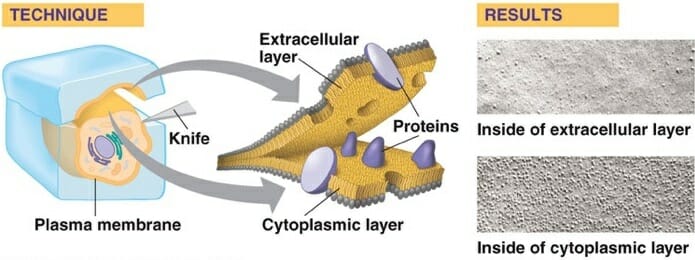
While gross anatomy provided the basis for many modern sciences, modern technology has revolutionized the study of microscopic anatomy. Starting with the invention of light microscopy and carrying through modern day inventions such as the electron microscope, the inner workings of cells and organisms are becoming increasingly understood. Entire new worlds of organisms, such as bacteria and single-celled eukaryotes, have been opened up for study. Cellular biology is an entire field dedicated to the study of cells, their organelles, and how they function. Microscopic anatomy is central to this study.


Microscopic anatomy covers everything from tissues, which are groups of similar cells, down to the inner workings of the molecules which direct the cell’s activities. A histologist studying muscle tissue, for example, would examine how the cells are held together in the tissue. Looking further into the cells using an electron microscope, he would see the complex arrangement of proteins in the cell which allow it to contract. He may also notice the nucleus, which contains the DNA coding for all of the proteins and products the cell produces.


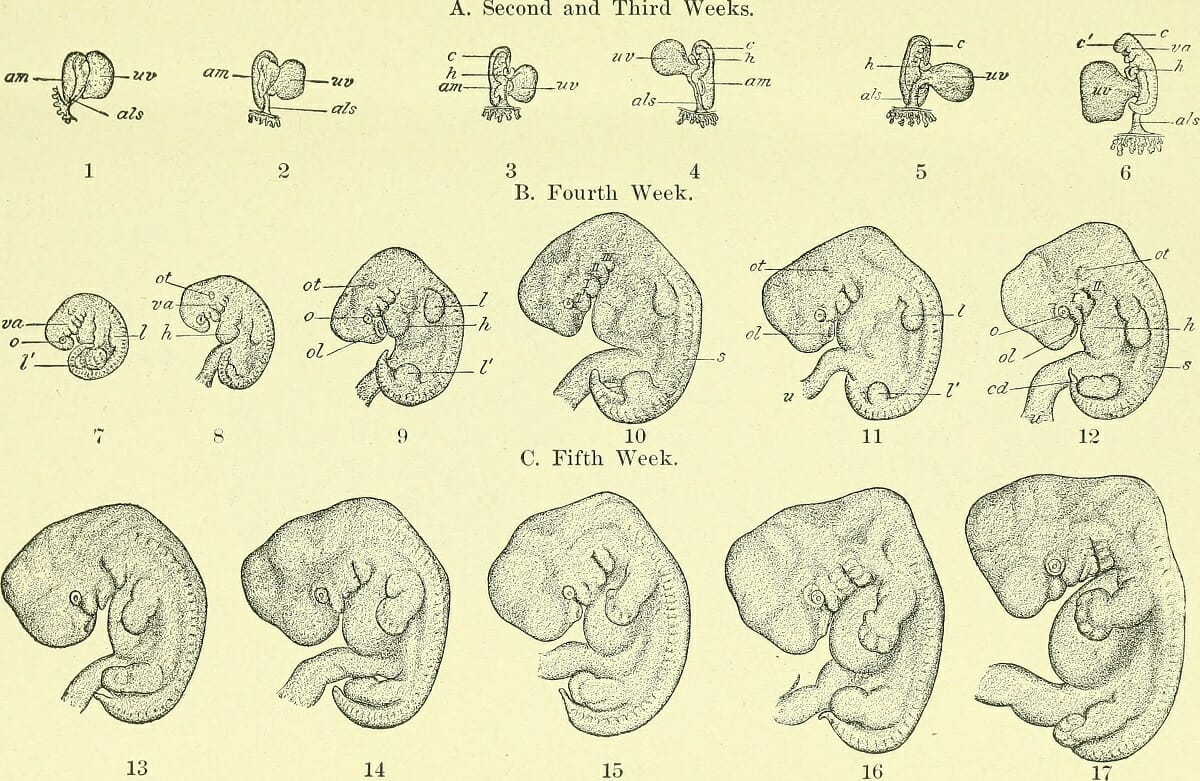
Microscopic anatomy is often paired with biochemistry, molecular biology, and other disciplines to fully understand the organism or tissues being studied. Science knew for decades that cells contained many organelles. However, it was not until recent advances in DNA processing and protein analysis that the function of the many different organelles was understood. Using microscopic anatomy, scientist can also study the cells during the development of an organism. This is called embryology, and has developed into a wide field covering everything from human development to evolutionary relationships of organisms based on their developmental processes.


History of Anatomy


Anatomy is a science older than science itself. The first anatomists where the first humans, categorizing and recognizing the other organisms in their environment using skills of gross anatomy. Vision is fundamental to humans, and is the basis of our understanding of the world. As we advanced in thought and organization, early thinkers began to try to classify organisms. Without any other information, anatomy was often the only evidence available to bind organisms into groups. Aristotle was among the first to attempt serious organization of living things and used many attributes of their anatomy to group them together. His two main groups were plants and animals, two groups we can still easily distinguish today based on their gross anatomies.


Early medicine advanced quickly once the moratorium on dissection was lifted. Often frowned upon in early society, early anatomists like Leonardo Da Vinci often received scrutiny from the public or the church for their scientific inquiry. However, an understanding of the human body arose from these early pioneers, upon which is built the medical knowledge of today. Many of the first works of human and animal anatomy were published during the Renaissance. Many authors showed an advanced, if slightly lacking or skewed view of anatomy as we know it today. But, without any way to understand the workings of the body further, gross anatomy was stranded by itself.


Fast forward several hundred years and the “Father of Taxonomy” Carl Linnaeus was still mainly focused on gross anatomy as a starting point for classification. Darwin’s idea of evolution and common ancestors became accepted at the end of the 1800’s. Still, there were not many methods to evaluate the relationships between animals further. With the advent of better imaging technology, the 1900’s brought the emergence of microscopic anatomy, and really started to change biology. Once it was understood that DNA was the principle mode through which organisms inherited traits, revolutions in many disciplines occurred. Medicine saw a rapid increase in understanding, thanks to the discovery that bacteria and other microbes can cause disease. The inner workings of the cell were being pieced together, and the functions of the many different organelles understood. Many aspects of evolutionary biology were rediscovered or overturned as microscopic anatomy and DNA revealed different relationships than were once assumed. This revolution continues today, as new developments in microscopic anatomy and physiology continually reshape our understanding of organisms.


Careers


Many careers in the biological sciences require some knowledge of both gross and microscopic anatomy. Some professionals, such as a doctor, require specific anatomical knowledge of one species: humans. Human anatomy is the study of both the macroscopic and microscopic portions of the human body. Human anatomy is essential for professionals in the medical field as they must be able to discern between the many types of tissues in the body, and understand their relationship to each other. Ergonomics is the study of the physical stresses on the human body, and relies on a detailed understanding of its various components.


Other scientists focus on the anatomy of other species, or groups of species. A mammologist understands mammal anatomy, where a herpetologist understands the anatomy of reptiles and amphibians. An evolutionary biologist must understand the complex anatomies of many groups, and uses the information to understand their hereditary relationships. Archeologists study mainly gross anatomy of fossilized organisms, whereas cellular biologists and bacteriologists must rely on microscopic anatomy as their organisms are unicellular.


Degrees in anatomy can be obtained at the bachelors, graduate, and doctoral levels, with a wide variety of concentrations. Many schools offer concentrations and courses in human anatomy as a prerequisite for medical school. Other schools and programs focus on general anatomy, necessary for veterinary science, zoology, advanced biology degrees and other specialties that may rely heavily on anatomy. As a professor of anatomy, one would study and teach about the various aspects of anatomy. Many colleges have researchers who incorporate different aspects of anatomy into their research.


If you are good at anatomy or are interested in career paths with anatomy involved, try to find which branch of anatomy you enjoy the most. If gross anatomy suits you, then you may want to pursue a job as a surgeon or evolutionary biologist. If microscopic anatomy is more up your alley, you could become a microbiologist or study internal medicine. Anatomy is extremely important in many fields, especially when it is coupled with other disciplines of science, such as chemistry and physics. It can yield great insights into the world in branches of biology as far ranging as human medicine and evolution.


Reference


  • De luliis, G., & Pulera, D. (2007). The Dissection of Vertebrates. Amsterdam: Academic Press.

  • McMahon, M. J., Kofranek, A. M., & Rubatzky, V. E. (2011). Plant Science: Growth, Development, and Utilization of Cultivated Plants (5th ed.). Boston: Prentince Hall.


Anatomy