Sunday, June 25, 2017

What Do Biogeochemical Cycles Connect?

The biogeochemical cycles on Earth connect the energy and molecules on the planet into continuous loops that support life. The basic building blocks of life like water, oxygen, carbon, sulfur, nitrogen and phosphorous are recycled and go back into their respective cycles repeatedly. The biogeochemical cycles also create reservoirs of these building blocks such as the water stored in lakes and oceans and sulfur stored in rocks and minerals.


The biogeochemical cycles have existed on Earth for billions of years and the elements that make up a modern-day human (or anything else on the planet) have been part of many other organisms and non-living molecules in the past. The process known as nucleosynthesis that occurred during and after the Big Bang was the ultimate source of all the materials that make up the universe. For example, the lighter elements hydrogen and helium formed within the first few minutes of the Big Bang and went on to help form the first stars. When the stars got old and eventually exploded, these elements went on to be incorporated into other cycles, processes, stars and planets in the universe. In addition to recycling elements and molecules, the explosion of stars also creates new materials like metals that are expelled into space and become part of the whole process.


Carbon cycle

The image above shows the carbon cycle, one of the biogeochemical cycles on Earth that recycles and stores the elements essential for life.


References


  • Nucleosynthesis. (n.d.). In Wikipedia. Retrieved June 25, 2017 from https://en.wikipedia.org/wiki/Nucleosynthesis

  • OpenStax, Biology. OpenStax. May 20, 2013. http://cnx.org/content/col11448/latest/


What Do Biogeochemical Cycles Connect?

The Importance of Decomposers to the Overall Biogeochemical Cycle

The organisms that occupy the decomposer trophic level of the food web on Earth are vital to the existence of life on the planet. Bacteria, fungi and worms take the dead and decaying material and break it down (decomposition) so that the components can be recycled through the biogeochemical cycles. If there were no decomposers, plants would not be able to get nitrogen from the atmosphere to use in their biological processes, and some of the trace elements essential for life would remain bound up in rotting organic material. This would cause a chain reaction that would interrupt the natural flow of energy and molecules through the biogeochemical cycles, and result in the death of producers and consumers in the food web as well.


Soil food web

The image above shows the interdependence of all the components in the soil food web. If one trophic level is impaired or eliminated, the entire system will be affected.


References


  • OpenStax, Biology. OpenStax. May 20, 2013. http://cnx.org/content/col11448/latest/


The Importance of Decomposers to the Overall Biogeochemical Cycle

The Path of Nitrogen through its Biogeochemical Cycle

The Earth’s nitrogen cycle captures and recycles nitrogen in a variety of forms such as ammonia, nitrate, nitrite, nitrous oxide and nitrogen gas. Throughout the process, bacteria and plants play a role in moving nitrogen through the cycle. Human activity adds nitrogen to the environment through the combustion of fossil fuels and the use of artificial fertilizers in the agriculture industry.


Nitrogen Fixation


Although nitrogen makes up about 78% of the Earth’s atmosphere, moving it into the living world can be a challenge. Plants and phytoplankton are not able to use nitrogen directly from the atmosphere. They rely on microorganisms in the soil that “fix” nitrogen by taking it from the atmosphere and combining it with hydrogen to make ammonia. The bacteria then convert the ammonia into other organic compounds that are taken up by plants and phytoplankton in the next step. Some of these bacteria live in the nodules on the roots of plants like alfalfa and peas. The bacteria trade some of the nitrogen they make with the plants in exchange for carbohydrates.


Assimilation


Plants are the major players in the assimilation phase of the nitrogen cycle. They absorb nitrogen from the soil by taking up ionic forms of nitrite, nitrate or ammonium through their root hairs. Plants are one of the main foods for terrestrial animals and are their main source of nitrogen. Animals assimilate the nitrogen from the food they eat and use it to make molecules that are essential for life such as amino acids.


Ammonification


This process comes into play when a plant or animal dies or when an animal expels waste. Initially, the nitrogen is bound to other molecules in an organic form. The molecules are broken down by bacteria and fungi and the nitrogen is released as ammonium.


Nitrification


Nitrification converts the ammonium created in the previous step into nitrate ions. This is accomplished primarily by bacteria from genera including Nitrosomonas and Nitrobacter.


Denitrification


Nitrogen is released back into the atmosphere as nitrogen gas in the denitrification step. Bacteria convert the nitrate ions formed in the nitrification step into nitrogen gas and the cycle begins again. This process is usually done under anaerobic conditions by bacteria of the genera Psuedomonas and Clostridium, although it occurs under aerobic conditions as well.


Nitrogen Cycle Steps

Nitrogen Cycle Steps


The image above shows an overview of the nitrogen cycle. Oceans have a similar nitrogen cycle that is carried out by different genera of bacteria and plants.


References


  • OpenStax, Biology. OpenStax. May 20, 2013. http://cnx.org/content/col11448/latest/

  • Nitrogen cycle. (n.d.). In Wikipedia. Retrieved June 22, 2017 from https://en.wikipedia.org/wiki/Nitrogen_cycle


The Path of Nitrogen through its Biogeochemical Cycle

Purpose of the Biogeochemical Cycles

The Earth is a primarily a closed system because it’s mass stays virtually constant even though meteors and radiation from the sun can penetrate the atmosphere. In contrast, the biogeochemical cycles that take place in, on and around the planet are open systems because of the constant exchange of matter and energy that goes on between them. The energy from sunlight continually flows through ecosystems and is eventually released as heat. However, the biogeochemical cycles function to conserve and recycle the matter that is part of living organisms. There are several biogeochemical cycles on Earth including water, carbon, nitrogen, oxygen, phosphorous, sulfur and rock. A new cycle that is currently being studied by scientists is the mercury cycle.


Phosphorus Cycle

The image above shows Earths phosphorous cycle, one of several cycles on the planet that conserve and recycle the chemical substances that make up living things.


References


  • OpenStax, Biology. OpenStax. May 20, 2013. http://cnx.org/content/col11448/latest/

  • Biogeochemical cycle. (n.d.). In Wikipedia. Retrieved June 19, 2017 from https://en.wikipedia.org/wiki/Biogeochemical_cycle


Purpose of the Biogeochemical Cycles

Binomial Nomenclature Rules

It is important to have rules for naming species using binomial nomenclature (also called binomial, binominal or binary names) so that everyone does it in a uniform way to create brief and unique names that can be used and understood worldwide. The International Code of Zoological Nomenclature and the International Code of Nomenclature for Algae, Fungi and Plants are the two main governing bodies that establish and enforce the rules for binomial nomenclature. There are some specific rules for certain naming situations, but below is a summary of the major ones.


General Rules for Creating Binomial Names


Binomial names consist of two parts, the genus and the species which are always written in modern Latin. Either the genus and/or species name can be derived from words in any language. When referring to the name of a species of organism, the genus and species names are both used. For example, the human species is referred to as Homo sapiens.


The Genus


The genus name is also called the generic name and always begins with a capital letter. For example, binomial name for cats is Felis catus. The genus name must be unique within each kingdom (see image below) but the same genus can be used in more than one kingdom.


The Species


The species within the genus is known by other names depending on the discipline. For general usage, the species is also called the specific descriptor. In botany, it is called the specific epithet and specific name in used in zoology. A species name can be used more than once within a kingdom.


The species name always begins with a lower case letter. It can be a noun or an adjective and it must agree with the Latin gender of the genus name; masculine, feminine or neuter. For example, if the gender of the genus is masculine, the species name should end in -us, -a or -um. An example of this is Passer domesticus, the house sparrow, in which Passer has a masculine gender. Endings for feminine species names include -is and -e and -or is used for neuter genders.


Writing Using Binomial Names


Binomial names are always written in italics

. In general practice, the font used for the italics should be different from the font used for the rest of the text. For binomial names written by hand, the genus and species are to be underlined separately. For example, Tyrannosaurus rex.


Using Abbreviations of Binomial Names


If a binomial name is repeated several times in a report or paper, the genus can be written in full the first time it is used and in an abbreviated form from that point on. The abbreviation is created using the first letter of the genus and a period. For example, after using the full binomial name of the blue whale, Balaenoptera musculus in the text, it can be written as B. musculus from that point on. This rule is also used if several species of the same genus are listed or discussed.


The abbreviations “spp.” (plural) or “sp.” (singular),” meaning “several species” and “species,” respectively, are used when the binomial name of a species does not need to be specified or cannot be specified. Italics are not used for the abbreviations. An example of this usage is writing “Acer spp.” to refer to any species of maple tree within the genus Acer. If a writer uses the singular form such as “Acer sp.,” this refers to an unspecified species in the genus Acer, or perhaps a newly discovered species of maple tree.


Taxonomic classification

The image above shows how the binomial nomenclature for on organism emerges out of the overall organization of taxonomic classification. Note that the species name of an organism is designated using the binomial name. The levels of organization above species are called taxa.


References


  • Binomial nomenclature. (n.d.). In Wikipedia. Retrieved June 17, 2017 from https://en.wikipedia.org/wiki/Binomial_nomenclature

  • Binomial nomenclature. (2017, June 17). In New World Encyclopedia online. Retrieved from http://www.newworldencyclopedia.org/entry/Binomial_nomenclature


Binomial Nomenclature Rules

Who Came Up With Binomial Nomenclature?

Binomial nomenclature is a system used to name species of animals, plants and insects using two Latin names called the genus and species. For example, in binomial nomenclature, humans are known as Homo sapiens which translates literally from Latin to “A wise man.” Some binomial nomenclature names are derived from the Classical Greek and Mongolian languages as well. In addition, the names of discoverers can be used to create the species names, such as Ablerus longfellowi, a wasp named in honor of the poet Henry Wadsworth Longfellow.


Gaspard and Johann Bauhin


An early form of the naming system was developed by the brothers Gaspard and Johann Bauhin, Swedish botanists who lived in from the mid-1500’s to the early 1600’s. In 1596, Gaspard published his book Pinax theatric botanici (“Illustrated Exposition of Plants”) that described and classified thousands of plants. His classification system was simple but was the first of its kind, grouping plants into “herbs,” “trees” and “shrubs” and further dividing the categories based on how the plant is used. The major contribution of this work, however, was his idea to describe plants using a genus and/or species. During the same time period, his brother Johann worked on his pioneering book Historia plantarum universalis (“General History of Plants”) which was published after his death. Although unfinished, it captured all the knowledge of botany that was known at the time including Gaspard’s unique naming convention.


Carl Linnaeus


The credit for creating the binomial nomenclature system is often given to another Swedish botanist, Carl Linnaeus (also known as Carl von Linné) who was born over 100 years after the deaths of the Bauhin brothers. In truth, Linnaeus adopted their work and is responsible for the formal introduction of the naming system into the scientific world through his 1735 publication Systema Naturae which listed around 10,000 species consisting of 6,000 plants and 4,236 animals. Later, Linnaeus’ 1753 publication Species plantarum was the first work to consistently use a binomial nomenclature system consisting of what Linnaeus called a “trivial name” followed by a generic name. The trivial name is now called the specific epithet or specific name of the species. Linnaeus used many of the genus names created by the Bauhin brothers to make the trivial names he used in his work.


Today, the binomial nomenclature system is regulated by two international codes of rules, the International Code of Zoological Nomenclature (ICZN) and the International Code of Nomenclature for Algae, Fungi and Plants (ICN).


Linnaeus - Regnum Animale (1735)

The image above shows the Regnum Animale published by Carl Linnaeus in 1735 as part of his work Systema Naturae which expanded on the nomenclature work of Gaspard and Johann Bauhin. It addition to Regnum Animale that described the animal kingdom, the other parts of the work are Regnum Vegetabile that described the plant kingdom and Regnum Lapideum. that described the “mineral kingdom.”


References


  • Binomial nomenclature. (n.d.). In Wikipedia. Retrieved June 16, 2017 from https://en.wikipedia.org/wiki/Binomial_nomenclature

  • Systema Naturae. (n.d.). In Wikipedia. Retrieved June 16, 2017 from https://en.wikipedia.org/wiki/Systema_Naturae


Who Came Up With Binomial Nomenclature?

Compare and Contrast Binary Fission and Conjugation

Binary fission is an asexual reproduction process that prokaryote organisms use to duplicate themselves. There is no exchange of genetic information between organisms during binary fission, so over time, populations can experience a lack of genetic diversity. To overcome this, one of the ways that prokaryotes like bacteria have developed to add genetic diversity to their DNA is conjugation. In this process, bacteria come into contact with each other by using hair-like structures called pili that are on the surface. Through a single pilus, one of the bacteria can transfer a DNA plasmid to the other. Two other methods that prokaryotes use to transfer DNA are transformation (they take up a fragment of DNA from the environment) and transduction (a piece of DNA is injected into the bacteria by a bacteriophage).


Comparison Chart


Binary FissionConjugation
What is it used for?Duplication of an organismDNA transfer between organisms
Used by prokaryotic organisms?YesYes
Used in sexual reproduction?NoNo
Recent adaptation?NoNo

Binary fission

The image above shows the process of binary fission.


Bacterial Conjugation

The image above shows how a bacterium uses a pilus to conjugate with another bacterium.


References


  • Fission (biology). (n.d.). In Wikipedia. Retrieved June 13, 2017 from https://en.wikipedia.org/wiki/Fission_(biology)

  • OpenStax, Biology. OpenStax. May 20, 2013. http://cnx.org/content/col11448/latest/


Compare and Contrast Binary Fission and Conjugation

Ovipositor

Ovipositor


An ovipositor is a tube shaped organ used by insects and most fish to deposit eggs.


The morphology of the ovipositor varies from species to species. It may be long, short, wide or needle thin; in insects it is usually composed of the hardened sclerites of the exoskeleton. Often it is concealed within the body and protracted for use, although sometimes the ovipositor is permanently extended outside of the body.


Abdomen of a cricket

The image shows the abdomen of a cricket. The upward pointing spike structure is the ovipositor.


Function of Ovipositor


Egg Depositing


The primary function of the ovipositor in the majority of insects and fish is to release mature eggs from inside of the female body.


Immature eggs, known as oocytes, develop from specialized stem cells within a part of the ovary called the germarium. The oocytes are then transported down through tapered structures within the ovary called ovarioles. At this point, the oocytes are surrounded by a layer of follicle cells, which enable the transport of substances from the hemolymph (the equivalent to blood in most invertebrates) into the oocyte cytoplasm.


When the oocytes are passed further down the ovarioles, the follicle cells go on to synthesize the chorion (the eggshell), which provides waterproofing and protection once the egg has been laid. Now lacking in follicle cells, the egg is free to move into the oviduct in a process called ovulation.


Ovulation is only initiated once the eggs have matured within the ovariole and the brain receives the physical stimulus associated with mating.


Once mating and fertilization, has occurred, muscle contractions facilitate the movement of eggs downward through the oviduct. They are then passed into the ovipositor, allowing the controlled placement of the eggs into a suitable environment. As the eggs are released, glands, called ‘accessory glands’ produce a cement-like substance, which attaches the eggs to each other and also to the substrate where they are laid.


The females must select an oviposition site that maximizes the survival chance of her offspring, considering factors such as food available to the larvae and chance of attack from predators. They can perform this selection using olfactory (smell) cues or visual cues based on the shape, size or spectral quality of a potential oviposition site.


Digging


Many insects use their ovipositor as a tool, which allows them to excavate through a substrate and deposit their eggs in a suitable and safe place.


A notable example is the ovipositor of a grasshopper. Extending beyond the tip of the abdomen, the ovipositor is comprised of two pairs of shovel-shaped structures. One pair is located dorsally (on top) and one pair ventrally (on the bottom), These are called ‘ovipositor valves’. During oviposition, muscle contractions in the abdomen allow the valves to be opened and closed, and for the ovipositor to be pushed and pulled in and out of the tip of the abdomen.


The action of the ovipositor opening and closing causes the grasshopper to dig a deep hole in the ground; to initiate the action the grasshopper pushes the tip of the abdomen firmly into the substrate and engages the motion of the valves. The grasshopper then stands on the substrate surface as the ovipositor digs into the ground below.


Once the hole has been dug, the abdomen is retracted slightly and the opening and closing action speeds up so that the eggs are released one by one, and are dropped into the bottom of the hole, accompanied by the secretion of a frothy substance. After the deposition of each egg, the valve is closed, which reverses the orientation of the egg, ensuring that each developing offspring will hatch with its head facing upward.


Once the last egg has been laid—there are usually around 50-100 laid in each oviposition—the abdomen is retracted slowly from the hole and a final portion of the frothy substance is secreted. The froth turns dark and hardens in order to cap off the ‘egg-pod’, protecting them from desiccation and predators.


Grasshopper

The image shows a grasshopper positioned with her ovipositor inside the tunnel where she will deposit her eggs. The shovel-shaped valves of the ovipositor have dug out the tunnel.


Piercing


Rather than laying their eggs onto a substrate, many insects insert their ovipositor directly into a host—usually either a plant or another insect.


Once a female has located a suitable host, the ovipositor acts as a needle, penetrating the skin or exoskeleton. Often, the ovipositor will release venomous substances to paralyze the host. The paralysis is often permanent although may also be temporary, in which case the host recovers and continues feeding with the parasitic eggs inside its body. In some parasitoid species, the larvae develop within the body, selectively feeding on the internal tissues of the host. The digestive tract and nervous system are left till last so that the host remains alive as long as possible.


The ovipositors of some wasp species have jagged ridges, which allow the wasp to cut through tough plant tissue and even wood, in order to reach hosts that are concealed within. In this case the ovipositors can be exceptionally long, sometimes up to eight times the body length of the parasitoid! In order to pass the egg along this extended tube, the eggs are often very small so that they can slide along the narrow tube, expanding in size once they are inside the host body.


Bracon brevicornis

The image shows a parasitoid wasp (Bracon brevicornis) inserting its ovipositor into its paralyzed host, a black-headed caterpillar (Opisina arenosella).


In some insects, especially the Hymenoptera, the stinger is actually a modified ovipositor. It is likely that the ovipositor was initially used to penetrate the tissues of plants—a behavior still observable in primitive sawflies. This provided an evolved pre-adaption to the parasitism of other insects, and many modern day wasps still utilize their venomous stings for this purpose.


Further down the line, bees evolved a reversion to a diet of pollen and nectar, so no longer needed to use the sting as a weapon to paralyze other insects; furthermore, the social colony structure of a bee population means that only the queen is required to lay eggs and that females are sterile. Despite having no use for an egg-laying ovipositor, the venomous capabilities within the stinger proved beneficial as a defensive weapon, and the stinger remained functional.


Counting


Scientists have found that some species of parasitoid insects, which use their ovipositors to lay their eggs within the bodies of other insects, can also use their ovipositors to count!


Because the parasitoid offspring feed from the body tissues of the parasitized insect, it is important that they have exclusive access to the host tissues, giving them the best chance of being able to feed once hatched. If the parasitoid lays its eggs within the body of a host that has already been parasitized (a scenario called ‘superparasitism’) the competition between the larvae of the first and second parasitoid will eventually result in the death of either one.


To avoid this happening, parasitoids have evolved the ability to detect when hosts have already been parasitized and avoid laying their eggs there. Further, they have been shown to discriminate between hosts with different numbers of eggs, and are less likely to lay eggs inside hosts that have already been heavily parasitized.


This is possible due to sensory receptors at the end of their ovipositor, which are connected to the neurons of the parasitoid. If there are already eggs inside the host, the pheromones that are used to mark eggs when deposited will be detected by the sensory receptors once the ovipositor penetrates the host body. For each egg detected within the parasitized host, a neuron response is triggered, allowing the parasitoid to accurately determine the number of foreign eggs within a host.


Quiz


1. Which of the following is not a function of the ovipositor?
A. Releasing Venom
B. Producing immature eggs
C. Digging
D. Detecting pheromones

Answer to Question #1

2. Which of the following statements is true?
A. Only females have ovipositors
B. All ovipositors look the same
C. All ovipositors produce eggs
D. All ovipositors contain venom

Answer to Question #2

References


  • Godfray, H. C. J. Parasitoid Natural History. (1994) In: Parasitoids: Behavioural and Evolutionary Ecology. Princeton University Press. Chichester, West Sussex.

  • Jans, N. (2002) Evolutionary ecology of oviposition strategies. Pp349-376. In M.Hilker, & T. Meiners (Eds), Chemoecology of insect eggs and egg deposition. Blackwell, Berlin.

  • Joeballenger2005 (2015) Why can’t male bees (or wasps) sting? Ask an entomologist. Retrieved from: https://askentomologists.com/2015/09/23/why-cant-male-bees-or-wasps-sting/

  • Parasitic Wasps (2016) Kansas State University. Department of Entomology. Retrieved from: http://entomology.k-state.edu/extension/insect-information/beneficial-organisms/parasitoids.html


Ovipositor

Calmodulin

Calmodulin Definition


Calmodulin, or calcium-modulated protein, is a calcium-binding protein found in the cytoplasm of all eukaryotic cells. It interacts with many other proteins in the cell, and acts as a regulator or an effector molecule in a wide variety of cellular functions. These functions include things as diverse as regulation of the cell cycle, intracellular signalling, fertilization, and muscle contraction. Calmodulin is in a family of proteins along with troponin C, another essential calcium-binding protein involved in muscle contraction. Calmodulin is an essential protein; mutations to any of the calmodulin-encoding genes or damage to the calmodulin binding sites often proves lethal.


Calmodulin Structure


Calmodulin is a protein made up of 148 amino acid residues. It is encoded by multiple genes; in humans: CALM1, CALM2, and CALM3 which are found on chromosomes 14, 2, and 19, respectively. Calmodulin forms two globular domains connected by a flexible central linker. Each domain binds two calcium ions in E-F hand motifs, a motif ubiquitous in calcium-binding proteins, so that calmodulin can bind a total of four Ca2+ ions. The calcium binding sites are 12 amino acids long and contain many negatively-charged or polar amino acid residues such as aspartate, glutamate, and asparagine. The side chains on these amino acids form ionic bonds with the Ca2+ ions. Other amino acid residues with side chains rich in oxygen atoms also attract the calcium cations. This promotes binding even at very low concentrations of Ca2+.


When calcium is bound to calmodulin a helix-loop-helix is formed along the backbone and a conformational change occurs. This conformational change, coupled with the flexibility of the protein due to the flexible connecting linker, allows calmodulin to interact with and bind to a wide variety of other proteins.


Calmodulin-Ca

This figure depicts the structure of calmodulin with four calcium ions bound.


Calmodulin Function


Calmodulin is a ubiquitous regulator protein that is involved in many calcium-mediated processes. When Ca2+ binds to calmodulin it forms the Ca2+/calmodulin complex which then interacts with other proteins in the cell. These proteins are enzymes and effector proteins involved in a variety of cellular and physiological processes. The Ca2+/calmodulin complex can also regulate processes directly.


One of the functions of the Ca2+/calmodulin complex is to activate calcium pumps. These pumps remove calcium from the cytoplasm by either pumping it out of the cell or storing it in the endoplasmic reticulum. By controlling the amount of calcium in the cell, the downstream responses are regulated.


Further examples of Ca2+/calmodulin complex function include binding to Ca2+/calmodulin kinases (CAMK) such as the myosin light chain kinase. This binding allows the CAMKs to phosphorylate effector proteins by transferring phosphates from ATP to serine and threonine residues on the receiving proteins. These proteins then go on to activate downstream processes such as intracellular signalling, smooth muscle contractions, neurotransmitter and hormone synthesis and release, and cell cycle regulation.


signaling events in the brain

This figure shows an example of how calmodulin (CaM) can be involved in a complex pathway in a post-synaptic neuron. The pathway shown here is the KEGG pathway in human drug addiction.


Calcium


It is becoming increasingly apparent that calcium plays a crucial role in a number of physiological processes. When not in use the concentration gradient of calcium ions between the inside and outside of the cell is very large; the concentration of extracellular calcium is approximately 1 mM while the concentration of free calcium ions within the cell is less than 0.1 μM. This is likely due to the fact that calcium will interact readily with many proteins.


The majority of calcium in the cell enters through gated calcium channels. It can also be stored in the endoplasmic reticulum. The calcium channels are large trans-membrane proteins that allow passage of ions into the cell when a specific stimuli is met. this usually occurs when the membrane is depolarized or a ligand is attached.


Quiz


1. Where is calmodulin located?
A. nucleus
B. plasma membrane
C. cytosol
D. outside of the cell

Answer to Question #1

2. How many calcium ions can bind to calmodulin?
A. none
B. one
C. two
D. four

Answer to Question #2

3. What processes is calmodulin involved in?
A. cell signalling
B. muscle contraction
C. phosphorylation pathways
D. all of the above

Answer to Question #3

References


  • Campbell, N. A., & Reece, J. B. (2005).Biology, 7th. ed. Ch. 49. San Francisco, CA: Benjamin Cummings. ISBN: 0-8053-7171-0.

  • King, R. C., Mulligan, P. K., & Stansfield, W. D. (2014 online). . Oxford, UK: Oxford University Press. eISBN: 9780199376865.

  • Martin, E., & Hine, R. (2008 online). . Oxford, UK: Oxford University Press. eISBN: 9780191726507.

  • Randall, D., Burggren, W., & French, K. (2002).Eckert animal physiology: mechanisms and adaptations, 5th. ed. Chs. 9 and 10. New York, NY: W.H. Freeman and Company. ISBN: 0-7167-3863-5.

  • Weaver, R. F. (2005).Molecular biology, 3rd. ed. Ch. 24. New York, NY: McGraw-Hill. ISBN: 0-07-284611-9.


Calmodulin

Cilium

Cilium Definition


A cilium, or cilia (plural), are small hair-like protuberances on the outside of eukaryotic cells. They are primarily responsible for locomotion, either of the cell itself or of fluids on the cell surface. They are also involved in mechanoreception. There is even a class of microorganisms named for these small structures. Ciliates are protozoans that possess cilia which they use for both locomotion and feeding.


Structure of Cilium


A cilium is made up of microtubules coated in plasma membrane. The microtubules are small hollow rods made of the protein tubulin. Each cilium contains nine pairs of microtubules forming the outside of a ring, and two central microtubules. This structure is known as an axoneme, and the arrangement as ‘9+2’, an arrangement ubiquitous in motile cilia. The microtubules are held together by cross-linking proteins. Between the nine outer pairs are motor proteins called dynein.


Cilia attach to the cell at a basal body. The basal body is made up of microtubules arranged as nine triplets. The triplets are formed as the doublets from the cilia are joined by an additional microtubule from the cell. The two central microtubules end before entering the basal body.


The motor proteins (dynein) are large flexible molecules that allow the cilia to be motile. The proteins hydrolyze ATP for energy. As the proteins are activated, they undergo conformational changes which allow for complex movements. The dynein molecules essentially crawl along the microtubules, pulling the neighboring doublet up and reattaching further down. As the doublets are attached to each other through the cross-linking proteins they can only slide a short distance along each other. This movement causes bending in the cilium.


Cilia are very small structures – measuring approximately 0.25 μm in diameter and up to 20 μm in length. Where present they are found in large numbers on the cell surface. The cilia act like oars, beating back and forth to create movement.


Cilium Function


Cilia play an important role in locomotion. This can include movement of the cell itself, or of other substances and objects past the cell. In some organisms known as ciliates, cilia are responsible for movement of the organism as a whole. For example, in the unicellular protist Paramecium, cilia cover the surface of the organism and are responsible for movement as well as feeding. In addition to covering the outside of the organism, cilia also line the oral groove, moving food into the organism’s “mouth”.


Cilia can help to remove contaminants from organs or tissue by helping to move fluids over the cell. The lining of the nasopharynx and the trachea are covered in cilia. These ciliated epithelial cells remove mucus, bacteria, and other debris from the lungs. Another example is the lining of the fallopian tubes. The cilia here are responsible for helping in fertilization by movement of the egg towards the uterus.


Kinocilia are a specialized type of cilia found on the apical ends of vertebrate hair cells. Along with stereocilia, non-motile collections of actin filaments related to cilia, they are involved in hearing and balance (mechanoreception).


Eukaryotic cilium

This figure depicts the internal structure of a cilium, showing the nine pairs of outer microtubules and the two central microtubules, connected by protein linkers and dynein molecules.


Animals columnar epithelium with cilia

This figure depicts epithelial cells covered in small hair-like cilia.


Quiz


1. What are cilia composed of?
A. Microfilaments
B. Microtubules
C. Keratin
D. Actin

Answer to Question #1

2. What type of organism does not have cilia?
A. Bacteria
B. Protists
C. Plants
D. Animals

Answer to Question #2

3. Which of the following is not a function of cilia?
A. Locomotion
B. Feeding
C. Reproduction
D. Fighting infection
E. None of the above

Answer to Question #3

References


  • Campbell, N. A., & Reece, J. B. (2005).Biology, 7th. ed. Chs. 6 and 28. San Francisco, CA: Benjamin Cummings. ISBN: 0-8053-7171-0.

  • Madigan, M. T., & Martinko, J. M. (2006).Brock biology of microorganisms, 11th. ed. Ch. 14. Upper Saddle River, NJ: Pearson Prentice Hall. ISBN: 0-13-144329-1.

  • Randall, D., Burggren, W., & French, K. (2002).Eckert animal physiology: mechanisms and adaptations, 5th. ed. Ch. 7. New York, NY: W.H. Freeman and Company. ISBN: 0-7167-3863-5.


Cilium

Cholinergic

Cholinergic Definition


Cholinergic is a term used to refer to the molecule acetylcholine. It is usually employed to define neurons, receptors or synapses that use acetylcholine. For instance, a cholinergic neuron is a neuron that releases acetylcholine, and a cholinergic receptor is a receptor to which acetylcholine binds. Acetylcholine is a signal molecule in the nervous system that is used by nerve cells to transfer information. It is widely present in the peripheral nervous system, which is involved in contracting skeletal and smooth muscle and in dilating blood vessels, among other functions. Acetylcholine plays a major role at the neuromuscular junction, i.e. at the joint between nerve cells and muscle. Additionally, acetylcholine is also present in the central nervous system, where it plays a role in cognitive processes such as memory, learning and arousal.


Unsurprisingly, its role in numerous processes of the peripheral and the central nervous systems have made the cholinergic system a target in the treatment of multiple. In turn, several cholinergic drugs have been developed for clinical as well as for cosmetic purposes. For example, some cholinergic drugs are used to treat severe muscle spasms, others to slow down the progression of Alzheimer’s disease and others to reduce wrinkles. However, in addition to the therapeutic and cosmetic effects, cholinergic drugs can also induce a series of side effects, including paralysis of the autonomic nervous system.


Acetylcholine Function


Acetylcholine is present in the peripheral and in the central nervous systems. In the peripheral nervous system, acetylcholine is largely implicated in muscle movement and in other functions such as blood vessel dilation. In the central nervous system, it is involved in cognitive functions.


Acetylcholine acts by binding to cholinergic receptors, the two main types of which are muscarinic and nicotinic. Muscarinic acetycholine receptors (mAChR) are G protein-coupled receptors (GPCR) that modulate the activity of the cell by activating cellular mechanisms involving second messengers. There are five identified types known as M1 to M5. M1, M3 and M5 muscarinic receptors are usually excitatory and are of the Gq type; thus, they exert their function by activating phospholipase C (PLC), which in turn activates the IP3 signal transduction cascade, allowing extracellular calcium to enter the cell. M2 and M4 receptors are usually inhibitory and are of the Gi or Go type, i.e. they act by reducing cyclic AMP (cAMP) in the cell. Nicotinic acetylcholine receptors (nAChR), the other main type, are ligand-gated ion channels that, when activated by acetylcholine, directly allow ions to enter (e.g. sodium) or exit the cell (e.g. potassium).


Acetylcholine in the Peripheral Nervous System


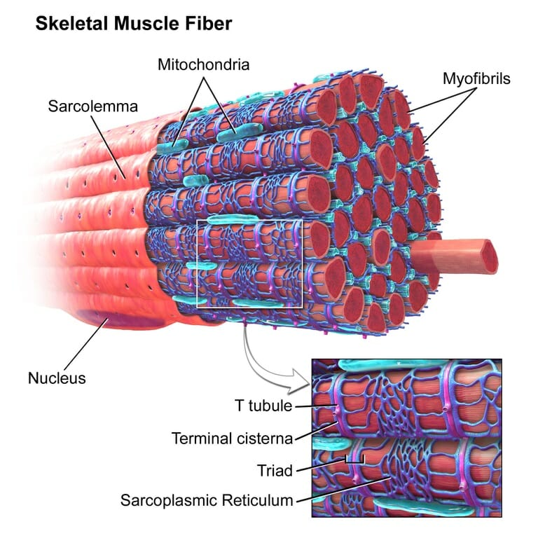
Acetylcholine is a major player at the neuromuscular junction. The neuromuscular junction is the site where a nerve cell (a neuron) and skeletal muscle (the kind of muscle that is voluntarily contracted) are connected. For a muscle to contract, the brain sends electrochemical signals from one neuron to another, until an action potential (electrical signal) is generated at the motor neuron, which is the neuron that contacts the muscle fiber. At the neuromuscular junction, acetylcholine is released by the motor neuron into the synaptic cleft, which then binds to nicotinic acetylcholine receptors present on the muscle fiber cell. Nicotinic acetylcholine receptors allow sodium to enter the muscle cell, after which a series of intracellular signals lead to the contraction of the muscle. Anomalies in peripheral cholinergic transmission have been linked to motor disorders such as myasthenia gravis, a disorder characterized by fatigue and muscle weakness.


Acetylcholine is also a widely used neurotransmitter in the autonomic nervous system—a part of the peripheral nervous system involved in the control of unconscious and involuntary bodily functions. Specifically, acetylcholine is released by neurons from central nervous system that project to neurons of the autonomic nervous system, the latter of which detect acetylcholine through nicotinic acetylcholine receptors. These neurons in turn project to body parts that do not belong to the nervous system, such as the gastrointestinal tract. In some cases, acetylcholine is also released at this junction between the peripheral nervous system and other body parts.


Acetylcholine in the Central Nervous System


In the central nervous system, cholinergic activity is related to arousal, awareness, learning, memory, attention and reward, among others. Not surprisingly, abnormal cholinergic transmission has been associated with Alzheimer’s disease, a neurodegenerative condition characterized by memory loss.


Cholinergic Drugs


The involvement of acetylcholine in diseases of the nervous system has naturally made the cholinergic system a target for therapeutic purposes. Drugs that activate (agonists) or inactivate (antagonists) acetylcholine receptors, as well as drugs that modulate cholinergic activity by facilitating or preventing the production, release or degradation of acetylcholine, have been developed with the aim to treat several neuropsychiatric conditions.


Agonists


Acetylcholine has a very short life: it does not last long in the bloodstream because it is degraded very fast. Therefore, acetylcholine itself is not used as a drug, but instead similar compounds that activate acetylcholine receptors are employed to activate them. These similar compounds that bind to and activate acetylcholine receptors are known as acetylcholine agonists.


An example of an agonist is pilocarpine, which activates muscarinic receptors and is usually applied in the pupil of the eye to treat a neurodegenerative disease that causes blindness called glaucoma. Another example of an agonist is nicotine, found in tobacco.


Antagonists


Many cholinergic drugs are acetylcholine receptor antagonists, which block acetylcholine receptors. Some antagonists are atropine, scopolamine, hexamethonium and trimethaphan. Atropine and scopolamine inactivate muscarinic receptors and are used to suppress bodily secretions (e.g. tears or mucus) and to relax smooth muscle (e.g. muscles in the gastrointestinal tract) during anesthesia, and to treat motion sickness. Hexamethonium and trimethaphan block nicotinic receptors and are used to reduce high blood pressure. Other agents that block nicotinic receptors are used because of their effects at the neuromuscular junction; these agents prevent skeletal muscles from contracting and are often employed during surgery to keep patients from making involuntary movements.


Other Drugs


In addition to cholinergic agonists and antagonists, other drugs can modulate acetylcholine activity by increasing or decreasing its production, release or degradation. For instance, inactivating acetylcholine transferase, which is an enzyme that breaks down acetylcholine, is employed to increase the levels of acetylcholine and to treat myasthenia gravis, a neuromuscular disorder. Similar drugs such as neostigmine and pyridostigmine do not cross the blood-brain barrier and are consequently employed to exert their effect at the neuromuscular junction and contract skeletal muscle.


Nonetheless, anticholinergics—drugs that reduce or block the effects of acetylcholine—are more widely used to treat numerous conditions. Some of these are involuntary movements, gastrointestinal disorders, incontinence and Parkinson’s disease. Another compound that blocks the release of acetylcholine is botulinum toxin—an agent produced by a type of bacterium—which paralyzes the skeletal muscle so that the organism is no longer able to move and which can even cause death. When applied locally, botulinum toxin relaxes muscles and is consequently utilized to treat severe muscle spasms. The same compound is used to reduce wrinkles by relaxing the muscles and skin; we know this under the trade name Botox.


Cholinergic Effects


The effects of activating cholinergic receptors include muscle contraction, heart rate deceleration, constriction of the iris (miosis) and of the lens, mucus secretion and broncho-constriction. Conversely, the effects of inactivating cholinergic receptors include muscle relaxation, heart rate acceleration, pupil dilation (mydriasis) and lens flattening (cyclopegia), dryness of the upper airway (of the respiratory system), inhibition of tear production, urine retention, mouth dryness, slowing down of mucociliary activity in the respiratory tract, constipation and muscle relaxation (skeletal muscle and smooth muscle).


Cholinergic Side Effects


Cholinergic drugs can help treat some disorders and ameliorate symptoms but they also have negative side effects. Most cholinergic drugs are anticholinergics, i.e. they reduce or block the effects of acetylcholine. For instance, the acetylcholine antagonists hexamethonium and trimethaphan, used to treat high blood pressure, can produce paralysis of the autonomic nervous system, producing effects such as blurred vision and inability to urinate. Anticholinergics in general can cause a raise in body temperature because they reduce the amount of sweating; they can also induce drowsiness, hallucinations, confusion, dry mouth, constipation, difficulty urinating and memory deficits. In older people, they can cause confusion, memory loss and cognitive decay. Mixing anticholinergics with alcohol have similar side effects as overdosing with anticholinergics, which include dizziness, fever, confusion, accelerated heart rate, trouble breathing, hallucinations, unconsciousness and even death. Caution is therefore warranted when taking cholinergic drugs.


Quiz


1. Where does acetylcholine play a major role?
A. At the neuromuscular junction.
B. At the corticospinal tract.
C. In all the peripheral nervous system.
D. In all the central nervous system.
E. All of the above.

Answer to Question #1

2. What are some effects of inactivating cholinergic receptors?
A. Heart rate deceleration and muscle relaxation.
B. Heart rate acceleration and muscle spasms.
C. Pupil dilation and muscle spasms.
D. Urine retention, dryness of the upper respiratory tract and muscle relaxation.

Answer to Question #2

3. Which disorders are treated with acetylcholine antagonists or anticholinergic drugs?
A. Gastrointestinal disorders, bipolar disorder and motion sickness.
B. Gastrointestinal disorders, Parkinson’s disease and high blood pressure.
C. Glaucoma, Alzheimer’s disease, Parkinson’s disease and low blood pressure.
D. Glaucoma, Alzheimer’s disease and high blood pressure.

Answer to Question #3

References


  • Anderson, K.-E. (2011). Muscarinic Acetylcholine Receptors in the Urinary Tract. In: Urinary Tract. Berlin-Heidelberg: Springer.

  • Changeux, J.-P., Edelstein, S.J. (2005). Nicotinic Acetylcholine Receptors: From Molecular Biology to Cognition. New York: Odile Jacob.

  • Encyclopedia Britannica (2014, December 18). Cholinergic drug. Retrieved from: https://www.britannica.com/science/cholinergic-drug


Cholinergic

Sex Linked Genes

Sex Linked Genes Definition


Sex linked genes are genes that are in the sex chromosomes and that are therefore inherited differently between males and females. In mammals, where the female has two X chromosomes (XX) and the male has one X and one Y chromosome (XY), recessive genes on the X chromosome are more often expressed in males because their only X chromosome has this gene, while females may carry a defective recessive gene on one X chromosome that is compensated by a healthy dominant gene on the other X chromosome. Common examples of sex linked genes are those that code for colorblindness or those that code for hemophilia (inability to make blood clots) in humans. In birds, on the other hand, where the female has two different chromosomes (ZW) and the male has two Z chromosomes (ZZ), it is the female who has higher chances of expressing recessive genes on the Z chromosome because they cannot compensate with the dominant gene on the W chromosome.


Sex Chromosomes


In species in which males and females are clearly differentiated, sex chromosomes determine the sex of the organism. In mammals, females have two X chromosomes (XX) and males have one X chromosome and one Y chromosome (XY) (see below for a different pattern of sex chromosome inheritance in birds). The other non-sex chromosomes (called autosomal chromosomes) are the same for males and females, i.e. they code for the same genes. The cells of each individual have two copies of each chromosome although each copy may contain different alleles. In other words, cells have pairs of chromosomes, each pair coding for the same genes (e.g. eye color) but each copy of the chromosome may have a different allele (e.g. one copy may code for blue eyes and the other copy for brown eyes). Humans have 23 pairs of chromosomes, i.e. 46 chromosomes: 22 pairs of autosomal chromosomes and 1 pair of sex chromosomes.


The way sex chromosomes are inherited is quite straightforward. Each organism has two copies of each chromosome; in the case of sex chromosomes this can be either XX (female) or XY (male). Females can thus only transfer X chromosomes to their offspring (because they only have X chromosomes), while males can transfer either one X chromosome or one Y chromosome to their offspring. From the offspring perspective, a female will have inherited one X chromosome from the mother (the only chromosome mothers can transfer to offspring) and the other X chromosome from the father; a male will have inherited one X chromosome from the mother and the Y chromosome from the father.


Sex chromosomes are different from autosomal chromosomes in that the X chromosome is larger than the Y chromosome and, not surprisingly, the distinct sizes entail that each sex chromosome contains different genes (even though there are some genes that are coded in both X and Y chromosomes, but these are not considered sex linked genes). This means that a gene that is coded on the Y chromosome will only be expressed in males, whereas a gene that is coded on the X chromosome could be expressed in males and in females.


Importantly, recessive genes—genes that need two copies to be expressed, otherwise the dominant gene is expressed—have specific consequences on each sex. When a recessive gene is expressed on the X chromosome, it more likely to be expressed in males than in females. This is because males have only one X chromosome, and will therefore express the gene even if it is recessive, whereas females have two X chromosomes and carrying a recessive gene may not be expressed if the other X chromosome carries another dominant gene. This is the reason these genes are called sex linked genes: because they are inherited differently depending on the sex of the organism. Let us look at one example that will make things easier to understand.


An Example: Colorblindness


An example of sex linked genes is colorblindness. Colorblindness is a recessive gene that is only expressed on the X chromosome (let’s use X* for the X chromosome carrying the recessive colorblind gene). If a male receives the colorblind gene from the mother, this individual will be colorblind (X*Y). If, on the other hand, a female receives one colorblind gene (either from the mother or the father) and another healthy gene (not colorblind, either from the mother or the father), then this female organism (XX*) will not be colorblind because the healthy gene is dominant and the recessive colorblind gene will not be expressed. She will be however a carrier, which implies that she can pass on the colorblind gene to her offspring. Finally, if a female receives a colorblind gene from the mother and another colorblind gene from the father, this female will be colorblind (X*X*).


In other words, females can be healthy (XX), carriers (XX*) without being colorblind, and colorblind (X*X*) while males can either be healthy (XY) or colorblind (X*Y). Therefore, the chances of males being colorblind are extremely higher than the chances of females being colorblind. In fact, around 1 in 20 men is colorblind and only 1 in 400 women is.


ZW System


In birds, the sex of the organisms is also determined by two different chromosomes but instead of the females having two equal chromosomes (XX) and males having to different chromosomes (XY), female birds have two different chromosomes (ZW) and male birds have two equal chromosomes (ZZ).


In pigeons, for instance, an example of a sex linked gene is the one that codes for the color of the feathers. This gene is coded on the Z chromosome, so that whichever allele (ash-red, blue or brown) is expressed on the Z chromosome will determine the feather color of the female. For males, it will depend on both Z chromosomes (ash-red is dominant to blue, and blue is dominant to brown).


Genetic Linkage During Homologous Recombination


When an individual has two copies of the same chromosome (any autosomal chromosome, two X chromosomes in the case of female mammals, or two Z chromosomes in the case of male birds), these chromosomes can recombine during meiosis in a processed called homologous recombination, resulting in swaps of some portions of the chromosomes. To put it simply, the two copies of a chromosome are cut at random places and the cut portion is swapped between both copies. If two genes sit physically close together on the chromosome, they are very likely to be inherited together because the cut during homologous recombination is not likely to happen in between them. Therefore, female mammals (XX) and male birds (ZZ) can show genetic linkage of sex linked genes.


An example of this would be feather color and color intensity in pigeons, both of which are always inherited together in females (ZW) and quite often in males too (ZZ). In males, because color and color intensity sit close together, they are likely to be inherited together because the chromosome cut during recombination is not likely to take place in between, although they can also be mixed and recombined.


Quiz


1. What are sex linked genes?
A. Genes that sit on any autosomal chromosome.
B. Any gene that sits on a sex chromosome.
C. Genes that sit on a sex chromosome and that are inherited differently in males and females.

Answer to Question #1

2. Can a colorblind mother and a healthy father have colorblind children?
A. Yes, but only colorblind daughters.
B. Yes, but only colorblind sons.
C. Yes, colorblind daughters and sons.
D. No.

Answer to Question #2

3. What are the sex chromosomes in birds?
A. X and Y, as in mammals: XX for females and XY for males.
B. X and Y: XY for females and XX for males.
C. Z and W: ZZ for females and ZW for males.
D. Z and W: ZZ for females and WW for males.
E. Z and W: ZW for females and ZZ for males.

Answer to Question #3

References


  • Genetic Science Learning Center. (2014, December 2). Sex Linkage. Retrieved from: http://learn.genetics.utah.edu/content/pigeons/sexlinkage/

  • Ohno, S. (2013). Sex chromosomes and sex-linked genes (Vol. 1). Springer Science & Business Media, Berlin, Germany.


Sex Linked Genes

Sarcomere

Sarcomere definition


A sarcomere is the functional unit of striated muscle. This means it is the most basic unit that makes up our skeletal muscle. Skeletal muscle is the muscle type that initiates all of our voluntary movement. Herein lies the sarcomere’s main purpose. Sarcomeres are able to initiate large, sweeping movement by contracting in unison. Their unique structure allows these tiny units to coordinate our muscles’ contractions.


Skeletal Muscle Fiber

The image depicts skeletal muscle fiber.


In fact, the contractile properties of muscle are a defining characteristic of animals. Animal movement is notably smooth and complex. Dexterous movement requires a change in muscle length as the muscle flexes. This calls for a molecular structure that can shorten along with the shortening muscle. Such requisites are found in the sarcomere.


Upon closer inspection, skeletal muscle tissue gives off a striped appearance, called striation. These “stripes” are given off by a pattern of alternating light and dark bands corresponding to different protein filaments. These stripes are formed by the interlocking fibers that comprise each sarcomere. Tubular fibers called myofibrils are the basic components that form muscle tissue. However, myofibrils themselves are essentially polymers, or repeating units, of sarcomere. Myofibrils are fibrous and long, and made of two types of protein filament that stack on top of each other. Myosin is a thick fiber with a globular head, and actin is a thinner filament that interacts with myosin when we flex.


Skeletal muscle

Depicted is a basic illustration of skeletal muscle’s underlying components, down to the sarcomere.


Sarcomere structure


When viewed under a microscope, muscle fibers of varied lengths are organized in a stacked pattern. The myofibril strands, thereby actin and myosin, form bundles of filament arranged parallel to one another. When a muscle in our body contracts, it is understood that the way this happens follows the sliding filament theory. This theory predicts that a muscle contracts when filaments are allowed to slide against each other. This interaction, then, is able to yield contractile force. However, the reason the sarcomere structure is so crucial in this theory is that a muscle needs to physically shorten. Thus, there is a need for a unit that is able to compensate for the lengthening or shortening of a flexing muscle.


The sliding filament theory was first posited by scientists who had used high-resolution microscopy and filament stains to observe myosin and actin filaments in action at various stages of contraction. They were able to visualize the physical lengthening of the sarcomere in its relaxed state, and the shortening in its contracted state. Their observations led to the discovery of sarcomere zones.


Sarcomere

The figure depicts the structure of a Sarcomere. (Each zone is labeled).


They first observed that the dynamic changes that were taking place were always happening in the same spots, or zones. They noticed that one zone of repeated sarcomere, later called the “A band,” maintained a constant length during contraction. The A band has a higher content of thick myosin filament, as expected by the area’s rigidity. The A band is the area in the center of the sarcomere where thick and thin filaments overlap. This gave researchers an idea of myosin’s central location. Within the A band is the H zone, which is the area composed only of thick myosin. Essentially, the A band can be thought to include “all” of the myosin including the myosin intertwined with actin at its bulbous head. Located on each end of the sarcomere’s length is the I band. The I bands are the two regions that exclusively contain thin filament. A quick way to remember this is that the I band has “thIn, actIn” filaments. The thick filaments are located not too far from the site of the I band; but on either side, their margins delineate where the thick filaments end. Likewise, the Z lines or discs that give sarcomeres a striped appearance under a light microscope actually delineate the regions between adjacent sarcomeres. The M line, or middle division, is found right in the middle of the Z lines and contain a less important third filament called myomesin.


Filament mental shortcut:


  • I is a thin letter, contains only thin filaments.

  • H is a wider letter, contains only thick filaments.


As mentioned before, contraction happens when the thick filaments slide along the thin filaments in quick succession to shorten the myofibrils. However, a crucial distinction to remember is that the myofilaments themselves do not contract. It is the sliding action that lends them their power to shorten or lengthen.


Sarcomere function


Filament sliding generates muscle tension, which is without question the sarcomere’s main contribution. This action lends muscles their physical force. A quick analogy of this is the way a long ladder can be extended or folded depending on our needs for it, without physically shortening its metal parts.


Thankfully, recent research gives us a good idea of how this sliding works. The sliding filament theory has been modified to include how myosin is able to pull on actin to shorten the length of the sarcomere. In this theory, myosin’s globular head is located close to actin in an area called the S1 region. This region is rich in hinged segments that can bend and thus facilitate contraction. The bending of S1 may be the key to understanding how myosin is able to “walk” along the length of the actin filaments. This is accomplished by myosin-actin cycling. This is the binding of the myosin S1 fragment, its contraction, and its eventual release.


When myosin and actin bind, they form extensions called “cross-bridges.” These cross-bridges can form and break with the presence (or absence) of ATP. ATP makes S1 contraction possible. When ATP binds to actin filament, it moves it into a position that exposes its myosin binding site. This allows myosin’s globular head to bind to this site to form the cross-bridge. This binding causes the phosphate group of the ATP to dissociate, and thus myosin initiates its power stroke. Myosin thus enters a lower energy state where the sarcomere can shorten. Moreover, ATP must bind myosin to break the cross-bridge, and allow myosin to re-bind actin and initiate the next spasm.


Quiz


1. Which zone of the sarcomere maintains a constant length during contraction?
A. Z lines
B. A band
C. I band
D. S zone

Answer to Question #1

2. Which of the following contains only actin filament?
A. A band
B. H band
C. I band
D. Z line

Answer to Question #2

3. Which of the following contains only myosin filament?
A. A band
B. H band
C. I band
D. Z line

Answer to Question #3

References


  • Krans, Jacob et al. (2010). “The sliding filament theory of muscle contraction.” Nature Education 3. 3(9):66.

  • MH Education (2017). “Animation: Sarcomere Contraction.” Human Anatomy: Mckinley O’Loughlin.” Retrieved on 2017-6-16 from http://www.macroevolution.net/sarcomere.html

  • Boundless (2017). “ATP and muscle contraction.” Boundless: The Musculoskeletal System. Retrieved on 2017-6-15 from https://www.boundless.com/biology/textbooks/boundless-biology-textbook/the-musculoskeletal-system-38/muscle-contraction-and-locomotion-218/atp-and-muscle-contraction-826-12069/


Sarcomere

Amniotic Fluid

Amniotic Fluid Definition


Amniotic fluid is the clear liquid that surrounds a developing fetus in the mother’s womb. It is formed from the mother’s plasma (or the fluid part of blood cells) as it diffuses past the fetal membranes and succumbs to the forces of osmosis and hydrostatic pressure. Visually speaking, the amniotic fluid will often have a yellow tinge, but it is always contained within an amniotic sac. The amniotic sac is the pouch that encloses the unborn fetus until its birth. The sac is made up of an amnion (inner) and a chorion (outer) membranes. Unborn babies are able to swallow or inhale the amniotic fluid before releasing it, as they have yet to breathe through their still developing lungs which will require oxygen from the outside environment.


Amniotic fluid levels notably fluctuate during pregnancy, as well. The highest levels of amniotic fluid are present at the thirty-four-week mark with an average of eight hundred milliliters. On the other hand, at full term, or forty weeks of pregnancy, the amniotic fluid stands at around six hundred milliliters. Amniotic levels are important to maintain, as an overflow or underflow of amniotic fluid can render disease in the infant and/or mother, as discussed below.


Development of Amniotic Fluid


To better understand amniotic fluid, it is important to discuss its origin. The space that comes to hold the amniotic sac is chosen at the time of embryo implantation during the first week of pregnancy. This cavity fills with fluid even before the embryo can be identified, and the fill rate surpasses the growth rate of the embryo, initially. In early fetal development, the volume of fluid increases linearly with the dimensions of the fetus. The water component of the amniotic fluid originates from the mother as it is pulled from her plasma. This is made possible by the bidirectional diffusion that takes place across the thin surfaces of the placenta or umbilical cord and the fetus’s skin, which has yet to harden with keratin. These thin surfaces are fully permeable to solutes and water. Two months into gestation, the fetus begins to make urine once its urethra opens, and the infant also begins to swallow. These forces more or less oppose each other and therefore do not have a great impact on the sac’s volume. By week twenty, however, the fetus’s skin begins to keratinize. This is when the linear relationship between size of the fetus and the volume of fluid stops. As the skin hardens, urine excretion begins to factor into the total volume of the amniotic sac, as does the efflux of fluid from the baby’s lungs and the reduction in his or her swallowing of fluid.


The actual composition of the amniotic fluid changes with gestational stage. At the beginning of the pregnancy, the amniotic fluid will contain some electrolytes and water. But around the fourteenth week mark, the amniotic fluid will become rich in proteins, sugars, lipids, and urea. All of these are nutrients that aid in the unborn baby’s growth. For a long time, amniotic fluid was thought to be entirely made up of fetal urine. This idea has changed in recent times. The consensus now is that amniotic fluid is a rich concoction of nutrients and growth factors that play a role in the infant’s growth and fosters antimicrobial protection. Obstetric physicians can take a sample of the fluid for early diagnostic purposes in a test called amniocentesis or AFT. Prenatal tests on amniotic fluid can reveal an infant’s sex, chromosomal abnormalities (i.e. down’s syndrome), fetal infections, and test for amniotic fluid embolism. The latter results from having amniotic fluid or fetal debris enter the mother’s circulatory system, which poses a lethal threat to the mother as it triggers a massive autoimmune response.


Amniocentesis

The picture above depicts how Amniocentesis is performed.


Amniotic fluid embolism

The image is a histological slide of an amniotic fluid embolism, caused by the presence of fetal cells in one of the mother’s pulmonary arterioles.


Amniotic Fluid Functions


Like most conserved processes, there has been an evolutionary drive to conserve amniotic fluid in fetal development. There are few evolutionary processes as important as successful fetal growth. The importance of amniotic fluid lies in its functions. The light, amorphous nature of amniotic fluid renders it the ideal medium for fetuses to move in. Motion is an essential part of the baby’s development as it encourages bone growth of the fetal limbs. Likewise, amniotic fluid envelops the fetus in a homeostatic environment where the temperature is constant and the fetus loses no warmth in the process. The liquid itself acts as a barrier between the fetus and its surroundings that effectively cushions the baby from external jolts or blows. Another body system that benefits from the aqueous nature of the amniotic sac is the nascent respiratory system, specifically the lungs. Fetal respiration bypasses the lungs completely for the entire pregnancy, in favor of diffusing nutrients and gases between the fetus and the mother through the placenta. It will take nine months, or the date of birth, before the infant can expand its lungs to take its first breath. But in the meantime, the lungs are allowed to grow and their sensitive linings are kept moist by the surrounding amniotic fluid.


Summary of amniotic fluid functions include:


  • Fetal cushioning, or protection

  • Homeostatic conditions are maintained

  • Fosters bone growth of fetal limbs

  • Lung development


Amniotic Fluid Disease


When the levels of amniotic fluid exceed the norm, or are less than normal, this results in disease. Polyhydramnios is when there is too much fluid, a common occurrence for women who are carrying twins or triplets or if the baby has congenital defects. Too little is called oligohydramnios, and this results from having late pregnancies, placental defects, or a broken membrane. Of course, abnormal levels of fluid cause some alarm for obstetric physicians. Thankfully, an amniocentesis can reveal developmental abnormalities early on in the pregnancy.


Quiz


1. Amniotic fluid is derived from which source?
A. Fetal plasma
B. Maternal plasma
C. Fetal lymph
D. Maternal fat cells

Answer to Question #1

2. What is the name of the inner membrane of the amniotic sac?
A. Chorion
B. Yolk sac
C. Exocoelom
D. Amnion

Answer to Question #2

3. Which diagnostic tool can obstetric physicians use to diagnose prenatal disease from amniotic fluid samples, as discussed above?
A. AFT
B. Ultrasound
C. Amniogenesis
D. AMF

Answer to Question #3

References


  • Medline Plus (2017). “Amniotic Fluid.” U.S. National Library of Medicine. Retrieved on 2017-06-14 from https://medlineplus.gov/ency/article/002220.htm

  • Study (2017). “What is amniotic fluid? – Levels, Functions, & Composition.” Anatomy & Physiology. Retrieved on 2017-06-13 from http://study.com/academy/lesson/what-is-amniotic-fluid-levels-function-composition.html

  • Underwood, M. et al. (2005). “Amniotic Fluid: Not just fetal urine anymore.” Journal of Perinatology. 25, 341-348. doi:10.1038/sj.jp.7211290

  • Moore, Lisa MD (2017). “Amniotic Fluid Embolism.” Medscape Obstetrics & Gynecology. Retrieve from 2017-06-14 from http://emedicine.medscape.com/article/253068-overview


Amniotic Fluid

Saturday, June 24, 2017

Salivary Glands

Salivary Glands Definition


A salivary gland is the tissue in our mouths that expels saliva. Anytime our mouth comes near a hot sandwich we feel them activate. Salivary glands are only found in mammals. As exocrine glands, they expel saliva into the epithelial surface of our mouths by way of ducts, rather than through the bloodstream. Each day, our glands produce as much as a quart of saliva. Saliva is a mixture of water, mucus, antibacterial substance, and digestive enzymes. One of the most recognizable digestive enzymes in the human body is alpha-amylase. This enzyme is able to break down the starch in our food to simpler and more easily digestible sugars like glucose and maltose. Whenever we chew, we are activating these glands in preparation for the safe breakdown of our meal.


Salivary Glands Function


Briefly, saliva itself serves many uses. As the only secretion of our salivary glands, it is helpful in creating the food bolus, or the finely packed ball of food that we roll inside our mouths. This shape facilitates its safe passage through our alimentary canal. Saliva has lubricating properties that are protective, as well. Saliva protects the inside of our mouths, our teeth, and our throats as we begin to swallow the bolus. It also cleanses the mouth after a meal, and dissolves food into chemicals that we perceive as taste.


Salivary glands come in three flavors, if you will: the parotid, sublingual, and submandibular glands. Each is aptly named after the area in the oral cavity in which it is located. Let’s begin this discussion with the parotid gland.


three types of salivary glands

The diagram depicts the three types of salivary glands in the mouth: 1) parotid glands, 2) submandibular gland, 3) sublingual gland.


Two parotid glands are located within each of our cheeks. Parotid glands are the largest type of salivary gland. They account for up to twenty percent of the saliva in our oral cavity. Their main role lies in facilitating mastication, or “chewing,” and in commencing the first digestive phase of our food. The parotid gland is notably labeled a serous type of gland. Serous glands are those that secrete protein-rich fluid, which in this case is an enzyme-rich suspension of alpha-amylase.


Next, the submandibular gland is located close to our mandible. This is the movable part of our jaw. In essence, this gland lies on the floor of our mouths. Since it is superficially located, we can feel it if we place our fingers about two inches above the Adam’s apple. It is the second-largest salivary gland, and produces the most saliva (up to 65%). It is considered a mixture of serous and mucus glands since the suspension is both rich in enzymes and gooey mucus that is released into the oral cavity through the submandibular ducts.


Lastly, sublingual glands are located under the tongue. They are the smallest and most dispersed salivary gland. Its secretion is mostly mucus, and exits directly through the Rivinus excretory ducts. Only a minimal (~5%) amount of saliva in the oral cavity comes from these.


Salivary Glands Innervation


Salivary glands are innervated by both branches of the autonomic, or “involuntary,” nervous system. This is commonly associated with the fight or flight response. When we see a bear, for instance, we trigger our sympathetic response. This threat triggers the release of norepinephrine, a rise in our heart rate, dilation in our eyes, and notably, slowed digestion and a dry mouth. This means that whereas sympathetic stimulation normally overstimulates its target, it inhibits the salivary gland. So, we produce less saliva. In contrast, parasympathetic stimulation of the salivary gland renders a heavy flow of saliva.


Salivary Glands Infection


There are symptoms that may signal a compromised salivary gland. This includes gland swelling, fever, a foul taste in the mouth, and dry mouth. Measures can be taken to lessen the effects of dry mouth (listed below).


Dry Mouth Treatment

The figure lists the ways in which the symptoms of dry mouth can be lessened.


Swollen glands are often caused by “salivary stones,” or buildup of crystalized saliva, that can clog the gland from releasing saliva. This causes pain, and unless the blockage is cleared it can infect the gland. Salivary glands can also be painfully blocked by bacterial infection. Staphylococcus aureus is the most common bacterium that infects it. If left untreated, the bacterium will cause fever, severe pain, and an abscess or pus. Other bacteria that commonly target the salivary gland via exposure or bad hygiene are: Streptococcus viridans, Haemophilus influenza, Streptococcus pyogenes, E. coli. Nevertheless, viruses can also infect the glands. Among them are viral infections in children that were not vaccinated for mumps, but it is also caused by influenza, HIV, and herpes. A tumor can likewise block salivary gland tissue. Most tumors are benign, but salivary gland cancer is a rare type of cancer that develops in 1 of 100,000 people each year.


Quiz


1. Which enzyme is the primary digestive enzyme in human saliva?
A. Lipase
B. Alpha-amylase
C. Pancreatin
D. Beta-amylase

Answer to Question #1

2. Which salivary gland type is the largest contributor to the saliva in our oral cavity?
A. Mucosal
B. Parotid
C. Sublingual
D. Submandibular

Answer to Question #2

3. Identify the type of innervation that stimulates saliva flow:
A. Autonomic
B. Parasympathetic
C. Somatic
D. Sympathetic

Answer to Question #3

References


  • Anatomy and Physiology (2017). “The Salivary Glands.” Anatomy and Physiology, a learning initiative. Retrieved on 2017-06-19 from http://anatomyandphysiologyi.com/salivary-glands/

  • WebMD (2017). “Salivary Gland Problems.” WebMD: Oral Care. Retrieved on 2017-06-20 from http://www.webmd.com/oral-health/guide/salivary-gland-problems-infections-swelling#1

  • Krucik, Greg (2015). “What is a salivary gland infection?” Healthline. Retrieved on 2017-06-19 from http://www.healthline.com/health/salivary-gland-infections

  • Segal, K et al. (1996). “Parasympathetic innervation of the salivary glands.” Operative Techniques in Otolaryngology-Head and Neck Surgery. 7(4): 333-338


Salivary Glands