Ectoderm Definition
The ectoderm is a germ layer, or tissue layer, that forms in an animal embryo during development. As the name suggests, the ectoderm is the germ layer that covers the outside of the embryo (‘ecto’ meaning outside). The ectoderm then goes on to give rise to a number of both internal and external structures. The ectoderm is one of the two tissue layers present in diploblasts, along with the endoderm, and one of the three layers found in triploblasts, along with the endoderm and mesoderm.
Ectoderm Formation
The ectoderm forms during gastrulation, the process in embryogenesis where cells rearrange and begin to differentiate. Early in development, when the embryo has undergone several cell divisions (cleavage) but has not yet begun gastrulation, cells in the upper animal region are already earmarked as future ectoderm cells.
Gastrulation proceeds differently in different organisms. In amphibians, the ectoderm is restricted to the animal region of the blastula until gastrulation, at which point it extends to cover the entire embryo in a process called epiboly. In fish and birds the future endoderm and mesoderm cells migrate inwards in a process called introgression, leaving the cells that remain on the outside surface as the future ectoderm cells. In placental mammals it is again different due to the extra-embryonic structures that are required for an embryo to develop in the womb. Prior to gastrulation the fertilized egg is composed of a trophectoderm and an inner cell mass; the inner cell mass will develop into the embryo proper while the trophectoderm will form extra-embryonic structures such as the placenta. The embryo is then differentiated into primitive endoderm and ectoderm, and the embryonic ectoderm begins gastrulation on the inner side of the embryo.
Ectoderm Function
The ectoderm is one of the primary layers of cells that exists in an embryo. The ectoderm cells differentiate into cells that form a number of external structures such as skin, sweat glands, skin sensor receptors, and hair follicles. In addition, the ectoderm forms the external surfaces of the eyes (cornea and lens), teeth (enamel), mouth, and rectum, as well as the pineal and pituitary glands.
The main function of the ectoderm is to form the central nervous system (brain and spinal cord). Following gastrulation, the mesoderm forms the rod-like notochord which signals the adjacent dorsal ectoderm to thicken and form the neural plate. The neural plate then folds in to form the neural tube in a process known as neurulation. The neural tube will ultimately become the spinal cord and the brain. The point where the neural tube separates from the dorsal ectoderm is the neural crest. The cells of the neural crest will eventually differentiate into a variety of cell types including peripheral nerves and skull bones.
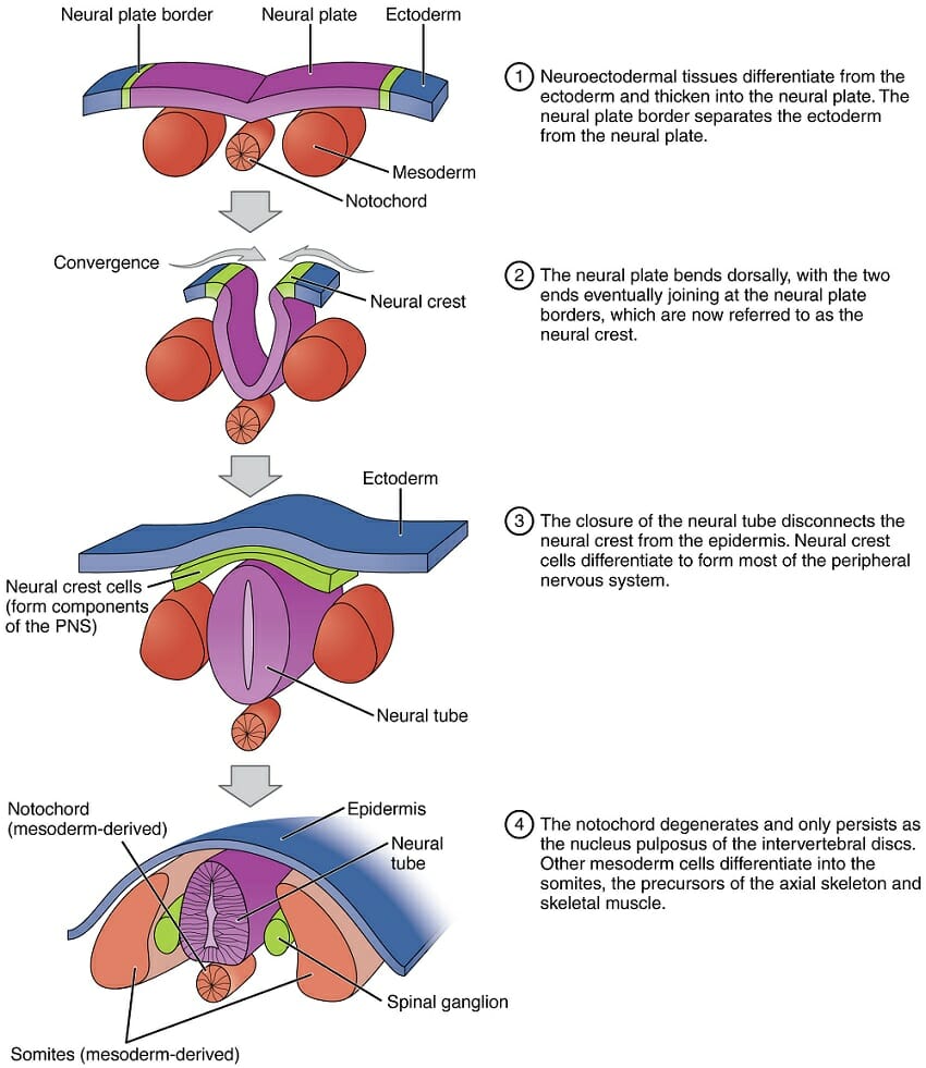
This figure depicts neurulation, the formation of the neural tube from ectoderm. It shows the dorsal ectoderm becoming the epidermis and the neural plate. The neural plate then forms the neural tube.
Quiz
1. What structure is not formed by ectoderm?
A. dermis of the skin
B. pituitary gland
C. tooth enamel
D. spinal cord
2. At what point are the cells differentiated as ectoderm cells?
A. cleavage
B. gastrulation
C. organogenesis
D. pattern formation
3. What structure goes on to form the central nervous system?
A. notochord
B. neural crest
C. neural tube
D. somites
References
- Campbell, N. A., & Reece, J. B. (2005).Biology, 7th. ed. Chs. 32 and 47. San Francisco, CA: Benjamin Cummings. ISBN: 0-8053-7171-0.
- Jessell, T., Lawrence, P., Meyerowitz, E., Robertson, E., & Smith, J. (2005).Principles of Development, 3rd. ed. Chs. 1 and 3. New York, NY: Oxford University Press. ISBN: 0-19-927537-8.
Ectoderm
No comments:
Post a Comment