Sunday, August 20, 2017

Leaf

Leaf Definition


The term leaf refers to the organ that forms the main lateral appendage on the stem of vascular plants. In general, leaves are thin, flat organs responsible for the photosynthesis of the plant. Although photosynthesis typically only occurs on the upper surface of the leaf, it can occur on both sides in some plant species. Leaves are typically comprised of a distinct upper and lower surface, stomata for gas exchange, waxy coating, hairs, and venation. Each side of the leaf differs in regarding the level to which these features are expressed. Although leaves are typically located above ground, some species have leaves which reside underground (e.g., bulb scales) or underwater (e.g., aquatic plant species). Moreover, the leaves of some plants may not be associated with photosynthesis (e.g., cataphylls). Leaves are typically oriented on a plant to avoid blocking the sunlight of the leaves situated underneath.


Function of the Leaf


As one of the most important constituents of plants, leaves have several essential functions:


Photosynthesis


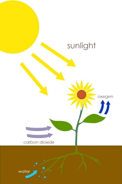
The primary function of the leaf is the conversion of carbon dioxide, water, and UV light into sugar (e.g., glucose) via photosynthesis (shown below). The simple sugars formed via photosynthesis are later processed into various macromolecules (e.g., cellulose) required for the formation of the plant cell wall and other structures. Therefore, the leaf must be highly specialized to combine the carbon dioxide, water, and UV light for this process. Carbon dioxide is diffused from the atmosphere through specialized pores, termed stomata, in the outer layer of the leaf. Water is directed to the leaves via the plant’s vascular conducting system, termed the xylem. Leaves are orientated to ensure maximal exposure to sunlight, and are typically thin and flat in shape to allow sunlight to penetrate the leaf to reach the chloroplasts, which are specialized organelles that perform photosynthesis. Once sugar is formed from photosynthesis, the leaves function to transport it down the plant via specialized structures called the phloem, which run in parallel to the xylem. The sugar is typically transported to the roots and shoots of the plant, to support growth.


Plants


Transpiration


Transpiration refers to the movement of water through the plant, and subsequent evaporation via the leaves. When the stomata open to accommodate the diffusion of carbon dioxide into the plant for photosynthesis, water flows out. This process also serves to cool the plant via evaporation of the water from the leaf, as well as regulate the plant’s osmotic pressure.


Guttation


Guttation refers to the excretion of xylem from the edges of leaves and other vascular plants due to increased levels of water in the soil at night, when the stomata are closed. The pressure caused at the roots results in the leakage of water from the xylem out of specialized water glands at the edges of leaves.


Storage


Leaves are a primary site of water and energy storage since they provide the site of photosynthesis. Succulents are particularly adept at water storage, as evidenced by the thick leaves. Due to the high levels of nutrients and water, many animal species ingest the leaves of plants as a source of food.


Defense


Some leaves have also evolved defense mechanisms to avoid being eaten or damaged. Some examples include the spines of cacti, cones of gymnosperms, respectively. In addition, hairs found on leaves prevent water loss in dry climates and sting animals that detour herbivores (e.g., Urticaceae). Moreover, the waxy coatings found on leaves serve to protect against water loss, rain, and forms of contamination. Oils and other secreted substances also detract from being consumed by herbivores.


Types of Leaf


In general, the types of leaf can be divided into six major types, although there are also plants with highly specialized leaves:


Conifer Leaf


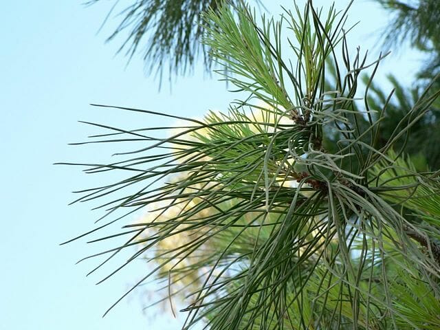
Conifer leaves are needle-shaped or in the form of scales. Conifer leaves are typically heavily waxed and highly adapted to colder climates, arranged to dispel snow and resist freezing temperatures. Some examples include Douglas firs and spruce trees. The images below illustrate this type of leaf.
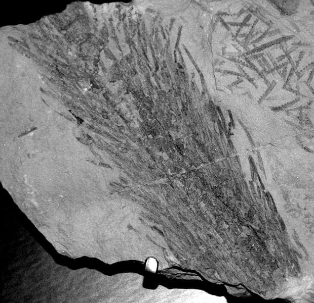

Conifer needle


Microphyll Leaf


Microphyll leaves are characterized by a single vein that is unbranched. Although this type of leaf is abundant in the fossil record, few plants exhibit this type of leaf today. Some examples include horsetails and clubmosses. The image below illustrates this type of leaf.


Baragwanathia fossil land plant


Megaphyll Leaf


Megaphyll leaves are characterized by multiple veins that can be highly branched. Megaphyll leaves are broad and flat, and generally comprise the foliage of most plant species. The image below illustrates this type of leaf.
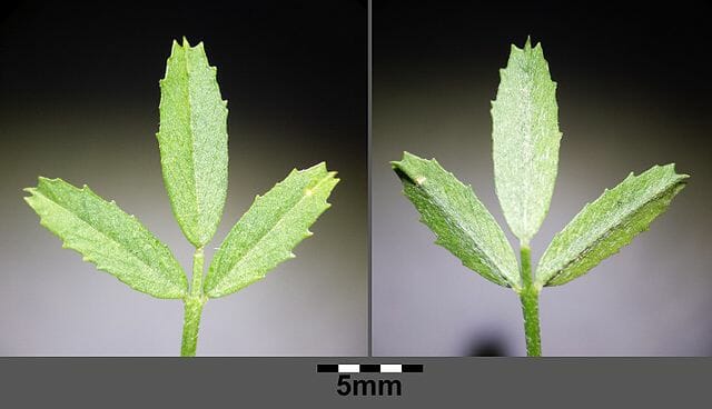

Setaria megaphylla leaves


Angiosperm Leaf


Angiosperm leaves are those found on flowering plants. These leaves are characterized by stipules, a lamina, and a petiole. The illustration below shows an example of an angiosperm leaves.
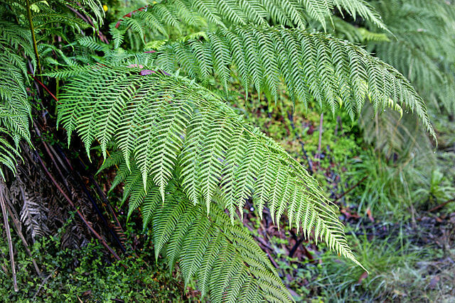

Medicago prostrata


Fronds


Fronds are large, divided leaves characteristic of ferns and palms. The blades can be singular or divided into branches. The image below presents an example of a frond.
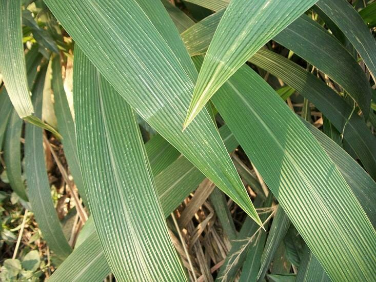

Fern


Sheath Leaf


Sheath leaves are typical of grass species and monocots. Thus, the leaves are long and narrow, with a sheathing surrounding the stem at the base. Moreover, the vein structure is striated and each node contains only one leaf. The image below presents an example of a sheath leaf.


Juncus bufonius


Quiz


1. The primary function of a leaf is:
A. Water evaporation for cooling
B. Photosynthesis
C. Provide shade to the shoot and root structures of the plant
D. Transpiration

Answer to Question #1

2. Which of the following statements is TRUE regarding guttation:
A. It typically occurs at night.
B. It occurs when the stomata are closed.
C. It results from increased water pressure in the soil.
D. All of the above

Answer to Question #2

References


  • Brodersen C and McElrone A. (2013). Maintenance of xylem Network Transport Capacity: A Review of Embolism Repair in Vascular Plants. Front Plant Sci.4:108.

  • El-Sharkawy, M. and Hesketh, J. (1965) Photosynthesis among species in relation to characteristics of leaf anatomy and CO2 diffusion resistances. Crop Science. 5(6):pp. 517-521.

  • Roth-Nebelsick A, Uhl D, and Kerp H. (2001). Evolution and Function of Leaf Venation Architecture: A Review. Ann Bot. 87(5): 553-566.

  • Sadras and Milroy. (1996). Soil-water thresholds for the responses of leaf expansion and gas exchange: A review. Field Crops Research. 47(2): 253-266.


Leaf

Pollen

Pollen Definition


Pollen refers to the powdery product synthesized by seed plants responsible for the production of the male gametes of the plant (shown below).


Pollen


The pollen grains are termed microgameteophytes, and consist of a sporopollenin coating which serves to protect the gameteophytes as they are transported from the stamens (male) or male cone to the pistil (female) or female cone in flowering and coniferous plants, respectively. When the pollen reaches the pistil or female cone, a pollen tube is formed, which transports the sperm to the ovule containing the female gametophyte. The term pollination refers to the transfer of pollen grains from the anther to the stigma of a flower. Cross-pollination involves the transfer of pollen from one flower to the stigma of another flower. In contrast, self-pollination involves the transfer of pollen from one flower to the stigma of the same flower. A diagram illustrating the location of the anther and stigma is shown below.


Stigma shapes


Pollen Structure


Pollen grains vary in size, shape, and surface characteristics depending on the plant species (shown below). In general, pollen grains have a double wall consisting of a thin inner wall composed of cellulose, termed the endospore, and a thick outer wall comprised of sporopollenin, termed the exospore. The shape and the external features of the exospore are highly variable, and are often used to distinguish pollen grain produced by different species. The purpose of this structure is to protect the male genetic material from the environment (e.g., UV radiation, compression, and water) during the transportation from the anther to the stigma. The surface of the pollen grain also contains various waxes and proteins which help repel moisture and interact with the stigma, respectively. However, such protein structures on the surface of pollen are often recognized by immune cells and are the source of the allergic reactions to pollen observed in humans.


Pollen colorized


Pollen Formation


In coniferous plants, pollen is formed in the microsporangia of the male cone, whereas it is produced in the anthers of flowering plants (also termed angiosperms). Each microsporocyte is diploid and forms four haploid cells, termed microspores, via meiosis. This process is termed microsporogenesis. The four microspores then form the double wall of the pollen grain within a structure made of callose. During this process, the callose is digested by callase and the pollen grains are able to grow and complete the formation of the endospore and exospore. The diagram below illustrates the release of pollen grains from the callose structures.


Anther schematic


Quiz


1. The transfer of pollen from the anther to the stigma of a flower is termed:
A. Endoporation
B. Germination
C. Pollination
D. Microsporogenesis

Answer to Question #1

2. The primary function of the exospore is:
A. Pollination
B. Protect the plant’s male genetic material
C. Protect the plant’s female genetic material
D. Endoporation

Answer to Question #2

References


  • Clarke A, Gleeson P, Harrison S, and Knox B. (1979). Cell Biology Pollen-stigma interactions: Identification and characterization of surface components with recognition potential. Proc. Nati. Acad. Sci.76(7):3358-3362.

  • Heberle-Bors E. (1985). In vitro haploid formation from pollen: a critical review. Theoretical and Applied Genetics. 71(3): 361-374.

  • Gullvag B. (1966). The fine structure of pollen grains and spores: a selective review from the last twenty years of research. Phytomorphology. 16: 211-27.

  • Ward M, Dick C, Gribel R, and Lowe A. (2005). To self, or not to self… A review of outcrossing and pollen-mediated gene flow in neotropical trees. Heredity.95(4): 246-54.


Pollen

Saturday, August 19, 2017

Overpopulation

Overpopulation Definition


Overpopulation refers to a population which exceeds its sustainable size within a particular environment or habitat. Overpopulation results from an increased birth rate, decreased death rate, the immigration to a new ecological niche with fewer predators, or the sudden decline in available resources. Therefore, overpopulation describes a situation in which a population in a given ecosystem limit the resources available for survival.


Overpopulation Effects


Overpopulation can have several effects on the environment, as well as other species within an ecological system. Indeed, human overpopulation has resulted in technological advances which have increased human lifespan and fertility, and consequently placed pressure on global resources. Such effects are such that the planet is currently in a novel geological epoch called the Anthropocene. In general, overpopulation results in an ecological disruption as resources are depleted. This disruption can lead to the decline of other populations which compete for the same resources. Typically, such effects result in the cycling between periods of population growth and periods of population decline until it can reach homeostasis within a particular ecological niche. Some examples of naturally regulated population growth are rodents, rabbits, and various insect populations (e.g., army worms and locusts).


In situations of overpopulation caused by the introduction of a foreign species for which they have no natural predators, they can become an invasive species. An example is the inadvertent introduction of zebra mussels to the North American water systems. Since zebra mussels are natively from the Black Sea and Caspian Sea, they have no natural predators in the foreign ecosystems of North America and parts of Europe. As such, zebra mussels quickly became an invasive species, clogging water treatment pipes, affecting power plants, and impacting the local freshwater fish populations. It is estimated that the overpopulation of zebra mussels has cost approximately $5 billion USD since their introduction. The image below illustrates an infestation of zebra mussels on a North American lock due to the overpopulation of zebra mussels in the North American waterways. Other economic effects of overpopulation include those caused by crop destruction, as seen with the overpopulation of rabbits in Australia. While the overpopulation of rabbits destroyed farmers crops, leading to poor yields, the continent also experienced a loss of native plant species, as well as the removal of precious topsoil due to erosion.


Another effect of the overpopulation of one species, is the increased population growth of the natural predators of such species. This effect is generally considered to be positive, as the predator population serves to control the overpopulated prey species. Such effects also serve to drive evolutionary changes as the prey species evolves to avoid increased predation.


Solutions for Overpopulation


Historically, there have been several situations for which overpopulated species could not be managed naturally. In these instances, issues with overpopulation have been overcome using a variety of methods. One of the most common causes of overpopulation is the introduction of foreign species to a new ecological niche for which they have no natural predators. A famous example is the introduction of rabbits to Australia in the 19th century, where they had no natural predators. In an attempt to control the overpopulation of rabbits in Australia, several different methods were employed. Poison, hunting, a rabbit-proof gate, and the introduction of predators (e.g., ferrets and cats) were some methods used in an attempt to control the rabbit population. However, after these methods failed, scientists released the myxoma virus into the rabbit population. Myxoma virus is a rabbit-specific virus that successfully reduced the rabbit population by approximately 500 million.


Causes of Overpopulation


The overpopulation of a species can result from a variety of factors. The most common include:


  1. The introduction of a foreign species for which it has no natural predators. Often, such species become invasive, as seen in the above examples of zebra mussels and the introduction of rabbits in Australia.

  2. An increased birth rate will result in population growth, which can lead to the overpopulation of a species if such growth exceeds the resources within a particular geographic area.

  3. Decreased mortality rates can result in the overpopulation of a species if the increased lifespan of a species results in limiting the available resources within an ecological niche.

  4. A reduction in available resources can result in overpopulation if the amount of available resources cannot sustain the population within that region. Some examples include desert environments or times of drought which make crops and other sources of food scarce.

Quiz


1. Which of the following is NOT a cause of overpopulation:
A. Increased mortality rate
B. Increased birth rate
C. Decreased mortality rate
D. Absence of predators

Answer to Question #1

2. Zebra mussels in North America and rabbits in Australia are examples of what effect of overpopulation?
A. Decreased food availability
B. Increased population growth of natural predators
C. Invasive species
D. None of the above

Answer to Question #2

References


  • Flowerdew J and Sumpton K. (1985). The ecological effects of the decline in Rabbits (Oryctolagus cuniculus L.) due to myxomatosis. Mammal Review. 15(4): 151-186.

  • Petersen B. (1972). Reproductive efficiency and overpopulation. Population Review. 16(1): 60-63.

  • Ratcliffe F. (1955). Review of Myxomatosis in Australia, 1950-1955. Journal of the Australian Institute of Agricultural Science. 21(3): 130-133.

  • Strayer D, Caraco N, Cole J, Findlay S, and Pace M. (1999). Transformation of Freshwater Ecosystems by Bivalves: A case study of zebra mussels in the Hudson River. BioScience. 49(1): 19-28.


Overpopulation

Tuesday, August 15, 2017

Euphoria

Euphoria Definition


Euphoria refers to an affective state characterized by feelings of intense pleasure, happiness, contentment, and excitement. A state of euphoria can be naturally induced (e.g., in response to exercise, social activities, romance/sexual response, and artistic endeavours), chemically induced (e.g., recreational drug use), or the result of a neurological condition (e.g., mania).


Types of Euphoria


There are several types of euphoria, each induced by different mechanisms and involve various neurological pathways. Such types of euphoria are described in greater detail below:


Exercise-Induced Euphoria


Euphoria can be induced by prolonged aerobic exercise, often referred to as a “runner’s high”. Exercise induces euphoria through the dopamine pathway through the synthesis of -endorphin (molecule pictured below), anandamide, and phenethylamine. As such, exercise in the form of running, cycling, and other aerobic activities is recommended as an adjunct treatment for addiction, neurodegenerative disorders (e.g., Parkinson’s and Alzheimer’s), and major depressive disorder. Studies have shown that the euphoric properties of exercise can aid in the recovery of drug addictions, function as an antidepressant, as well as improve overall cognition and brain health.


Beta-endorphin


Drug-Induced Euphoria


Several common recreational drugs exhibit addictive properties due to the reward response they induce in the brain. Some of the most common drugs that induce a state of euphoria include, stimulants (e.g., amphetamine, methamphetamine, and cocaine), depressants (e.g., alcohol, barbiturates, and benzodiazepines), cannabinoids (e.g., THC in cannabis), gases (e.g., nitrous oxide), and opioids (e.g., heroine, fentanyl, codeine, morphine, and oxycodone). Some of these drugs directly induce the dopamine response (i.e., reward response), while others engage the opioid receptors (responsible for the management of stress, pain, appetite, emotion, and attachment behaviors.


Neuropsychiatric Euphoria


There are several neuropsychiatric conditions which also induce a state of euphoria in affected individuals. Such disorders include, bipolar disorder, epilepsy, and migraine headaches. In individuals with bipolar disorder (and some other conditions), euphoria is associated with periods of mania, which involve rapid speech, delusions of grandeur, and increased flight of ideas. In some individuals with epilepsy or affected by migraine headaches, euphoria may be associated with the onset or after the resolution of symptoms. Although the precise mechanisms remain unknown, a euphoric state associated with these conditions is thought to be attributed to the disrupted brain chemistry that occurs during the progression of either a migraine headache or epilepsy.


Quiz


1. Which of the following is NOT associated with the induction of euphoria?
A. Exercise
B. Cocaine
C. Bipolar disorder
D. Major depressive disorder

Answer to Question #1

2. Which of the following statements is TRUE regarding euphoria:
A. Listening to music can induce euphoria.
B. Sexual stimulation can induce euphoria.
C. Euphoria can be associated with migraine headaches.
D. Euphoria is associated with the reward/pleasure response in the brain.
E. All of the above.

Answer to Question #2

References


  • Barbanti P, Fofi L, Aurilia C, and Egeo G. (2013). Dopaminergic symptoms in migraine. Neurol Sci. 34 Suppl 1:S67-70.

  • Bearn J and O’Brien M. (2015). “Addicted to Euphoria”: The History, Clinical Presentation, and Management of Party Drug Misuse. Int Rev Neurobiol.120:205-33.

  • Grossman et al. (1984). The role of opioid peptides in the hormonal responses to acute exercise in man. Clin Sci (Lond). 67(5):483-91.

  • Harber VJ and Sutton JR. (1984). Endorphins and exercise. Sports Medicine. 1(2):154-71.

  • Henry JL. (1982). Circulating opioids: possible physiological roles in central nervous function. Neurosci Biobehav Rev. 6(3):229-45.

  • Koukopoulos A and Ghaemi SN. (2009). The primacy of mania: a reconsideration of mood disorders. Eur Psychiatry. 24(2):125-34.

  • Landtblom AM, Lindehammar H, Karlsson H, and Craig AD. (2011). Insular cortex activation in a patient with “sensed presence”/ecstatic seizures. Epilepsy Behav. 20(4):714-8.

  • Mellion MB. (1985). Exercise therapy for anxiety and depression. 2. What are the specific considerations for clinical application? Postgrad Med. 77(3):91-3, 95, 98.


Euphoria

Abdomen

Abdomen Definition


The abdomen refers to the region between the pelvis (pelvic brim) and the thorax (thoracic diaphragm) in vertebrates, including humans. The space constituting the abdomen is termed the abdominal cavity. The borders of the abdominal cavity are comprised of the posterior peritoneal surface, the anterior abdominal wall, the inferior pelvic inlet, and the superior thoracic diaphragm. The abdomen functions to house the digestive system and provides muscles essential for posture, balance, and breathing.


Abdomen Anatomy


The abdomen is comprised primarily of the digestive tract and other accessory organs which assist in digestion, the urinary system, spleen, and the abdominal muscles (shown below). The majority of these organs are encased in a protective membrane termed the peritoneum. While the digestive organs and assessor organs are located within the peritoneum, the kidneys, ureters and urinary bladder are located outsider of the peritoneum, and thus, are considered by some scientists to be pelvic organs.


Abdominal Organs Anatomy


Digestive Tract


The organs of the digestive tract consist of the small and large intestines, the stomach, cecum, and the appendix. The stomach is located between the esophagus and the small intestine in the upper left region of the abdomen. The stomach is responsible for the secretion of digestive enzymes and gastric acid required to digest food products. The small intestine is situated between the stomach and large intestine and consists of the three segments (duodenum, jejunum, and ileum), each exhibiting distinct functional properties. The duodenum is situated around the top of the pancreas and receives the digested stomach contents known as gastric chyme. The duodenum functions to neutralize the acid contained in the gastric chyme, as well as break down proteins and fat via enzymes and bile. The jejunum is the middle segment of the small intestine and is responsible for the absorption of sugar, amino acids, and fatty acids into the bloodstream. The final segment of the small intestine is the ileum, which connects to the large intestine. The ileum is responsible for the absorption of vitamin B12, as well as any remaining nutrients. The large intestine consists of the cecum, colon, rectum, and anus and stretches the entire width of the abdominal cavity. The primary function of the large intestine is to absorb water and store the remaining food material as feces until it can be excreted from the body via defecation.


Accessory Digestive Organs


The organs which assist in digestion consist of the pancreas, liver, and gallbladder. These organs secrete various hormones (i.e., insulin), enzymes, and bile via specialized ducts to aid in digestion. In particular, the pancreas functions as an endocrine organ which secretes a variety of digestive enzymes as well as hormones which aid in the digestion of food passing through the digestive tract. The pancreas is located behind the stomach. The liver is located in the upper right quadrant of the abdomen and functions to produce bile, which is responsible for breaking down fats. The liver also functions to produce hormones, regulate the storage of glycogen, and detoxification of the blood. The gallbladder is responsible for the storage of bile produced by the liver until it is released into the small intestine. The gallbladder is situated in the abdomen just under the right lobe of the liver.


Spleen


The spleen functions as a secondary lymphoid organ and is responsible for the removal of red blood cells via active filtration. The spleen also acts as a reservoir of red blood cells and metabolizes hemoglobin obtained from old red blood cells. The spleen is located in the upper left quadrant of the abdomen.


Urinary System


The urinary system consists of the kidneys, ureters, and urinary bladder, which are responsible for the filtration and excretion of waste in the form of urine from the body. Since these organs are located outside of the peritoneum, they can also be considered pelvic organs by some researchers. In particular, the kidneys function to filter the blood of waste products, regulate blood pressure, and control the blood pH. The ureters are connected to the kidneys and are used to drain urine into the urinary bladder. The urinary bladder serves as to store the accumulated urine until it can be excreted via urination.


Abdomen Function


The primary functions of the abdomen consist of digestion, breathing, posture and balance, as well as movement. The major organs located in the abdomen are associated with digestion, for which the functions are described above. The abdomen is also required for breathing via the accessory muscles of respiration. Such muscles are also involved in postural support, movement, balance, coughing, urination, vomiting, singing, childbirth, and defecation.


Respiration


Although the diaphragm controls respiration under steady-state conditions, the accessory muscles of respiration assist in respiration when greater effort is required. These muscles include the scalene and sternocleidomastoid muscles which serve to raise the ribcage. When these muscles are engaged, it is typically a sign of respiratory distress, such as that observed during an asthma attack.


Movement and Posture


Abdominal muscles are also required for the maintenance of posture and balance, as well as movement. The transverse abdominis muscle and internal obliques affect posture by providing spinal support during rotation and lateral flexion, and stabilize the spine when standing. Both of these muscles are situated deep within the abdomen. The external obliques also function to support the lateral flexion and stabilize the spine when standing. Finally, the rectus abdominis functions to bend the spine forward.


Abdominal Muscles


The abdominal muscles consist of three distinct layers residing within the abdominal wall and extend to the pubis, iliac crest, lower ribs, and vertebral column. The muscle fibers merge at the midline, surround the rectus abdominus, and join on the other side at a point known as the linea alba. The abdominal muscle fibers criss-cross each other for added strength, with the transverse abdominal muscle extending horizontally forward, and the internal and external obliques running upward and downward, respectively towards the front (shown below).


Muscles of the Abdomen


Rectus Abdominis


The muscles comprising the rectus abdominis are long and flat, with three tendinous intersections crossing over the muscle. As described above, the three muscles forming the lateral abdominal wall enclose the rectus abdominis in a sheath. The rectus abdominal muscles begin at the pubis bone, line the sides of the linea alba and attach to the lower ribs. The inguinal canal passes through the lower layers of the rectus abdominis muscles in the groin accommodate the attachment of the uterus in females and the dissention of the testes from the abdominal wall in males.


Transverse Abdominal Muscle


The transverse abdominal muscle is a flat, triangular muscle composed of horizontal fibers that is situated between the internal oblique and transverse fascia. The transverse abdominal muscle attaches at the inner lip of the ilium, the lumbar fascia, and the inner surface of cartilage on the six lower ribs. The transverse abdominal muscle passes behind the rectus abdominis to meet the linea alba.


Pyramidalis Muscle


The pyramidalis muscle is a small, triangular-shaped muscle situated in front of the rectus abdominis in the lower portion of the abdomen. The pyramidalis muscle stretches from the pubic bone to the linea alba, joining before the umbilicus. The pyramidalis muscle functions to contract the linea alba (shown below).


Anatomy of the Human Body


Quiz


1. Which of the following statements is NOT true regarding the pancreas?
A. The pancreas is situated behind the stomach.
B. The pancreas secretes insulin.
C. The pancreas is an endocrine gland.
D. The pancreas is a secondary lymphoid organ.

Answer to Question #1

2. A primary function of the spleen is:
A. Regulation of blood pressure.
B. The production of digestive enzymes.
C. Secondary lymphoid organ.
D. All of the above are primary functions of the spleen.

Answer to Question #2

3. The major muscles providing spinal support required for posture in humans are:
A. Rectus abdominus
B. Transverse abdominus
C. Linea alba
D. Pyramidalis

Answer to Question #3

4. Which of the following abdominal organs is NOT required for digestion?
A. Liver
B. Gallbladder
C. Spleen
D. Pancreas

Answer to Question #4

References


  • Bilal M, Voin V, Topale N, Iwanaga J, Loukas M, and Tubbs RS. (2017). The Clinical anatomy of the physical examination of the abdomen: A comprehensive review. Clin Anat. 30(3):352-356.

  • Ghamkhar L and Kahlaee AH. (2015). Trunk muscles activation pattern during walking in subjects with and without chronic low back pain: a systematic review. PM R. 7(5):519-26.

  • Stensby JD, Baker JC, and Fox MG. (2016). Athletic injuries of the lateral abdominal wall: review of anatomy and MR imaging appearance. Skeletal Radiol. 45(2):155-62.


Abdomen

Sunday, August 6, 2017

Intron

Intron Definition


An intron is a long stretch of noncoding DNA found between exons (or coding regions) in a gene. Genes that contain introns are known as discontinuous or split genes as the coding regions are not continuous. Introns are found only in eukaryotic organisms.


Pre-mRNA

Here we see the structure of a pre-mRNA (or hrRNA) and a mature mRNA following mRNA processing (splicing, the addition of a 5′-cap and a poly-A tail).


Intron Discovery


Introns were discovered in 1977 with the introduction of DNA sequencing. While it was known that mature eukaryotic mRNA molecules were shorter than the initial transcripts, it was believed that the transcripts were simply trimmed at the ends. When the two molecule types were sequenced it was revealed that this was not the case; much of the removed transcript came from internal regions rather than the extreme ends. This prompted extensive research into how introns were removed from transcripts, and what their role might be.


Intron Structure


In general, introns are much longer than exons; they can make up as much as 90% of a gene and can be over 10,000 nucleotides long. Introns are prevalent in genes; over 90% of human genes contain introns with an average of nine introns per gene.


An intron is a stretch of DNA that begins and ends with a specific series of nucleotides. These sequences act as the boundary between introns and exons and are known as splice sites. The recognition of the boundary between coding and non-coding DNA is crucial for the creation of functioning genes. In humans and most other vertebrates introns begin with 5′ GUA and end in CAG 3′. There are other conserved sequences found in introns of both vertebrates and invertebrates including a branch point involved in lariat (loop) formation.


Consensus intron

Here we see a consensus sequence for a vertebrate intron. The intron begins with GUR and ends in a polypyrimidine tract followed by YAG.


Intron Function


While introns were initially – and to an extent still are – considered “junk DNA”, it has been shown that introns likely play an important role in regulation and gene expression. As introns cause an increase in gene length, this increases the likelihood of crossing over and recombination between sister chromosomes. This increases genetic variation and can result in new gene variants through duplications, deletions, and exon shuffling. Introns also allow for alternative splicing. This allows a single gene to encode multiple proteins as the exons can be assembled in multiple ways.


Splicing


During transcription RNA polymerase copies the entire gene, both introns and exons, into the initial mRNA transcript known as pre-mRNA or heterogeneous nuclear RNA (hrRNA). As introns are not transcribed, they must then be removed before translation can occur. The excision of introns and the connection of exons into a mature mRNA molecule occurs in the nucleus and is known as splicing.


Introns contain a number of sequences that are involved in splicing including spliceosome recognition sites. These sites allow the spliceosome to recognise the boundary between the introns and exons. The sites themselves are recognised by small nucleolar ribonucleoproteins (snRNPs). There are a number of snRNPs involved in mRNA splicing which combined create a spliceosome.


Splicing occurs in three steps:


  1. Cleavage of the phosphodiester bond between the exon and the GU at the 5′ end of the intron. One snRNP (U1) contains a complementary sequence to the 5′ splice site and binds there to initiate splicing.

  2. Formation of a lariat or loop structure. The free 5′ end of the intron connects to a branch site, a conserved sequence near the 3′ end of the intron. A second snRNP (U2) binds to the branch site and attracts U1 to initiate the lariat. The lariat is then formed by a phosphodiester bond between the free 5′ G and an A at the branch site.

  3. Cleavage of the phosphodiester bond between the second exon and the 3′ AG of the intron.

RNA splicing reaction

This figure shows the splicing of an intron through formation of a lariat. The intron is then removed leaving the two exons connected.


It is unknown how the snRNPs and the spliceosome identify which recognition sites to bind to given the that the introns can be thousands of base pairs long and there are many cryptic splice sites where the recognition sequences are found elsewhere in the gene. It is believed that certain proteins (for example, SR proteins), enhancers, and silencers are involved. Splicing silencers have also been implicated in human diseases.


Alternative splicing


Introns and the splicing mechanism also allow for alternative gene products in a process known as alternative splicing. Each discontinuous gene is made up of two or more exons, allowing for multiple ways in which the exons can be assembled. Alternative splicing can result in two to hundreds of different mRNAs. Alternative splicing is common in some species but rare in others; it is found in over 80% of human genes but there are only three known cases in Saccharomyces cerevisiae (yeast).


Alternative splicing can occur in a number of ways:


  • Exon skipping: one (or more) exons are not included in the final mRNA

  • Intron retention: part of the intron is not properly spliced and remains in the final mRNA

  • Alternative splice site: the spliceosome removes part of one (or more) exon as well as the intron


Alternative splicing

Different alternative splicing mechanisms


rRNAs and tRNAs


Introns can also be found in both pre-rRNAs and pre-tRNAs. Introns in rRNAs are rare, with examples so far found only in lower eukaryotes. Unlike introns in other molecules, some rRNA introns have a unique characteristic – they are self-splicing. Self-splicing introns fall into a category known as Group I introns. Rather than relying on an external enzyme to perform the excision the introns themselves act as an enzyme known as a ribozyme. Ribozymes were discovered in the ciliate Tetrahymena in 1982 and revolutionized the way scientists viewed enzymes.


Introns in tRNAs are more common than those in rRNAs but much less prevalent than in mRNAs, particularly in vertebrates (i.e., 6% of human tRNAs). Introns in tRNAs are relatively short, ranging from 14 to 60 base pairs in length. The introns form part of the stem and loop structure of the tRNA, binding to a section of the anticodon arm. Removal of pre-tRNA introns is done by a single endonuclease.


Quiz


1. Which organisms do not have introns?
A. bacteria
B. fungi
C. protozoa
D. plants

Answer to Question #1

2. Where does splicing occur?
A. cytosol
B. ribosomes
C. nucleus
D. chloroplasts

Answer to Question #2

3. What molecules contain introns?
A. pre-mRNA
B. pre-rRNA
C. pre-tRNA
D. all of the above

Answer to Question #3

References


  • Brown, T. (2012).Introduction to genetics: a molecular approach Chs. 3, 5, and 6. New York, NY: Garland Science, Taylor & Francis Group, LLC. ISBN: 978-0-8153-6509-9.

  • Wong, G. K.-S., Passey, D. A., Huang, Y.-Z., Yang, Z., & Yu, J. (2000). “Is “junk” DNA mostly intron DNA?”. Genome Research 10: 1672-1678.

  • Wong, J. J.-L., Au, A. Y. M., Ritchie, W., & Rasko, J. E. J. (2015). Intron retention in mRNA: no longer nonsense. Bioessays 38: 41-49.


Intron

Exon

Exon Definition


An exon is a coding region of a gene that contains the information required to encode a protein. In eukaryotes, genes are made up of coding exons interspersed with non-coding introns. These introns are then removed to make a functioning messenger RNA (mRNA) that can be translated into a protein.


Exon Structure


Exons are made up of stretches of DNA that will ultimately be translated into amino acids and proteins. In the DNA of eukaryotic organisms, exons can be together in a continuous gene or separated by introns in a discontinuous gene. When the gene is transcribed into pre-mRNA the transcript contains both introns and exons. The pre-mRNA is then processed and the introns are spliced out of the molecule. Mature mRNAs can be a few hundred to several thousand nucleotides long.


The mature mRNA consists of exons and short untranslated regions (UTRs) on either end. The exons make up the final reading frame which consists of nucleotides arranged in triplets. The reading frame begins with a start codon (usually AUG) and ends in a termination codon. The nucleotides are arranged in triplets as each amino acid is coded for by a three-nucleotide sequence.


Mir-155 gene

The figure depicts a gene made up of three exons. The resulting gene is 1317 bp in length despite an initial gene length of over 13,000 bp.


Exon Function


Exons are pieces of coding DNA that encode proteins. Different exons code for different domains of a protein. The domains may be encoded by a single exon or multiple exons spliced together. The presence of exons and introns allows for greater molecular evolution through the process of exon shuffling. Exon shuffling occurs when exons on sister chromosomes are exchanged during recombination. This allows for the formation of new genes.


Exons also allow for multiple proteins to be translated from the same gene through alternative splicing. This process allows the exons to be arranged in different combinations when the introns are removed. The different configurations can include the complete removal of an exon, the inclusion of part of an exon, or the inclusion of part of an intron. Alternative splicing can occur in the same location to produce different variants of a gene with a similar role, such as the human slo gene, or it can occur in different cell or tissue types, such as the mouse alpha-amylase gene. Alternative splicing, and defects in alternative splicing, can result in a number of diseases including alcoholism and cancer.


Splicing Slide

The figure depicts possible alternative splicing mechanisms which can result in alternative proteins being translated.


Human slo gene


An example of extreme alternative splicing is the human slo gene which encodes a transmembrane protein involved in regulation of potassium entry in the hair cells of the inner ear, resulting in frequency perception. The gene consists of 35 exons which can combine to form over 500 mRNAs through the excision of one to eight exons. The different mRNAs control which sound frequencies can be heard.


Mouse alpha-amylase gene


The mouse alpha-amylase gene encodes two different mRNAs – one in the salivary glands and one in the liver. Which of the mRNA transcripts is formed is controlled by different promoters specific to the tissue type. In this case the processed mRNA contains the same two exons, resulting in the same protein, but it is regulated by a tissue-specific promoter.


Quiz


1. A protein is coded for by how many exons?
A. 1
B. 2
C. 10
D. All of the above

Answer to Question #1

2. How can new genes be formed?
A. Alternative splicing
B. Exon shuffling
C. Splicing
D. None of the above

Answer to Question #2

3. What sequence is commonly found at the beginning of an exon?
A. AUG
B. UAG
C. UAA
D. UGA

Answer to Question #3

References


  • Brown, T. (2012). Introduction to genetics: a molecular approach Ch. 5. New York, NY: Garland Science, Taylor & Francis Group, LLC. ISBN: 978-0-8153-6509-9.

  • Klug, W. S., & Cummings, M. R. (1994). Concepts of genetics, 4th ed. Ch. 17. Englewood Cliffs, NJ: Prentice Hall, Inc. ISBN: 0-02-364801-5.

  • Warden, A. S., & Mayfield, R. D. (2017). Gene expression profiling in the human alcoholic brain. Neuropharmacology 122: 161-174.


Exon

Pyrimidine

Pyrimidine Definition


Pyrimidines are simple aromatic compounds composed of carbon and nitrogen atoms in a six-membered ring. The term pyrimidine is also used to refer to pyrimidine derivatives, most notably the three nitrogenous bases that, along with the two purines, are the building blocks of both deoxyribonucleic acid (DNA) and ribonucleic acid (RNA). The pyrimidine nitrogenous bases are derived from the organic compound pyrimidine through the addition of various functional groups. The three pyrimidines are thymine which is only found in DNA, uracil which is only found in RNA, and cytosine which is found in both DNA and RNA.


Pyrimidine Structure


Pyrimidine is a simple aromatic ring composed of two nitrogen atoms and four carbon atoms, with hydrogen atoms attached to each carbon. The carbon and nitrogen atoms are connected via alternating double and single bonds. This bond structure allows for resonance, or aromaticity, causing the ring to be very stable. There are many derivatives of this structure through the addition of one or more functional group. These derivatives all retain the simple six-membered ring, but the modifications can range from addition of a few atoms in nucleic acids to complex structures in drugs and vitamins.


Pyrimidine 2D aromatic

This figure depicts the 2-dimensional structure of a pyrimidine molecule. The atoms can be numbered counter-clockwise from the bottom N.


Tetrodotoxin 2D skeletal

This figure depicts the complex structure of tetrodotoxin, a pyrimidine derivative. The pyrimidine ring is found in the lower left.


Structure of Nitrogenous Bases


The three pyrimidine nitrogenous bases, thymine (T), cytosine (C), and uracil (U), are modified forms of the aromatic compound pyrimidine. They consist of a six-membered ring with two nitrogen atoms and four carbon atoms, but instead of being an aromatic ring with alternating double and single bonds they all have a ketone (carbonyl group) on the 2′ carbon atom (the carbon between the two nitrogen atoms). The addition of this double bond removes a bond from the ring, resulting in two double bonds and four single bonds.


In addition to the carbonyl group, the three nitrogenous bases also have a functional group attached to the 4′ carbon (a ketone for T and U, and an amino group for C), and T has a methyl group attached to the 5′ carbon as well. The addition of another ketone in T and U removes another double bond from the ring, leaving only one double bond in U and T, and two double bonds in C. In all three there are only two bonds to the 1′ nitrogen; this is where the nitrogenous base attaches to the sugar in the nucleic acid to form a nucleoside (or a nucleotide when phosphorus is attached).


Purine and Pyrimidine

This figure depicts the structure of the five nitrogenous bases separated into purines and pyrimidines. The colored line is where the base attaches to the ribose sugar.


A number of modified pyrimidines can also be found in both DNA and RNA. The nucleotides can be altered through oxidation, methylation, amination, or the addition of other functional groups such as aldehydes, thioketones, and alcohols These modifications often result in deleterious effects such as altering gene expression or disrupting replication. Modifications are more prevalent in RNA than DNA, particularly in small nuclear RNA (snRNA).


Pyrimidine Function


The aromatic compound pyrimidine, and its derivatives, are ubiquitous in nature. They are found in nucleic acids, vitamins, amino acids, antibiotics, alkaloids, and a variety of toxins. These derivatives play a variety of functions, from production of amino acids and proteins, contributing to an organisms’ health, providing vital nutrients, boosting the immune system, or antagonising and destroying cells. For example, the neurotoxin tetrodotoxin is a pyrimidine derivative. It is found in a number of species including the Japanese puffer fish, the blue-ringed octopus, and the orange-bellied newt. Tetrodotoxin prevents the transmission of nerve signals and can result in paralysis and death.


Pyrimidine derivatives also play an important role in drug development, either in concert with other compounds or on their own. They have been used in a wide variety of pharmaceuticals including general anesthetics, anti-epilepsy medication, anti-malaria medication, drugs for treating high blood pressure, and HIV medication.


Function of Nitrogenous Bases


The three pyrimidine nitrogenous bases, along with the two purine bases, act as the genetic material in all living organisms. Their function is two-fold: to pass information from parent to offspring through replication, mitosis, and meiosis, and between different organisms through horizontal gene transfer; and to encode genes and regulatory information.


Before DNA can be passed from parent to offspring, it must first be passed on to daughter cells. The nucleic acids pass on information via semi-conservative replication. This takes advantage of the fact that there are strict rules in the way in which the nitrogenous bases pair with each. In what is known as Chargaff’s rules, the pyrimidines, which are single-ringed molecules, will each bind with a double-ringed purine. This allows any double-stranded DNA to maintain a constant width along the length of the molecule. The pairings are even more specific than a pyrimidine with a purine – cytosine will only bind with guanine, and thymine and uracil will both only bind with adenine. This is because cytosine and guanine both have the ability to form three hydrogen bonds, while the other three bases can only form two hydrogen bonds. These hydrogen bonds are what holds the bases, and thus the strands, together. During DNA replication a parent molecule acts as a template. It is then copied by the formation of an anti-parallel strand that forms according to Chargaff’s rules.


The nitrogenous bases, and the nucleotides which they are a part of, form strands of DNA and RNA which are composed of coding and non-coding regions. The coding regions can be translated into amino acids which form proteins. This is done through transcription, or the formation of an RNA intermediary, followed by translation, the reading of the messenger RNA (mRNA) to form peptide chains. While the non-coding regions are not transcribed, they have a variety of important functions including regulation, and encoding molecules such as ribosomal RNA (rRNA) or transfer RNA (tRNA), both of which are further involved in translation and gene expression.


Quiz


1. How many carbon atoms are in a pyrimidine ring?
A. two
B. three
C. four
D. six

Answer to Question #1

2. Which is not a function of pyrimidine?
A. hereditary material
B. energy source
C. anti-epilepsy drugs
D. vitamin B

Answer to Question #2

3. What pyrimidine is not found in DNA?
A. thymine
B. adenine
C. cytosine
D. uracil

Answer to Question #3

4. Which nitrogenous base does uracil bind to?
A. thymine
B. adenine
C. cytosine
D. guanine

Answer to Question #4

References


  • Brown, T. (2012).Introduction to genetics: a molecular approach Ch. 2. New York, NY: Garland Science, Taylor & Francis Group, LLC. ISBN: 978-0-8153-6509-9.

  • Lagoja, I. M. (2005). “Pyrimidine as constituent of natural biologically active compounds”. Chem. Biodiversity 2: 1–50.


Pyrimidine

Biofilm

Biofilm Definition


A biofilm is a thick layer of prokaryotic organisms that have aggregated to form a colony. The colony attaches to a surface with a slime layer which aids in protecting the microorganisms. There are a number of reasons why biofilms are formed, all of which promote growth and survival or the microorganisms. Biofilms are found in almost all environments, and can have negative effects.


Biofilm Structure


A microbial biofilm is made up of many prokaryotic organisms that combine to form a colony. The colony is adhered to a surface and coated with a polysaccharide layer (or slime layer). The slime consists of many porous layers with channels which allow the cells in the centre of the colony to receive nutrients and remove waste products.


Mixed bacterial biofilm of staphylococci and bacilli

This figure depicts a biofilm composed of both gram positive and gram negative bacteria.


A biofilm is formed and maintained via cell-to-cell communication. A biofilm first forms when one or a few cells attach to a surface. These first cells produce proteins that act as signals to nearby cells. The signals are detected by neighboring cells and essentially recruit new cells into the colony. As the nearby cells detect the chemical cues they aggregate and begin to form the biofilm. These cells then send out additional signals, recruiting more cells to the colony and growing the biofilm. The proteins also signal the development of polysaccharides that will form the slime layer. This slime layer forms over and around the growing colony.


The formation of a biofilm in vitro

The formation of a biofilm in vitro


Biofilm Function


The microorganisms in a biofilm aggregate to form a colony for metabolic cooperation. This cooperative method of growth increases the cells’ survival through improved defense, increased availability of nutrients, and better opportunities for cellular communication and transfer of genetic material.


Cellular defense is important to combat physical threats such as displacement by a flowing fluid or removal by the immune system. The polysaccharide coating on the biofilm acts as an adhesive to attach the colony to a surface. This prevents removal of the cells by physical force. It also prevents penetration of the biofilm by the immune system or antibiotics. Biofilms can be difficult to remove and can cause risks to human health. For example, with cystic fibrosis a biofilm can form in the lungs leading to adverse symptoms. Dental plaque is another example of a bacterial biofilm; this can lead to cavities and gum disease. A number of other bacterial conditions may also be caused by biofilms including cholera, tuberculosis, and Legionnaire’s disease.


The biofilm provides a favorable environment for the microorganisms. The cells adhere to a surface with increased nutrient source, retaining the cells in an optimal niche. The cells are in close proximity which allows for ease of cellular communication through signal molecules. The proximity also provides increased opportunity for horizontal gene transfer, or exchange of genetic material among cells.


Quiz


1. Which of the following is not a reason for biofilm formation?
A. reproduction
B. defense
C. communication
D. growth

Answer to Question #1

2. What is the slime layer made of?
A. protein
B. fat
C. sugar
D. none of the above

Answer to Question #2

3. Where are biofilms found?
A. in the ocean
B. in animals
C. on metal surfaces
D. all of the above

Answer to Question #3

References


  • Madigan, M. T., & Martinko, J. M. (2006).Brock biology of microorganisms, 11th. ed. Ch. 19. Upper Saddle River, NJ: Pearson Prentice Hall. ISBN: 0-13-144329-1.


Biofilm