What Are Seashells
A seashell is a hard, protective exoskeleton formed by invertebrate animals who live in the sea and are often found washed up on beaches throughout the world. The most common animals which produce a seashell are mollusks, crabs, oysters, barnacles, brachiopods, annelid worms, and sea urchins. While most seashells are external, some species (e.g., cephalopods) exhibit internal seashells. Since the seashell is a part of the animal, empty shells signify that the animal has died by natural causes or has been consumed by another animal. Seashells are comprised of calcium carbonate and a small quantity of protein.
Seashell Formation
Seashells are typically formed in distinct layers via the extracellular secretion of proteins which are then covered by calcium carbonate. Therefore, the shell grows from the bottom upwards, with the constant secretion of new material at the margin between the animal and the shell. The tissue responsible for shell formation is called the mantle. The mantle resides at the interface between the body of the animal and the shell. As the animal grows, the shell also grows and becomes increasingly strong, to accommodate the larger size of the animal and provide adequate protection. There are three distinct layers of the shell produced by the mantle:
Outer proteinaceous periosteum
The outer proteinaceous periosteum is the non-calcified layer on the outer surface of the shell. It is composed of a thin, hard layer of dark protein which serves to protect the edge of the shell as it grows. This layer also provides the structural foundation on which the subsequent layers can be built and allows for the accumulation of calcium ions, which promotes crystallization.
Prismatic layer
The prismatic layer forms the middle layer of the shell, which is comprised of a hard, prismatic calcium carbonate which exhibits a chalky appearance. The prismatic and the periosteum layers are formed by the same specialized mantle cells.
Inner pearly layer (nacre)
The inner pearly layer is also calcified, but is a pearly lamellar substance which is formed by the epithelial cells of the mantle surface. Nacre is also referred to as “Mother of Pearl” and is known for its exceptional strength. Nacre is formed by the “brick-like” arrangement of calcium carbonate sheets interspersed with biopolymers which provides the shell with elasticity, strength, and resistance to cracking.
Types of Seashells
There are a wide variety of seashells, each distinguished by the species from which they are derived. By far the most common types of seashell encountered on beaches are those produced by mollusks. In addition to mollusks, various other species produce seashells, including sea urchins, corals, arthropods, and brachiopods. Each can be distinguished by their characteristic morphology.
Mollusk Shells
While there are both marine and freshwater mollusk species, there is a far greater number of marine species. The most common mollusks are the gastropods (i.e., snails) and bivalves (e.g., clams), which vary in color and size. In addition to these species, mollusks also encompass cephalopods (e.g., squid) which have an internal shell; chitons. Chitins have a shell comprised of eight separate plates which overlap but flex to allow the organism to move, while simultaneously providing protection; and scaphopods, which exhibit small, tusk-shaped shells.
Bivalves
Bivalves are the most common type of seashell to wash up onto beaches and encompass both salt and freshwater species. Bivalves are comprised of two shells connected via a flexible hinge, which house and protect the organism which lives inside. Bivalves are typically filter-feeders, have eyes, an open circulatory system, and are often harvested for pearls. Some common bivalve species include oysters, mussels, clams, and scallops. An example of bivalves can be observed below: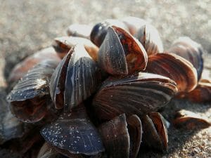
Gastropods
Gastropods include species of sea snails, the majority of which exhibit a spiral or conical seashell. Typically, the shells of gastropods are small, but vary in the overall shape and size. Gastropod seashells are also the most common type of shells utilized by hermit crabs. The shells of gastropods also typically have a thick layer of nacre, which makes these shells exceptionally durable. The different types of shells can be observed in the image below: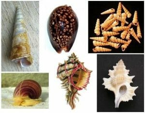
Arthropods
Arthropods include crustacean species (e.g., shrimp, crabs, and lobster) which have a hard exoskeleton comprised of chitin in addition to calcium carbonate, which is shed as the animal grows. The exoskeleton is usually composed of several plates, which form a carapace. Since the exoskeleton must be shed, the animal is vulnerable during the molting process until the new exoskeleton hardens. The shed exoskeleton is often washed ashore onto beaches, and can be classified as a “seashell” in a broad sense of the word. Crab shells that are commonly found near the beach are illustrated in the picture below.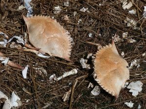
Annelids
Annelids include various species of marine worms, which exhibit a hard seashell-like tube composed of calcium carbonate. Unlike mollusk shells, annelid tubes are comprised of only two layers, a protein and a calcium carbonate layer. Some species anchor their tube in the substrate or form burrows. The tube of a marine annelid is shown below: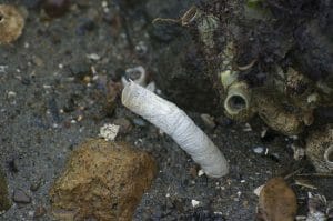
Brachiopods
The most common species of brachiopod is the lamp shell, which has a similar appearance to clams. Brachiopods vary in size and contain two shells called “valves” which protect the dorsal and ventral surfaces of the organism and are either linked by muscle or a hinge. The valves are composed of three layers, similar to mollusk shells; the outer layer is composed of proteins, the middle layer is comprised of calcium carbonate, and the inner layer consists of a mixture of calcium and protein. Similar to mollusks, brachiopods exhibit a mantle located close to the hinge, which secretes the various components which form the shell. A picture of fossilized brachiopods is presented below: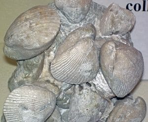
Sea Urchins
Species of sea urchin have a hard shell called a “test” composed of calcium carbonate, as well as a dermis and epidermis that encloses the organs. The test contains five distinct grooves interspersed by five regions, each containing two rows of calcium carbonate plates. Tubercles cover the plates, and form an attachment for the characteristic spines exhibited by sea urchins. The test also contains an inner lining called a peritoneum. An example of a sea urchin test is shown below: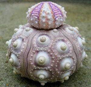
Corals
Both hard and soft corals can wash ashore and consist of a calcium carbonate skeleton or skeletal components, respectively. Hard coral polyps form a skeletal tube consisting of vertical plates termed the septocostae (shown below) joined by a coenosteum and a thin coating called an epitheca. In contrast, soft corals produce spiny sclerites formed by the cross-linkage of protein chains and/or the formation of calcium carbonate.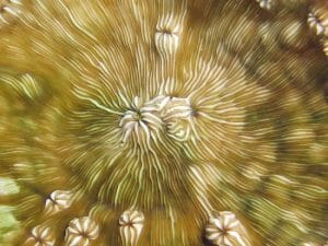
Coccolithophore
Coccolithophores are a type of phytoplankton characterized by the formation of “coccolith” exoskeletons comprised of calcium carbonate. Coccoliths can be shed and reproduced as the animal grows and are typically transparent so as to allow for the passage of light required for photosynthesis. While the precise purpose of this type of seashell remains poorly understood, it is hypothesized that coccoliths provide protection from both predators, as well as the marine environment. An electron micrograph of a coccolith is shown below: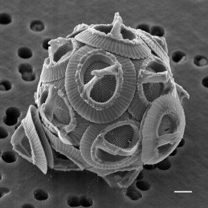
Quiz
1. The exceptional strength exhibited by nacre is derived from:
A. Prism-like sheets of calcium carbonate
B. Brick-like sheets of calcium carbonate interspersed with biopolymers
C. Minerals in the water
D. Nacre is not strong, it is characterized by brittleness
2. What is the major difference between bivalves and brachiopods?
A. Bivalves contain a hinge structure which separates the two shells.
B. Bivalve shells do not contain calcium carbonate.
C. Brachiopod shells do not contain calcium carbonate.
D. The two shells of brachiopods are separated by a hinge structure.
3. The seashell formed by sea urchins is called a:
A. Test
B. Coccolith
C. Tube
D. Sclerites
References
- Allison PG, Seiter JM, Diaz A, Lindsay JH, Moser RD, Tappero RV, and Kennedy AJ. (2016). Gastropod (Otala lactea) shell nanomechanical and structural characterization as a biomonitoring tool for dermal and dietary exposure to a model metal. J Mech Behav Biomed Mater. 53:142-50.
- Ameye L, De Becker G, Killian C, Wilt F, Kemps R, Kuypers S, and Dubois P. (2001). Proteins and saccharides of the sea urchin organic matrix of mineralization: characterization and localization in the spine skeleton. J Struct Biol. 134(1):56-66.
- Gilbert SF. (2000). Developmental Biology. 6th edition. Sinauer Associates: Sunderland (MA).
- Guggolz T, Henne S, Politi Y, Schütz R, Mašić A, Müller CH, Meißner K. Histochemical evidence of β-chitin in parapodial glandular organs and tubes of Spiophanes (Annelida, Sedentaria: Spionidae), and first studies on selected Annelida. J Morphol. 276(12):1433-47.
- Kanold JM, Guichard N, Immel F, Plasseraud L, Corneillat M, Alcaraz G, Brümmer F, Marin F. (2015). Spine and test skeletal matrices of the Mediterranean sea urchin Arbacia lixula–a comparative characterization of their sugar signature. FEBS J. 282(10):1891-905.
- Marin F, Luquet G, Marie B, and Medakovic D. (2008). Molluscan shell proteins: primary structure, origin, and evolution. Curr Top Dev Biol. 80:209-76.
- Marin F, Le Roy N, and Marie B. (2012). The formation and mineralization of mollusk shell. Front Biosci (Schol Ed). 4:1099-125.
- Nudelman F. (2015). Nacre biomineralisation: A review on the mechanisms of crystal nucleation. Semin Cell Dev Biol. 46:2-10.
- Ozaki N, Sakuda S, and Nagasawa H. (2007). A novel highly acidic polysaccharide with inhibitory activity on calcification from the calcified scale “coccolith” of a coccolithophorid alga, Pleurochrysis haptonemofera. Biochem Biophys Res Commun.357(4):1172-6.
- Tentori E and Allemand D. (2006). Light-enhanced calcification and dark decalcification in isolates of the soft coral Cladiella sp. during tissue recovery. Biol Bull. 211(2):193-202.
- van der Wal P, de Jong EW, Westbroek P, de Bruijn WC, and Mulder-Stapel AA. Polysaccharide localization, coccolith formation, and Golgi dynamics in the coccolithophorid Hymenomonas carterae. J Ultrastruct Res. 85(2):139-58.
- Weiss IM, Lüke F, Eichner N, Guth C, and Clausen-Schaumann H. (2013). On the function of chitin synthase extracellular domains in biomineralization. J Struct Biol. 183(2):216-25.
Seashell
No comments:
Post a Comment