Flow Cytometry Definition
Flow Cytometry is a tool used to analyze single-cell characteristics, even when we are presented with a rich heterogeneous cell population. It can find parameters like cell size, health, and phenotype but is so refined that even individual signaling proteins and their downstream transcription factors can be traced. As a result, we have a way of analyzing a cell’s diverse inner workings and signaling networks.
While flow cytometry can use either laser light or electrical impedance (an electronic measure of the opposition of current in an AC circuit), it runs on the same principle. Cells are suspended in a stream of fluid and, one at a time, are passed through a focus of exciting light. Since the proteins or cell components of interest have been fluorescently labeled with antibodies, the emitting light will scatter into distinct wavelengths that are read by an electronic detection apparatus. This then translates the scattered light into quantifiable physical and chemical traits of the particles in the fluid. This ability earns flow cytometry its place as an essential biomedical instrument.
Flow Cytometry Steps
While laboratories continue to optimize its protocols, flow cytometry conventionally includes the following steps:
- First, cells are fixed with formaldehyde to immobilize the proteins of interest and their transient signaling events.
- Methanol or detergent is added to the test tube to make cells permeable to antibodies that can then enter the intracellular spaces.
- Fluorescently tagged antibodies are added, and must be chosen carefully to allow optimal targeting of our epitopes and proper antigen detection when staining multiple surface and intracellular proteins, simultaneously.
- The test tube is placed in the flow cytometer and the fluid is allowed to reach and then exit through the flow chamber, one cell at a time.
- As each cell crosses the laser beam, the light that bounces off each cell is transmitted to light/color detectors.
- The data retrieved from this experiment can finally undergo multiplex analysis to unravel the unique facets of the cells themselves and their patterns of cell signaling.
The antibody staining can vary, as well.
- In direct staining, cells are incubated with an antibody directly conjugated to a fluorochome (e.g. FITC). This is a one-step incubation and is particularly useful for intracellular staining
- In indirect staining, the primary antibody is not labeled, but instead is detected by a fluorochrome-labeled secondary antibody. This method means unconjugated primary antibodies can be raised against many different targets, which widens the choice of target proteins for the researcher
- Intracellular staining refers to staining for intracellular antigens
- Finally, detection of secreted proteins is also possible with a Golgi block, followed by intracellular staining.
Flow Cytometry Applications
Flow cytometry has a great variety of uses. In its simplest application, it can count cells as each crosses the laser beam. Flow cytometry can also sort cells from heterogeneous mixtures by correlating the light signals they emit with certain known cell morphology and gene expression patterns. A lot can be said about the emitting particles themselves. Flow cytometry can reveal a particle’s size, granularity, and fluorescent intensity. It is also useful in detecting biomarkers that are important for understanding disease. In fact, we can use more than one cell-type specific marker in conjunction with phosphorylated-epitopes to provide a unique way of understanding signaling biomarkers in cell mixtures.
More basic applications of flow cytometry include studies in:
- Cell tracking
- Apoptosis
- Phenotyping
- Transduction
- Cell cycle and DNA synthesis
Decoding signal transduction patterns from this tool also carries great therapeutic significance. Deciphering normal signaling patterns can reveal how drugs work and how disruptions in normal signaling can lead to disease. This lends flow cytometry perhaps its most important role in diagnostic testing.
Flow Cytometry in Clinical Diagnostics
Flow cytometry has become something of a golden standard in pathology. It has been used in fields as diverse as marine science and plant biology, but has gained more popularity for its clinical applications in oncology, immunodeficiency disorders and prenatal diagnostics.
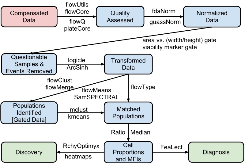
Flow cytometry automated data analysis pipeline
Flow cytometry is heavily important in modern cancer research. By staining for specific surface antigens, flow cytometry allows us to discern between different cancer cell types in lymph node, bone marrow, and blood samples collected from a patient. Likewise, this tool can evaluate the risk of recurrence of bladder, breast, and prostate cancers by measuring the amount of light-reactive DNA in cancer cells. This is clearly a crucial tool for the millions of cancer survivors who are in remission each year.
Its applications in diagnosing primary immunodeficiency (PI) diseases are just as potent. PI diseases are a group of rare, chronic disorders that compromise the affected patient’s immune system from fighting infections. These infections are recurring and may affect the patient’s skin, lungs, throat, brain, or even gastrointestinal and urinary tracts. While diagnosing PI disorders requires several tests, flow cytometry is a key part of the initial workup and subsequent management.
All in all, the diverse applications of flow cytometry make its use very promising, and it is predicted to hold a future in routine hospital and clinic visits in years to come.
Quiz
1. Define the main purpose of Flow Cytometry:
A. Amplifies short sequences of targeted DNA or RNA
B. Captures and evaluates colors in a solid or liquid to ensure color accuracy
C. Analyzes light to determine cell or protein properties
D. Replicates cells suspended in fluid
2. Which “tag”/conduit does Flow Cytometry use to identify its targets?
A. Glucose
B. Plasmids
C. Protein scaffolds
D. Antibodies
3. Which 2 sources of energy does Flow Cytometry use, as mentioned in the article?
A. Lasers and vibrational energy
B. Infrared beams and wind
C. Thermal energy and heat
D. Lasers and impedance
References
- Abcam (2017). “Introduction to Flow Cytometry” Abcam.com. Retrieved 2017-05-07 from http://www.abcam.com/protocols/introduction-to-flow-cytometry
- Cancer Treatment Centers of America (2017). “Flow Cytometry.” Diagnostics. Retrieved 2017-05-07 from http://www.cancercenter.com/treatments/flow-cytometry/
- Hedley DW, et al. “Pharmacodynamic monitoring of molecular-targeted agents in the peripheral blood of leukemia patients using flow cytometry.” Toxicol Pathol. 2008;36(1):133.
- Ibrahim SF, Van Den Engh G. (2007). “Flow Cytometry and Cell Sorting.” Adv Biochem Eng Biotechnol. 106:19-39.
- Illoh OC (2004). “Current applications of flow cytometry in the diagnosis of primary immunodeficiency diseases”. Arch Pathol Lab Med. 128(1);23-31.
- Oregon State University (2017). “Flow Cytometry – How Does It Work? “ Unsolved Mysteries of Human Health, Oregon State. Retrieved 2017-05-07 from http://unsolvedmysteries.oregonstate.edu/flow_06
- Robertson, Sally (2014). “What is Flow Cytometry?” News Medical Life Sciences. Retrieved 2017-05-06 from http://www.news-medical.net/life-sciences/What-is-Flow-Cytometry.aspx
- Roshini S. and Aubert, G (2016). “Flow Cytometry, a Versatile Tool for Diagnosis and Monitoring of Primary Immunodeficiencies” American Society for Microbiology: Clinical and Vaccine Immunology. vol. 23 no. 4 254-271
Flow Cytometry
No comments:
Post a Comment