Thursday, September 7, 2017

Eyes

Eyes Definition


Eyes are animal organs that are specialized for sight. These may be as simple as proteins or cells which can tell light from darkness – like the “eyes” found in many microorganisms – or they may be complex assemblies of lenses, filters, light-sensitive tissues, nerves, and support structures.


Most animals, including humans, have complex and highly specialized visual systems. Different animals have found several different ways to gather light and use it to accomplish complex visual processing.


Mammals, for example, have a single lens and retina which gather light and turn that light into information that the brain can read; insects, on the other hand, have “compound eyes” which use many separate lenses to gather light and put together a mosaic-like view of the world.


Here, we will focus on the details of the human eye.


Eye Parts and Functions


The eye has many parts which work together to accomplish vision, and to keep the structures required for vision safe from infection and injury. These parts include:


The Conjunctiva


The surface of the eye and of the inner eyelids is covered by a clear, protective membrane called the “conjunctiva.”


This is where the word “conjunctivitis” – the scientific name for “pink eye” – comes from. Conjunctivitis means simply “inflammation of the conjunctiva.”


The conjunctiva is lubricated by several substances produced by the body to keep the eye in good working order. These substances, which include mucuous, oils, and a watery solution, prevent the eye from drying out and protect it from surface irritants.


The Sclera


The sclera is also known as the “white of the eye.” It is – as you may have guessed – the white part of the eye that surrounds the iris and pupil.


The sclera does not collect visual data itself. Instead it acts as a tough, protective membrane for the eyeball. Only the outer part of the sclera is white; the interior of the membrane is brown, and wraps around the clear inner chambers of the eye which allow light to pass through.


The Cornea


Light starts its journey into the eye by passing through the cornea. This layer of transparent tissue sits on top of the iris and pupil. It helps to focus light to produce a clear image on the retina, and acts as an additional protective layer for the eye.


Although the cornea looks curved, it is usually actually a flat sheet of uniform thickness. The rounded bulge is the anterior chamber, which will be discussed next.


The cornea can be seen in this diagram:
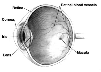

Human eye cross-sectional view


When you use contact lenses, these lenses essentially augment or re-shape your cornea to focus light properly. Some people also undergo laser surgery to change the shape of the cornea so that it will focus light better.


Because it is such a valuable part of the eye, the body wants to know when the cornea has been injured! For that reason it has many nerve fibers running through it, and may hurt a lot of it is scratched, irritated, dried out, or infected.


Because the cornea has almost no blood vessels in it – these would get in the way of light passing through – it can take a long time to heal from injury, and can have a difficult time fighting infection.


For that reason, people who suspect they may have an eye injury or infection should see a doctor right away. Eye injuries and infections can permanently damage vision if they are not treated properly.


Anterior Chamber


The anterior chamber of the eye refers to a small pocket of fluid that lies between the cornea and the iris. This fluid is “aqueous humor” a watery solution that helps the cornea and pupil to focus light.


Just like focusing light through a glass of water or a solid transparent lens, the aqueous humor helps the eye to form an image by refracting light at a constant rate.


The aqueous humor is made from blood plasma, using a special filtering process that removes proteins and other impurities that may cloud vision.


Posterior Chamber


The posterior chamber refers to the aqueous fluid-filled chamber behind the iris and pupil. The posterior chamber sits between the iris and the lens, which completes the job of focusing light.


It can be seen here:
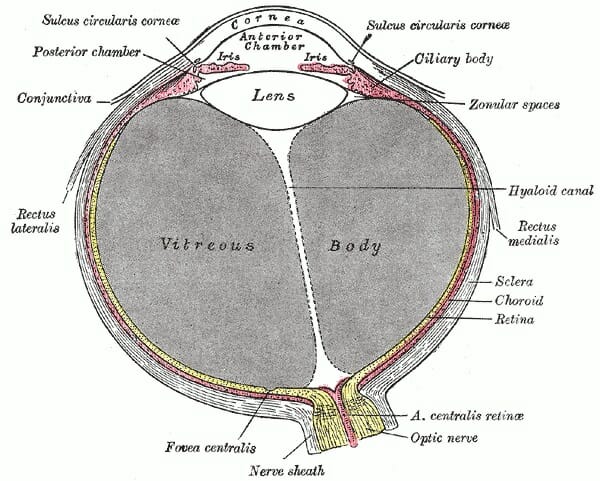

Conjunctiva


Glaucoma – a condition which leads to gradually impaired eyesight, and eventually blindness if untreated – occurs when aqueous fluid cannot drain properly from the anterior and posterior chambers.


When aqueous humor is unable to drain, the fluid’s pressure builds until permanent damage is caused to parts of the eye essential for vision.


Iris


The iris is the colored ring around the pupil. Different people have different amounts of pigment in their iris, resulting in eye colors ranging from blacks to very pale blues and greens.


Interestingly, there is actually no blue or green pigment produced by the human eye. All human eyes have brown pigment melanin, the same pigment that is found in our skin. But those with very small amounts of melanin reflect a lot of light, which is scattered as it reaches the surface of the eye.


Light that is scattered through a transparent substance tends to appear blue, because more red and green wavelengths are absorbed by the apparently transparent medium, while blue light tends to scatter and reflect. This scattering of blue light is the same reason that the sky is blue, that water in swimming pools looks blue, and the same reason that your veins look blue under your skin even though they are actually dark red.


Green eyes occur when someone with a very small amount of pigment in their iris – producing a blue color through scattering – also produces a yellow pigment that mixes with the blue color.


The iris has a sphincter muscle, which allows it to expand or contract, making the pupil larger or smaller. This is important for controlling the amount of light our eyes receive. If you ever have your pupils artificially dilated by an eye doctor, you’ll notice that having an overly-dilated pupil causes blurred vision, and can make bright lights painful.


Pupil


The pupil is the opening to the inner chamber of the eye. Pupils appear black because light passes through them and does not return. The pupil, then, is our actual “window to the world.”


Once it has passed through the pupil, light is focused by the lens. It then travels through the rest of the eyeball to the retina, which lies at the back of the eye. The retina turns the light into signals our brain can understand.


Lens


The lens of the eye lies immediately behind the pupil. Some people think that the lens of the eye is found on the outside, where the cornea is – perhaps because of the use of the word “contact lenses.” But the lens that performs the final focusing of light is found inside the eye, behind the pupil.


The lens is a complex structure. It is made of an elastic capsule containing proteins and water, which refract light at a constant rate just like the lenses used in glasses. It has layers of soft tissue surrounding a firm “nucleus.”


The softness of its outer layers allow the lens to change shape when pushed or pulled by the surrounding ciliary muscles, making it an “adjustable” lens that can change the way it focuses light depending on how close or far away an object is.


Many people’s lenses lose the ability to change shape around the age of 50. This is why many older people need reading glasses in order to focus light to read small print.


Vitreous Humor


The vitreous humor is a thick, gelatinous fluid that fills most of the eyeball. Like the aqueous humor, it refracts light at a constant rate – but unlike the aqueous humor, it is thick and jelly-like.


The jelly-like thickness of the vitreous humor helps the eye to retain its round shape. The precise maintenance of this shape is essential for vision, because light is focused by the cornea and lens with the intent of hitting the retina a set distance away. If the retina moves closer or further to the lens due to changes in eye shape, the light will not be properly focused when it reaches the retina.


Eyes that have “elongated” or “squashed” shapes are the causes of nearsightedness and farsightedness. Nearsighted eyes are elongated, causing light to focus on a point in front of the retina instead of on the retina itself. Likewise, farsighted eyes are too short, causing light to focus on a point behind the retina.


Glasses correct for nearsightedness or far sightedness by adjusting the focus of light before it enters the eye, so that light is properly focused when it hits the retina.


The focus point of light in a nearsighted eye, along with a nearsighted eye with a corrective lens, can be seen below:
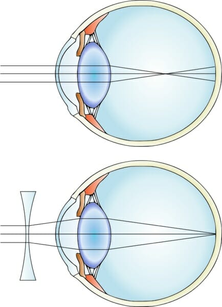

Myopia color

Myopia color


Retina


The retina is a light-sensitive layer of tissue that covers the back of the inner eyeball. It contains light-sensitive cells which can determine light, dark, and color to assemble images of the world. The retina then converts that color information into neural information and sends it to the brain for processing.


The retina contains two major types of light receptors: cone cells, and rod cells.


Cone cells allow us to see color. There are three types of cone cells (or more, in some people with rare mutations). Each type of cone cell responds to a certain wavelength – or color – of light.


S-type cone cells respond to short wavelengths of light, and allow us to see the colors blue and violet. M-type cone cells respond to medium wavelengths, and allow us to see the color green. L-type cone cells respond to long wavelengths of visible light – the red and orange wavelengths.


The color yellow is produced by the activation of both green M-type cone cells and red L-type cone cells. The color pink is produced by the activation of both blue S-type cone cells, and red L-type cone cells. The color white occurs when all cone cells are activated equally, indicating an object that is reflects all wavelengths in the visual spectrum.


Colorblindness occurs when a mutation prevents one or more types of cone cells from working properly. Often, these cone cells do respond to light – but not at the normal wavelength. This can lead to gaps in color perception.


Colorblindness can sometimes be treated using special glasses that filter out wavelengths of color which may confuse mutant cone cells, causing different colors to look the same. While wearing these glasses, many people with colorblindness report seeing all colors clearly and vibrantly.


Because cone cells only respond to a portion of the visual spectrum, they do not work well in low-light conditions. We see in the dark using rod cells, which cannot distinguish color, but which are more sensitive to overall light levels.


Rod cells respond to all wavelengths of visible light. They can tell us how much light is coming at us – but not what wavelength it is. That’s why we don’t see color in the dark; we are getting all of our information from cone cells, which cannot distinguish between different colors.


The retina can only extract information from the light that hits it. This means that in order for the retina to see a clear image of the world, the light that hits it must have been properly focused by the other parts of the eye. As discussed above, failure to focus light properly can lead to blurry vision and other impairments.


Optic Nerve


The optic nerve is a bundle of neural fibers that travel from the retina to the brain. Each optic nerve encodes the image data recorded by the retina in the form of neural signals that can be read by the brain.


The brain then reads the data and performs complex processing, including looking for associations with known objects. This is how we’re able to identify faces and other objects in our environment.


Interestingly, although the eyes are at the front of the head, the brain’s processing of visual signals occurs at the “occipital lobe” in the back of the head.


This means that the optic nerve must travel back into the brain, and then through special channels all the way through it. In the process, the optic nerves “cross over” – meaning that the left side of the occipital lobe interprets visual data from the right eye, and vice versa.


The “crossing over” point can be seen in this image from a brain scan, which has been artificially colored to show the path taken by the optic nerve. Note the red “X” that is formed where the optic nerves cross over each other behind the eyes:


Optic nerve optic tract optic radiation


Damage to the optic nerve or visual processing areas of the brain can result in permanent blindness, even if the eye itself is fine. Conversely, people with intact optic nerves can sometime be allowed to “see” by visual prostheses that stimulate the optic nerve, even if the rest of the eye is missing or nonfunctional.


It is hoped that as technology advances, our ability to artificially stimulate the optic nerve will continue to advance so that people with damaged eyes can have near-normal vision.


The Irreducible Complexity Argument


The complexity of the human eye has often been singled out as evidence for “irreducible complexity.”


The idea of irreducible complexity holds that some structures found in nature could not have evolved through mutation and natural selection, because they would not have functioned at all if even one piece were missing. Proponents of irreducible complexity have asked how the eye could have evolved through random mutation, since slight changes to its structure or the removal of any of its parts would render the eye useless.


However, in recent years new insight into the evolution of eyes has been discovered, which suggests a natural way in which all the structures of the eye could have come about through random mutation.


The historical tendency for things “science can’t explain” to later be explained by new discoveries has led many scientists who are also religious to warn against use of the “irreducible complexity” argument.


In his book “The Language of God,” geneticist Francis Collins warns readers that citing “things science can’t explain” as evidence of God creates a “God of the gaps” idea, in which the existence of God is seen to rely on the existence of things science can’t explain. As these “gaps” in scientific understanding continue to shrink, Francis argues, so to will the importance of a “God of the gaps.”


As a result, Collins argues that it’s better for science and religion alike to see the two fields as compatible, than to have a religious view that requires science to fail in order to work. Himself a devout believer in God, Collins has urged his fellow religious people to embrace scientific theories and see these as a means to learn more about God rather than as challenges to dogmatic ideas about God.


Quiz


1. Which of the following is NOT true of eyes?
A. All eyes must have, at minimum, a lens, a retina, and an optic nerve.
B. All eyes must contain cells or proteins that are responsive to light.
C. Almost all animals have some sort of eyes.
D. None of the above.

Answer to Question #1

2. Which of the following is a gel-like substance that helps the eye keep its shape?
A. Aqueous humor
B. The conjunctiva
C. Vitreous humor
D. None of the above

Answer to Question #2

3. Which of the following is NOT true of the cornea?
A. It has almost no direct blood supply, so it is vulnerable to injury and infection.
B. It has almost no nerves, so it is hard to tell when it is injured or infected.
C. It is a clear, flat membrane of uniform width.
D. None of the above.

Answer to Question #3

References


  • Land, M. (1992). The Evolution Of Eyes. Annual Review of Neuroscience, 15(1), 1-29. doi:10.1146/annurev.neuro.15.1.1

  • Montag, E. D. (n.d.). Parts of the Eye. Retrieved August 10, 2017, from https://www.cis.rit.edu/people/faculty/montag/vandplite/pages/chap_8/ch8p3.html

  • Liz Segre; eye illustration by Stephen Bagi. (n.d.). Human Eye Anatomy – Parts of the Eye Explained. Retrieved August 10, 2017, from http://www.allaboutvision.com/resources/anatomy.htm

  • The New Encyclopaedia Britannica. (1987). Chicago: Encyclopaedia Britannica.


Eyes

Methanol

Methanol Definition


Methanol, sometimes called “wood alcohol,” is a clear liquid with the chemical formula CH3OH. It is clear liquid with polar properties, making it a good solvent. It is also highly flammable, and highly toxic to humans if ingested.


Historically, methanol was created when cellulose, the main sugar in wood and some other plants, was fermented by bacteria. This fermentation process led to a substance that was deadly to drink, but useful as a solvent for scientific and industrial purposes.


After scientists discovered methanol and its uses, humans began to produce methanol for industrial purposes using a much faster process of combining carbon monoxide, carbon dioxide, and hydrogen gases along with a copper-based catalyst that prompts these raw materials to combine to form methanol.


Methanol is used industrially as an ingredient in antifreeze, various chemical solvents, certain fuels, the creation of many plastics, and in blends of alcohol intended for medical or industrial use instead of consumption. “Denatured” alcohol used in medicine and industry often includes both ethanol (the same grain alcohol found in beer and wine) and methanol, which makes it toxic to consume.


Because it can be produced from the fermentation of plant matter, methanol has been the causes of many fatal cases of poisoning from drinking illegally produced alcohol.


During prohibition in the United States, amateur attempts at distilling liquor sometimes led to blindness, neuropathy, and death as a result of drinking methanol. In other countries today, major poisoning incidents still happen when unscrupulous sellers try to profit by selling home-brewed alcohol at lower prices than those offered by major retailers.


Like formaldehyde, methanol is a simple enough organic substance that it can be created by inorganic chemical reactions. For that reason, methanol has been discovered by telescopes in some regions of deep space where no life exists.


What is Methanol Used For?


Methanol has many industrial and scientific uses.


One of the most common uses of methanol is as an ingredient for formaldehyde. This chemical which can be derived from methanol is used extensively in the production of plastics, including those used in construction materials, car parts, paints, explosives, and wrinkle-resistant artificial fabrics. Formaldehyde is also used by morticians and scientists to preserve corpses and laboratory specimens.


Methanol can be used to make other useful solvents including acetic acid, dimethyl ether, and propylene, which is used in anti-freeze. Methanol itself can also be an ingredient in anti-freeze.


Fuel for both gasoline-powered and biodiesel vehicles can include methanol. Its highly flammable nature and usefulness as a solvent allows it to assist other fuels in blending and igniting.


Pure methanol has even been used by itself as a fuel for race cars. It produces high speeds – but also led to a devastating fire which killed two American race car drivers.


Methanol fires are especially dangerous because they are extremely easy to spark, and the flames are almost invisible. This allows the fires to spread out of control and catch other materials very quickly.


Today, both the United States and Europe have safety regulations on how much methanol car fuel is allowed to contain.


Methanol Structure


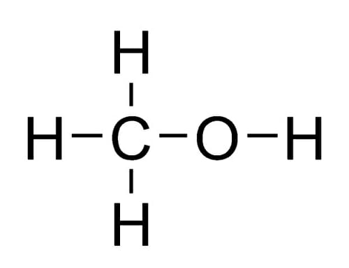
Methanol consists of an “OH” alcohol group attached to a single carbon atom. The carbon atom’s remaining bonding spots are occupied by three hydrogen atoms. This structure is illustrated below:


Methanol flat structure

Methanol flat structure


You may recognize that “methanol” shares a root word with the gas “methane.” The “meth” in both substances refers to this single carbon which is saturated with hydrogen atoms. In “methanol,” this carbon is attached to an alcohol group; in “methane,” the carbon with four hydrogens stands by itself.


Methanol is closely related to ethanol, or “grain alcohol.” Ethanol is the alcohol found in beer, wine, and liquor.


Where “meth” refers to a single carbon saturated by hydrogens, the prefix “eth” refers to a chain of two carbons saturated by hydrogens. Ethanol, then, has a chain of two carbons where methanol has one.


This extra carbon makes a very big difference to how our bodies metabolize the alcohols. While ethanol is safe to drink in moderation, methanol is broken down by our livers into formaldehyde – a highly toxic product that can cause blindness, nerve damage, and death.


Because methanol and ethanol are produced through similar chemical and microbial processes, great care must be taken when fermenting and distilling not to contaminate drinking alcohol with methanol.


Methanol Formula


The formula for methanol is CH3OH.


It is most commonly created by reacting precursor gases such as CO and CO2 with H2 hydrogen gas.


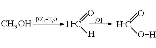
In the presence of a copper-based catalyst under the right conditions, the hydrogen atoms will bond to the carbon and oxygen atoms, displaying the double bonds between the C and O and resulting in a molecule that’s fully saturated with hydrogen.


This reaction can be seen here:


Reaction of production of formic with methanol oxidation


In the case of reacting CO2 with hydrogen, water is also created as a byproduct of the extra oxygen being saturated by hydrogen atoms.


Methanol Safety


One major risk of working with methanol is fire. Liquid methanol burns easily and can be set aflame by any stray spark or excessive heat. Methanol is also very dangerous to ingest.


Like formaldehyde, methanol is produced in tiny quantities by the activity of our own cells. However, also like formaldehyde, methanol is highly toxic to our cells and must be constantly removed and excreted through the liver and kidneys.


When ingested, methanol is metabolized by the liver into formaldehyde, and then into formic salts. These are highly toxic to the nervous system and can permanently destroy the optic nerve, causing blindness. The neurotoxic effects can also cause coma and death.


When methanol poisoning is treated properly, permanent damage can often be prevented. However, when left untreated, death from the neurotoxic effects of methanol can occur within hours of ingestion.


Quiz


1. What does the “meth” in “methanol” stand for?
A. “Meth” indicates that the chemical has a single carbon atom, just like the “meth” in “methane.”
B. “Meth” indicates that the substance is addictive, like the “meth” in “methamphetamine.”
C. “Meth” indicates that the substance is an alcohol.
D. None of the above.

Answer to Question #1

2. Which of the following is NOT a hazard of methanol?
A. It is highly toxic if consumed.
B. It is highly flammable.
C. It produces a thick, foggy vapor which can obscure your vision.
D. None of the above.

Answer to Question #2

3. What would you expect to happen if you reacted carbon dioxide with hydrogen gas and a copper catalyst?
A. Methanol would be created, with no other byproducts.
B. Methanol would be created, with water as a byproduct.
C. Methanol would be created, with formaldehyde as a byproduct.
D. None of the above.

Answer to Question #3

References


  • Methyl Alcohol (Methanol). (2012, March 15). Retrieved August 03, 2017, from https://www.cdc.gov/niosh/topics/methyl-alcohol/

  • References. Retrieved August 03, 2017, from http://www.marinemethanol.com/about-methanol/methanol-production

  • Lazonby, J. (n.d.). Methanol. Retrieved August 03, 2017, from http://www.essentialchemicalindustry.org/chemicals/methanol.html


Methanol

Formaldehyde

Formaldehyde Definition


Formaldehyde is a simple organic compound with the formula CH2O. It is of interest to doctors and scientists, as well as to many other industries, because of its unique chemical properties.


Formaldehyde is produced naturally by living things and some inorganic chemical reactions. It has been found via spectroscopy in interstellar space, and is produced in small quantities by our own bodies. However, like many of our body’s own waste products, it is toxic to us in high concentrations! Our bodies produce just enough formaldehyde to be handled safely by the liver and kidneys.


Breathing formaldehyde fumes can cause lung and sinus irritation, sometimes severe. Long-term formaldehyde exposure is correlated to an increased risk of certain cancers. Ingesting formaldehyde (drinking it) can be fatal.


However, as humans have found more and more uses for formaldehyde, we have begun artificially producing it in large quantities. The formaldehyde industry is booming due to the chemical’s vital role in industries such as car manufacture and the manufacture of building materials.


It is thought that long-term exposure to formaldehyde may increase the risk of cancer, and many regulatory agencies enforce rules about the legally allowable amount of formaldehyde in living and workspaces. The European Union has banned some products that are made with, or contain, formaldehyde.


Formaldehyde Structure


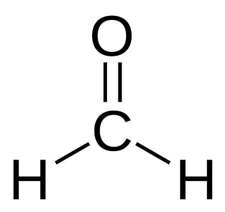
Formaldehyde consists of one carbon atom that shares a double bond with an oxygen atom. The carbon’s remaining electron-sharing slots are occupied by two hydrogen atoms.


Structure of formaldehyde (methanal)

Structure of formaldehyde (methanal)


The polar nature of its carbon-oxygen bond makes this a highly reactive compound. The oxygen atom attracts electrons more strongly than the carbon atom, resulting in a partial negative charge at one end of the compound and a partial positive charge at the other. This causes it to “stick” to other polar molecules and gives it some ability to accept and donate electrons.


Its small molecular size enables it to penetrate tissues and other substances easily, while the polar nature of its carbon-oxygen bond makes it an excellent solvent. This ability to dissolve and react with many compounds is the reason it is used for many industrial and medical applications.


In its pure state at room temperature, formaldehyde is a gas. It is most useful to science and industry in solution as a liquid. This is one reason why formaldehyde fumes are common where formaldehyde is used; it is quick to come out of solution and return to its gaseous state when exposed to air.


Formaldehyde Uses


Because it is highly chemically reactive, formaldehyde has many uses in science and industry. These uses include:


Formaldehyde Uses in Biology


Formaldehyde is often used in biology to preserve tissue specimens. Formaldehyde is useful for this purpose as it kills all bacteria and fungi, and can preserve the shape of a specimen by bonding with proteins and DNA.


For the same reason, formaldehyde it is often used in embalming fluids intended to delay decay in human corpses, as well as in the preservation of animal specimens for dissection.


Formaldehyde delays, but does not permanently prevent decay of tissue. For long-term preservation of samples, scientists must use processes like plasticization, which replace the specimen’s tissues with durable polymers.


In today’s era of genomic analysis, scientists also sometimes prefer methods of tissue preservation that preserve nucleic acids. Formaldehyde does not preserve nucleic acids and so is not ideal for preserving tissue samples that will undergo DNA or RNA analysis. These often require special treatments to de-activate enzymes which break down DNA and RNA within cells.


Formaldehyde Uses in Medicine


  • Used as an antiseptic, as it kills most bacteria and fungi.

  • Used in the treatment of warts and some parasites.

  • Used in the production and sterilization of some vaccines.

  • A formaldehyde precursor is sometimes used as an alternative to antibiotics in the treatment of urinary tract infections. The kidneys turn this precursor into formaldehyde, which is then excreted into the urinary tract instead of circulating in the blood.

  • Used in some personal hygiene products to prevent bacterial growth.


Formaldehyde Uses in Industry


  • Used as a reactant to produce many artificial materials such as resins, plastics, and other industrial chemicals.

  • Used to treat clothes to make fabrics crease-resistant.

  • Used to produce materials used in numerous parts of car manufacture.

  • Used in the production of plywood, carpeting, and building insulation.

  • Used in the production of sanitary paper products such as napkins, paper towels, and tissues.

  • Used to make chemicals used in paints and explosives.

  • Used to prevent bacterial and fungal growth in animal feed for commercial farming.

  • Used in the development of some types of photography film.


Formaldehyde Safety


Unfortunately, the same properties of formaldehyde that make it an excellent solvent, antiseptic, and preservative can also make it dangerous to human health. It is toxic to the human body if ingested, and can cause irritation of the skin, lungs, and sinuses which can sometimes lead to long-term problems.


The U.S. government lists formaldehyde as a “known carcinogen,” meaning it has is known to increase the risk of cancer with repeated exposure. People whose jobs require regular work with formaldehyde are recommended to use safety gear to limit exposure.


Exposure to formaldehyde fumes may also make the development of asthma more likely, and can result in temporary or permanent sensitivity of the sinus passages and skin. In the United States, buildings are monitored to ensure that their air does not contain high levels of formaldehyde, which can sometimes be a risk for newly constructed buildings as formaldehyde is used in producing and finishing many building materials.


The European Union has banned the import of some formaldehyde-containing and formaldehyde-treated products due to safety concerns.


Quiz


1. Which of the following is NOT a risk of formaldehyde exposure?
A. Long-term exposure can increase the risk of cancer.
B. Ingesting formaldehyde can be fatal.
C. Inhaling formaldehyde fumes for long periods of time can lead to asthma and other lung and sinus ailments.
D. None of the above.

Answer to Question #1

2. Which of the following industries does NOT use formaldehyde?
A. Construction and building materials
B. Car manufacture
C. Science & medicine
D. None of the above

Answer to Question #2

3. Which of the following is NOT a useful attribute of the formaldehyde molecule?
A. Its carbon-oxygen bond is polar, allowing it to bond with and dissolve many substances.
B. It is small, allowing it to penetrate other substances quickly and easily.
C. It is a complex molecule with long, branching arms.
D. None of the above.

Answer to Question #3

References


  • OECD SIDS FORMALDEHYDE – INCHEM. (n.d.). Retrieved July 28, 2017, from http://www.inchem.org/documents/sids/sids/FORMALDEHYDE.pdf

  • Haynes, W. M. (2014). CRC handbook of chemistry and physics. Boca Raton, FL: CRC Press.

  • Harder, J. (2015, June 29). What if you drank embalming fluid? Retrieved July 28, 2017, from http://science.howstuffworks.com/science-vs-myth/what-if/what-if-drank-embalming-fluid.htm


Formaldehyde

Skeleton

Skeleton Definition


The skeleton is the supporting framework of an organism. It is typically made out of hard, rigid tissue that supports the form of the animal’s body and protects vulnerable organs.


For land-dwelling animals, skeletons are also necessary to support movement, since walking and flying rely on the ability to exert force on rigid levers such as legs and wings.


Arthropods such as insects have an “exoskeleton” – an outer covering of a hard material called chitin that protects their internal tissues and allows them to walk, jump, and fly.


Vertebrates such as humans have internal skeletons, made of a tissue called bone that gives the limbs their rigidness and protects vital organs such as the heart and brain.


The term “vertebrate,” in fact, comes from a specific part of the internal skeleton – “vertebrae” are small bones that encase and protect the spinal cord, a vital tissue that acts as the information channel between the brain and the rest of the body.


The skeletons of most vertebrates, including humans, are made of bones. Bones are complex structures consisting of many different types of tissues, which perform both structural and biological functions.


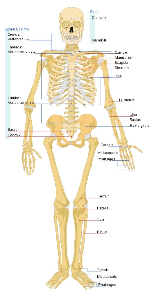
The image below shows the human skeleton with some of the most important bone groups labeled:


Human skeleton

Human skeleton


For anatomy students and medical students, it’s important to note that this skeleton’s right forearm is rotated forward to show how the arm bones look from a different angle. This is not the standard positioning found in most anatomy diagrams, so keep in mind that in most diagrams, both arms are positioned like this skeleton’s left arm.


Here we’ll talk more about the functions of the skeleton and the structures, functions, and classifications of its bones.


Function of Skeleton


For vertebrates such as humans, the skeleton performs many essential functions. Some are directly related to the purpose of all skeletons of providing structural support, protection, and support for locomotion. Others are biological functions unrelated to structural support that have been adopted by vertebrate bone tissues over time.


The functions of the skeleton include:


Structural Support


The skeleton serves the vital purpose of giving form to an animal’s body. Some animals that live in water, such as the octopus, have no skeleton. This is possible because their tissues are partially supported by the water that surrounds them, which is much heavier than air and allows some of an animal’s body structures to float. You’ll notice that octopi don’t do so well on dry land!


For land animals, it’s essential to have a skeleton that fights the force of gravity, which might otherwise prevent movement and even crush organs. That’s why all mobile land animals have either an exoskeleton, like those of insects and spiders, or an internal skeleton, like those of humans and other vertebrates.


Locomotion


Almost all forms of on-land locomotion require the ability to push rigid levers against our environment. When we walk, our leg bones are levers that exert force on the ground to propel us forward. When birds fly, their wing bones are levers that push against air molecules to allow them to move.


The role of bones in locomotion is the major reason why broken bones can be a death sentence for animals in the wild. Without intact levers to push against, animals can be unable to move quickly or at all, which in turn renders them unable to find food or escape from predators.


Protection


In addition to supporting the body’s structure against the force of gravity and allowing locomotion, the skeleton plays the vital role of protecting important organs from injury. Some important protective bones in the human body include:


  • The skull is a thick covering of bone that protects the brain from injury.

  • The spinal column – made of “vertebrae,” from which “vertebrates” get their name – protects the spinal cord, which is the major nerve cord that allows the brain to communicate with the body.

  • The ribcage forms a protective barrier around the lungs and heart, without which the body would not be able to supply blood to the brain and would soon die.


The vertebrate body must make compromises between protection and mobility. Our lower abdominal organs such as the intestines, for example, are not protected by the ribcage. But this lack of a hard covering around our abdomen allows us to bend climb, and shift our weight in a way that greatly enhances our mobility!


Blood cell production


For animals with internal skeletons, the bones also perform other vital biological functions that are not directly related to their role as structural support. In humans, one of the most important of these roles is blood cell production.


Our bones are made out of living tissue. Their outer tissues are hard and rigid, but their inner tissues are soft and serve other purposes. On the inside of our bones – in the part called the “bone marrow” – the stem cells that create our red and white blood cells can be found.


Without healthy bone marrow, our bodies would stop replacing their blood cells – and would soon lose the ability to transport oxygen and fight infection!


This is why bone marrow transplants are sometimes prescribed for people with “blood cancers.” In blood cancers such as leukemia, the cancerous cells actually originate in the bone marrow. These cancerous cells produce large numbers of blood cells, but the blood cells they produce do not work properly.


As a result, people with cancer of the stem cells that produce white blood cells may have very high white blood cell counts – but they have difficulty fighting infection, because these white blood cells produced by cancerous stem cells do not work properly.


In bone marrow transplants, doctors attempt to kill off the stem cells in a patient’s own bone marrow, and then replace some of the marrow with marrow from a healthy donor that can produce healthy blood cells.


Storage


Bones can store calorie-rich fat and minerals that other body tissues might need at a later date.


The hard part of bone tissue is rich in calcium, which in emergencies the body can release from the bones to serve other purposes.


Yellow bone marrow tissue is composed primarily of fat, which can act as a storage point for calories and nutrients.


Red bone marrow tissue is rich in iron, a necessary ingredient for red blood cells. Iron deficiency is a common cause of anemia – a condition in which insufficient production of red blood cells can lead to weakness, fatigue, dizziness, and even fainting.


Endocrine regulation


Bone cells release a hormone called osteocalcin, which has effects on blood sugar, fat storage, and male sex hormones.


The release of osteocalcin by bone cells prompts the pancreas to release more insulin, resulting in lowered blood sugar and increased consumption of sugar by cells. It also causes fat cells to release a hormone called adiponectin, which prompts the breakdown of fat for energy.


Osteocalcin directs the male testes to produce more testosterone, and is also thought to encourage the body to produce more bone cells.


The complex interplay between hormones in the human body is not well-understood. In this case, it’s possible that by prompting the release of insulin and the breakdown of fats for energy, it may be freeing up additional energy that the body can use to grow more bone cells.


Bone Types


While all bones are made of similar tissue, there are a few different kinds of bones that have different characteristics and growth patterns, which allow them to serve their different functions in the human body. These are:


Long Bones


Long bones are those that play a vital role in locomotion and in supporting our weight against the force of gravity. These include the long bones of the arms, legs, hands, and feet.


Long bones lengthen substantially as a person grows, and have a “growth plate” or “epiphyseal plate” at their ends, where new bone is formed during growth.


In children, most blood cells are produced by red bone marrow in the long bones. In adults, much of the red marrow in long bones is replaced by yellow marrow, and blood cell production takes place mostly in the flat bones.


Doctors can sometimes tell a person’s approximate age from looking at their epiphyseal plates, as these shrink and change as a person ages and stops growing. This approach is sometimes used to estimate the age of a person or animal at time of death by forensic investigators, archaeologists, and paleontologists.


Short Bones


Short bones are roughly cube-shaped bones that offer support and mobility in complex structures like the wrist and ankles.


Both our wrists and ankles require a complex large range of motion – but also require extreme strength and stability, especially our ankles which must support our weight.


To solve this problem, the body uses a series of interlocking cube-shaped bones, held together by strong ligaments. These provide a solid structure, but can also be shifted against each other to produce large or small changes in the shape and position of our hands and feet.


Flat Bones


Flat bones serve the primary purpose of protecting important organs. These bones to not require the same range of motion like the short bones, and to not undergo pronounced growth like the long bones.


In adults, most blood cell production occurs in the red bone marrow of the flat bones.


Examples of the flat bones include the skull, sternum, ribs, and the scapulae, which protect our lungs and heart from the back.


Irregular Bones


Irregular bones are bones that have complex shapes, which allow them to serve highly specific purposes. They often serve to both protect internal organs and give structure to the body.


Examples of irregular bones include the vertebrae themselves, whose complex shape allows them to shield the spinal column from all sides while allowing our spines to be mobile and flexible.


The pelvic girdle, which protects some internal organs while also providing a structural base for our legs, is another example of an irregular bone.


Sesamoid Bones


Sesamoid bones are bones that are embedded in tendons, and which provide additional shielding and cushioning for high-stress, high motion areas of the body.


One example of a sesamoid bones is the patella – a small, round bone which covers the kneecap and protects the tendons underneath. Sesamoid bones are also found in the hands, knees, and feet.


Bone Structure


The structure of bones is best exemplified by looking at long bones, which undergo the most growth and which contain distinct cavities for bone marrow. Long bones contain several types of tissues, each of which assist with the functions our bones must perform.


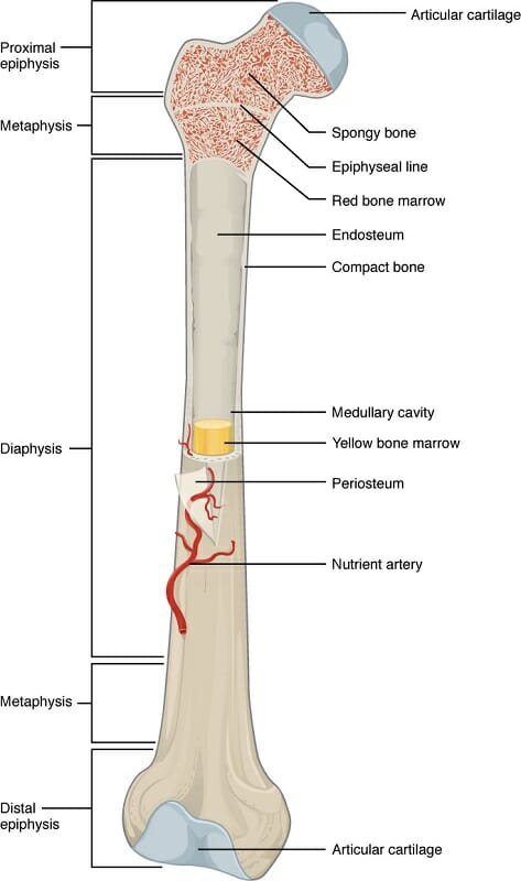
Anatomy of a Long Bone

Anatomy of a Long Bone


Tissues found in our bones include:


Compact Bone


Compact bone, also called “cortical bone,” is the hard outer shell of all bones. It consists of “osseous tissue” made of “osteocytes,” or bone cells.


In osseous tissue, bone cells are surrounded by a solid matrix of minerals and proteins. The most important mineral is hydroxyapatite, a calcium- and phosphorous-rich mineral also found in tooth enamel and in some naturally-occurring rocks.


Hydroxyapatite is hard and solid, but also prone to shattering, so it is interlaced in compact bone tissue with collagen fibers. Collagen is same tough, strong type of protein found in skin. The result is a matrix that is hard and solid, but also has flexibility and resilience.


The basic structural units of cortical bones are “osteons” – microscopic cylinders of bone tissue. Through the center of each cylinder runs a cord of blood vessels and nerves.


This structure of tiny cylinders ensures that bone cells receive the oxygen and nutrients they need to survive.


Spongy Bone


Spongy bone, also known as “cancellous bone,” is bone that has a “spongy” structure that consists of fibers of hard bone tissue interlaced with softer tissues such as blood vessels and bone marrow.


Spongy bone is structurally weaker than more dense types of bone tissues, but provides an excellent site for important biological functions such as the exchange of calcium ions with the blood and the production of red blood cells.


Spongy bone is found near the ends of long bones, and inside of vertebrae.


Articular Cartilage


Articular cartilage is, as the name suggests, cartilage – the same tough but flexible substance that makes up our noses and ears. At the ends of bones, cartilage provides cushioning and flexibility, allowing bones to slide past each other and permitting movement.


The name of “articular cartilage” comes from the verb “articulate,” which means “to unite by a joint.” This means that two structures are connected, but are able to move relative to each other because of the joint between them.


Pain and injury can result when this articular is torn or worn away, causing the hard, rigid surfaces of bones to rub against each other directly. Torn cartilage in the knee is one common sports injury that can occur when the knee joint is hit hard or wrenched violently.


In some cases, surgical repair of the cartilage is necessary to avoid damage to the hard outer shells of leg bones from rubbing against each other directly.


Epiphyseal Plate, or Epiphyseal Line


As mentioned above, “epiphyseal” refers to the region of bone where the bone can grow by producing new bone cells.


In children and teens, an “epiphyseal plate” is a site of active growth and new bone production; in adults, must of this growth tissue disappears, and what is left is an “epiphyseal line” that shows where growth once occurred. These epiphyseal regions are most obvious at the ends of long bones, which undergo extensive growth during a person’s lifetime.


The epiphyseal plate is made of cartilage – that same tough-but-flexible substance we heard about earlier. Bones grow by laying down a layer of cartilage, and then using that cartilage as a matrix for the growth of new bone cells. In that way new bone growth starts out flexible but grows strong and solid, and the cartilage helps to direct the shape and growth patterns of the new bone cells.


Soft areas that allow bone growth can also be found in other types of bone, such as the “fontanelle” of the skull. The fontanelle is the “soft spot” on a baby’s head, where soft tissue has not yet been replaced by bone cells because the skull still needs to have flexibility to accommodate the growing brain.


In adults the fontanelle, like the epiphyseal plate, disappears, leaving only a trace of its existence in the form of the seam where the bone plates of the skull fused.


Red Bone Marrow


Red bone marrow, unsurprisingly, is where red blood cells are made. It is also the site of production of white blood cells and platelets.


At birth, most bone marrow in the body is red marrow; as a person ages, about half of that red marrow is converted into yellow marrow, leaving red marrow mostly near the ends of long bones and inside the flat bones such as the pelvis and sternum.


This iron-rich tissue is vital to the health of the entire body as it is responsible for producing the vehicles that carry oxygen to the brain and other organs, and the cells that prevent and fight infection.


Diseases that destroy red bone marrow or leave it unable to function can lead to severe complications, including death.


Yellow Bone Marrow


Yellow bone marrow is a fatty tissue which contains stem cells that produce fat, cartilage, and bone. It does not produce blood cells, but rather functions mainly to store fat and provide the right environment to keep the surrounding bone healthy.


In cases of extreme blood loss or illness, yellow marrow can convert back into red marrow to help replace lost blood cells.


Periosteum


The periosteum is the membrane that covers the outer surface of bones. The only exception is the joints of the long bones, which are covered by cartilage cushioning.


The periosteum is made of connective tissue rich in collagen. It also contains nerve endings that can feel pain. This allows the periosteum to serve the double purpose of protecting our bones by resisting trauma, and informing us through pain when something is wrong.


The periosteum is also the site of new bone growth by bone cells called “osteoblasts” as bones grown and become thicker.


Even as new bone is laid down by osteoblasts beneath the periosteum, cells called “osteoclasts” digest bone tissue from the inside, widening the bone’s internal marrow-containing cavity and preventing the bone shell from becoming too thick.


Nutrient Artery


A nutrient artery is a part of the circulatory system responsible for delivering oxygen, nutrients, and other vital materials to the bone.


These arteries enter the bones through canals called “foramina,” which are holes in the bone that exist for the purpose of allowing arteries to enter the bone tissue.


From the nutrient artery, blood vessels branch down to the level of the osteons, where microscopic blood vessels run through the center of tiny cylinders of bone cells.


If nutrient arteries become blocked or damaged, bone tissue death and infection can result. Although bones may look like solid, “dead” tissue, they require the activities of living cells to stay strong and to avoid becoming prey for dangerous pathogens.


Endosteum


The endosteum is the membrane that covers the inside of the bone’s medullary cavities – the hollow spaces that are typically filled with marrow.


The endosteum greatly resembles the periosteum, consisting of a thin layer of very tough fibrous tissue, which also contains nerve cells.


During periods of starvation, the body can absorb and digest the endosteum along with some of the fat from bone marrow. This results in a weakening of the bones, but can help the body continue to function for longer until nutrients can be found.


The endosteum is also the site of bone reabsorption as bones grow and become thicker. To prevent the hard shell of compact bone from becoming too thick, bone tissue is absorbed and digested by cells called “osteoclasts” on the inside of the medullary cavity at the same time that new layers are being laid down on the outside of the bone, under the periosteum.


This ingenious mechanism allows bones to grow in both length and thickness while expanding the medullary cavity in tandem with the growth of the compact bone tissue.


Quiz


1. Which of the following is NOT a major function of the skeleton?
A. Provides structural support
B. Enables movement
C. Cleanses toxins from the blood
D. Produces blood cells

Answer to Question #1

2. Which of the following is NOT a change that occurs in the skeleton between birth and adulthood?
A. Epiphyseal plates shrink and become epiphyseal lines
B. About half of red marrow is converted into yellow marrow
C. Several bone plates in the skull fuse together, resulting in one solid bone
D. None of the above

Answer to Question #2

3. Which of the following combinations incorrectly matches an example of a bone to its bone type?
A. Skull – Flat Bone
B. Vertebra – Irregular Bone
C. Femur – Long Bone
D. Metatarsal – Short Bone

Answer to Question #3

References


  • Gray, Henry. Anatomy of the human body. London, England, Bounty, 2012.

  • Musculoskeletal system / Anatomy, physiology, and metabolic disorders. Summit, NJ, Ciba-Geigy, 1987.

  • Zimmermann, Kim Ann. “Skeletal System: Facts, Function & Diseases.” LiveScience,</em Purch, 11 Mar. 2016, www.livescience.com/22537-skeletal-system.html. Accessed 26 July 2017.

  • OpenStax College, Anatomy and Physiology. OpenStax CNX. http://cnx.org/contents/14fb4ad7-39a1-4eee-ab6e-3ef2482e3e22@8.103.

  • Bone Marrow – What Does Bone Marrow Do? http://www.medicalnewstoday.com/articles/285666.php

  • Steele, David Gentry, et al. The anatomy and biology of the human skeleton. College Station, Texas A&M University Press, 2007.


Skeleton

Teeth

Teeth Definition


Teeth are hard, mineral-rich structures which are used to chew food. They are not made of bone like the rest of the skeleton, but have their own unique structure to enable them to break down food.


Tooth enamel is the most mineralized tissue in the body, consisting mainly of the rock-hard mineral hydroxyapatite. Hydroxyapatite is also found in some rocks and makes up part of the mineral/protein matrix of bones.


Some animals have the ability to replace their teeth throughout their lifespan, as teeth may be lost due to injury or disease. Humans have two sets of teeth: baby teeth which loosen and fall out prior to adulthood, and adult teeth which stay in place throughout adult life.


The shape and number of an animal’s teeth vary according to what they eat. Here we’ll discuss the functions of different types of teeth, as well as the structure and different tissues which make up human teeth.


Function of Teeth


Teeth assist animals in obtaining food and breaking it down for efficient digestion. Animals who lose their teeth are generally unable to ingest enough nutrients to survive.


The specific mechanical functions that teeth must perform depends on the animal’s food source. The animal may need to pierce skin and tear meat, grind up fibrous vegetables, or do some combination of the two.


Predators often boast sharp, pointed or even serrated teeth to assist them in taking down prey and eating raw meat.


Herbivores on the other hand typically have long, sharp incisors at the front of their mouths to assist in cutting bite-sized pieces off of plants and well as molars with broad, textured surfaces in the back of the mouth which grind and break down plant matter for easier digestion by the stomach.


Humans eat both meat and a variety of plant matter. As a result, we have several types of teeth which are good for breaking down different types of food:


  • Front teeth, or incisors, which cut food into bite-sized chunks.

  • Sharp, pointed “canine” teeth, which can be used to tear chunks off of meat and other foods.

  • Molars and pre-molars which crush vegetable matter and other foods into a pulp.


Structure of the Tooth


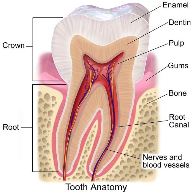
Tooth Anatomy

Tooth Anatomy


The structure of a human tooth includes the following tissues:


  • Enamel – The hard, calcified outer covering which is used to break down food. Enamel consists primarily of a matrix of hydroxyapatite – a mineral made of crystalline calcium phosphate which is created by the body’s cells during tooth development. Hydroxyapatite can also be found in some rocks, and in the mineral/protein matrix that makes up the hard outer shells of our bones.

  • Dentin – A softer, more vulnerable material which serves as a last line of defense for the tooth pulp in the event that enamel is broken or dissolved.

  • Cementum – a bone-like tissue that includes both hydroxyapatite and connective proteins. This tissue attaches the tooth to the periodontal ligaments, which hold the tooth firmly in place within the jawbone.

  • Pulp, which contains blood vessels and nerves used to keep the tooth healthy and alert the organism to dangerous tooth injuries and infections.

The tooth is often broken down by dentists into the following major regions:


  • The root, which consists of dentin and pulp, with a covering of cementum that anchors the tooth to the jaw. The root is especially vulnerable to injury and infection because it lacks protective enamel.

    Infections of the tooth’s root can spread into the bloodstream or the surrounding jaw and tissues, which is why infections of a tooth’s root require prompt and thorough medical attention.

  • The neck, which is the place where the cementum of the root meets the enamel of the tooth’s crown. The root has a thin layer of enamel and a thick layer of dentin protecting the root.

  • The crown, which consists of a thick enamel surface that is used to cut and grind food. A thick layer of dentin lies under the enamel, between it and the pulp of the tooth.

Quiz


1. Why is it important to floss?
A. To prevent potentially dangerous bacteria from reaching the root of the tooth.
B. To prevent bacteria from eating away at the enamel of the tooth’s neck and crown.
C. To prevent infections from spreading from the tooth to other parts of the body.
D. All of the above.

Answer to Question #1

2. What is likely the diet of an animal with sharp, shearing incisors in the front of the mouth and flat, grinding molars in the back?
A. A carnivorous diet of meat.
B. An omnivorous diet with both plants and meat.
C. A herbivorous diet of plants.
D. None of the above.

Answer to Question #2

3. What is one possible consequence of injury or infection to a wild animal’s teeth?
A. The infection may spread to other parts of the body, potentially resulting in death.
B. The animal may not be able to ingest enough nutrients to survive.
C. In some species, the teeth may be replaced by a new set of teeth.
D. All of the above.

Answer to Question #3

References


  • Romer, A. S., & Parsons, T. S. (1990). The vertebrate body. Fort Worth: Harcourt Brace Jovanovich College Publ.

  • Clemente, C. D. (2011). Anatomy: a regional atlas of the human body. Philadelphia: Wolters Kluwer/Lippincott Williams & Wilkins Health.

  • Dental Cervix – National Library of Medicine – PubMed Health. (n.d.). Retrieved July 17, 2017, from https://www.ncbi.nlm.nih.gov/pubmedhealth/PMHT0025298/


Teeth

Sunday, August 20, 2017

Leaf

Leaf Definition


The term leaf refers to the organ that forms the main lateral appendage on the stem of vascular plants. In general, leaves are thin, flat organs responsible for the photosynthesis of the plant. Although photosynthesis typically only occurs on the upper surface of the leaf, it can occur on both sides in some plant species. Leaves are typically comprised of a distinct upper and lower surface, stomata for gas exchange, waxy coating, hairs, and venation. Each side of the leaf differs in regarding the level to which these features are expressed. Although leaves are typically located above ground, some species have leaves which reside underground (e.g., bulb scales) or underwater (e.g., aquatic plant species). Moreover, the leaves of some plants may not be associated with photosynthesis (e.g., cataphylls). Leaves are typically oriented on a plant to avoid blocking the sunlight of the leaves situated underneath.


Function of the Leaf


As one of the most important constituents of plants, leaves have several essential functions:


Photosynthesis


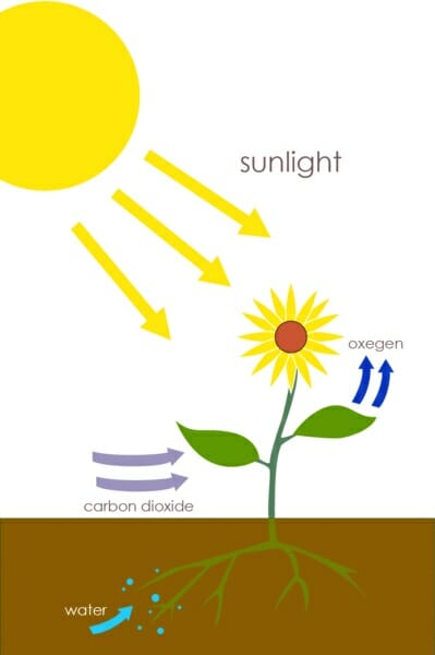
The primary function of the leaf is the conversion of carbon dioxide, water, and UV light into sugar (e.g., glucose) via photosynthesis (shown below). The simple sugars formed via photosynthesis are later processed into various macromolecules (e.g., cellulose) required for the formation of the plant cell wall and other structures. Therefore, the leaf must be highly specialized to combine the carbon dioxide, water, and UV light for this process. Carbon dioxide is diffused from the atmosphere through specialized pores, termed stomata, in the outer layer of the leaf. Water is directed to the leaves via the plant’s vascular conducting system, termed the xylem. Leaves are orientated to ensure maximal exposure to sunlight, and are typically thin and flat in shape to allow sunlight to penetrate the leaf to reach the chloroplasts, which are specialized organelles that perform photosynthesis. Once sugar is formed from photosynthesis, the leaves function to transport it down the plant via specialized structures called the phloem, which run in parallel to the xylem. The sugar is typically transported to the roots and shoots of the plant, to support growth.


Plants


Transpiration


Transpiration refers to the movement of water through the plant, and subsequent evaporation via the leaves. When the stomata open to accommodate the diffusion of carbon dioxide into the plant for photosynthesis, water flows out. This process also serves to cool the plant via evaporation of the water from the leaf, as well as regulate the plant’s osmotic pressure.


Guttation


Guttation refers to the excretion of xylem from the edges of leaves and other vascular plants due to increased levels of water in the soil at night, when the stomata are closed. The pressure caused at the roots results in the leakage of water from the xylem out of specialized water glands at the edges of leaves.


Storage


Leaves are a primary site of water and energy storage since they provide the site of photosynthesis. Succulents are particularly adept at water storage, as evidenced by the thick leaves. Due to the high levels of nutrients and water, many animal species ingest the leaves of plants as a source of food.


Defense


Some leaves have also evolved defense mechanisms to avoid being eaten or damaged. Some examples include the spines of cacti, cones of gymnosperms, respectively. In addition, hairs found on leaves prevent water loss in dry climates and sting animals that detour herbivores (e.g., Urticaceae). Moreover, the waxy coatings found on leaves serve to protect against water loss, rain, and forms of contamination. Oils and other secreted substances also detract from being consumed by herbivores.


Types of Leaf


In general, the types of leaf can be divided into six major types, although there are also plants with highly specialized leaves:


Conifer Leaf


Conifer leaves are needle-shaped or in the form of scales. Conifer leaves are typically heavily waxed and highly adapted to colder climates, arranged to dispel snow and resist freezing temperatures. Some examples include Douglas firs and spruce trees. The images below illustrate this type of leaf.


Conifer needle


Microphyll Leaf


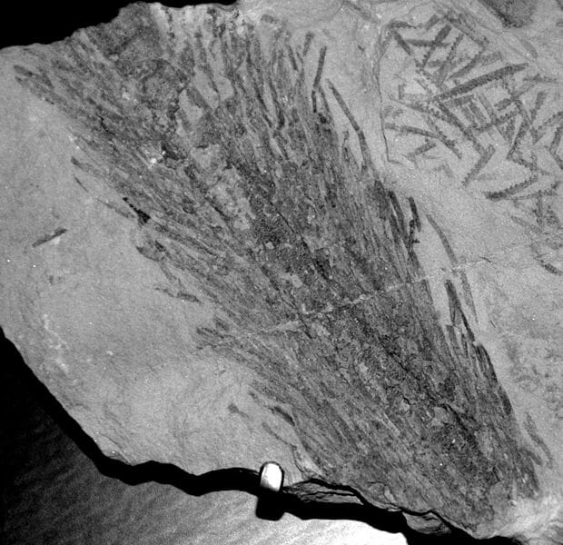
Microphyll leaves are characterized by a single vein that is unbranched. Although this type of leaf is abundant in the fossil record, few plants exhibit this type of leaf today. Some examples include horsetails and clubmosses. The image below illustrates this type of leaf.


Baragwanathia fossil land plant


Megaphyll Leaf


Megaphyll leaves are characterized by multiple veins that can be highly branched. Megaphyll leaves are broad and flat, and generally comprise the foliage of most plant species. The image below illustrates this type of leaf.


Setaria megaphylla leaves


Angiosperm Leaf


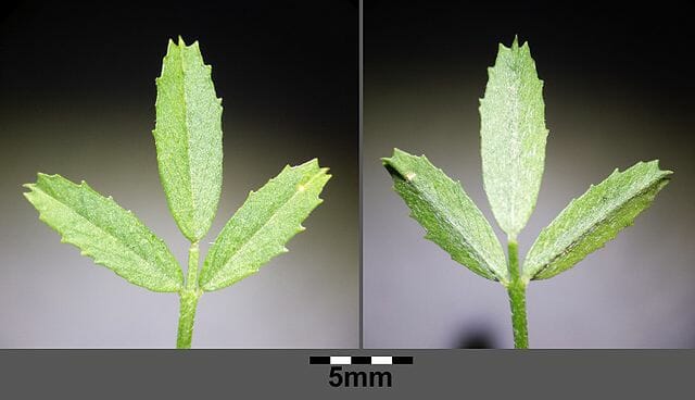
Angiosperm leaves are those found on flowering plants. These leaves are characterized by stipules, a lamina, and a petiole. The illustration below shows an example of an angiosperm leaves.


Medicago prostrata


Fronds


Fronds are large, divided leaves characteristic of ferns and palms. The blades can be singular or divided into branches. The image below presents an example of a frond.


Fern


Sheath Leaf


Sheath leaves are typical of grass species and monocots. Thus, the leaves are long and narrow, with a sheathing surrounding the stem at the base. Moreover, the vein structure is striated and each node contains only one leaf. The image below presents an example of a sheath leaf.


Juncus bufonius


Quiz


1. The primary function of a leaf is:
A. Water evaporation for cooling
B. Photosynthesis
C. Provide shade to the shoot and root structures of the plant
D. Transpiration

Answer to Question #1

2. Which of the following statements is TRUE regarding guttation:
A. It typically occurs at night.
B. It occurs when the stomata are closed.
C. It results from increased water pressure in the soil.
D. All of the above

Answer to Question #2

References


  • Brodersen C and McElrone A. (2013). Maintenance of xylem Network Transport Capacity: A Review of Embolism Repair in Vascular Plants. Front Plant Sci.4:108.

  • El-Sharkawy, M. and Hesketh, J. (1965) Photosynthesis among species in relation to characteristics of leaf anatomy and CO2 diffusion resistances. Crop Science. 5(6):pp. 517-521.

  • Roth-Nebelsick A, Uhl D, and Kerp H. (2001). Evolution and Function of Leaf Venation Architecture: A Review. Ann Bot. 87(5): 553-566.

  • Sadras and Milroy. (1996). Soil-water thresholds for the responses of leaf expansion and gas exchange: A review. Field Crops Research. 47(2): 253-266.


Leaf

Pollen

Pollen Definition


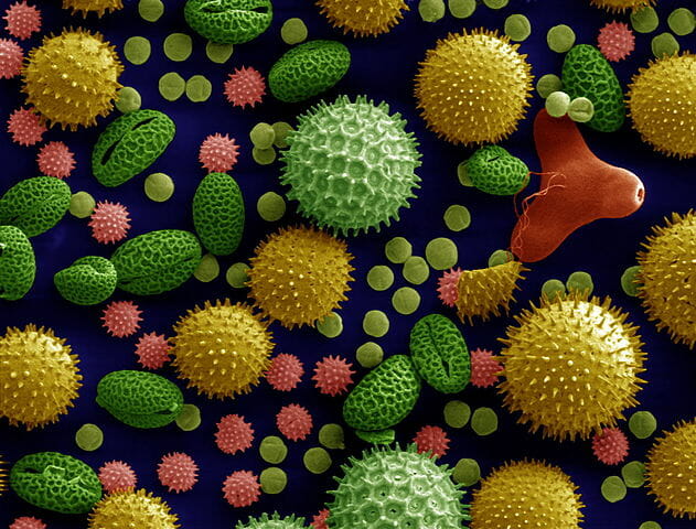
Pollen refers to the powdery product synthesized by seed plants responsible for the production of the male gametes of the plant (shown below).


Pollen


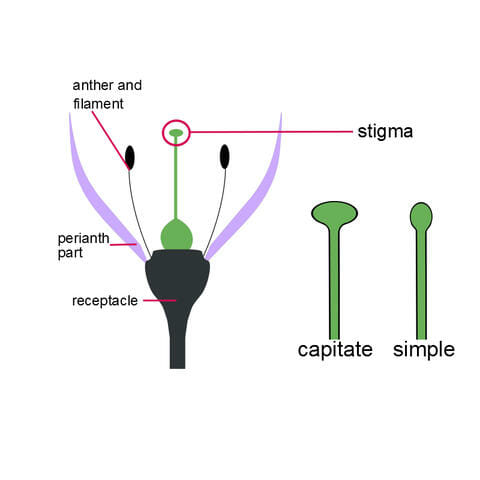
The pollen grains are termed microgameteophytes, and consist of a sporopollenin coating which serves to protect the gameteophytes as they are transported from the stamens (male) or male cone to the pistil (female) or female cone in flowering and coniferous plants, respectively. When the pollen reaches the pistil or female cone, a pollen tube is formed, which transports the sperm to the ovule containing the female gametophyte. The term pollination refers to the transfer of pollen grains from the anther to the stigma of a flower. Cross-pollination involves the transfer of pollen from one flower to the stigma of another flower. In contrast, self-pollination involves the transfer of pollen from one flower to the stigma of the same flower. A diagram illustrating the location of the anther and stigma is shown below.


Stigma shapes


Pollen Structure


Pollen grains vary in size, shape, and surface characteristics depending on the plant species (shown below). In general, pollen grains have a double wall consisting of a thin inner wall composed of cellulose, termed the endospore, and a thick outer wall comprised of sporopollenin, termed the exospore. The shape and the external features of the exospore are highly variable, and are often used to distinguish pollen grain produced by different species. The purpose of this structure is to protect the male genetic material from the environment (e.g., UV radiation, compression, and water) during the transportation from the anther to the stigma. The surface of the pollen grain also contains various waxes and proteins which help repel moisture and interact with the stigma, respectively. However, such protein structures on the surface of pollen are often recognized by immune cells and are the source of the allergic reactions to pollen observed in humans.


Pollen colorized


Pollen Formation


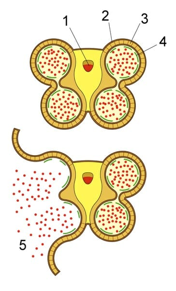
In coniferous plants, pollen is formed in the microsporangia of the male cone, whereas it is produced in the anthers of flowering plants (also termed angiosperms). Each microsporocyte is diploid and forms four haploid cells, termed microspores, via meiosis. This process is termed microsporogenesis. The four microspores then form the double wall of the pollen grain within a structure made of callose. During this process, the callose is digested by callase and the pollen grains are able to grow and complete the formation of the endospore and exospore. The diagram below illustrates the release of pollen grains from the callose structures.


Anther schematic


Quiz


1. The transfer of pollen from the anther to the stigma of a flower is termed:
A. Endoporation
B. Germination
C. Pollination
D. Microsporogenesis

Answer to Question #1

2. The primary function of the exospore is:
A. Pollination
B. Protect the plant’s male genetic material
C. Protect the plant’s female genetic material
D. Endoporation

Answer to Question #2

References


  • Clarke A, Gleeson P, Harrison S, and Knox B. (1979). Cell Biology Pollen-stigma interactions: Identification and characterization of surface components with recognition potential. Proc. Nati. Acad. Sci.76(7):3358-3362.

  • Heberle-Bors E. (1985). In vitro haploid formation from pollen: a critical review. Theoretical and Applied Genetics. 71(3): 361-374.

  • Gullvag B. (1966). The fine structure of pollen grains and spores: a selective review from the last twenty years of research. Phytomorphology. 16: 211-27.

  • Ward M, Dick C, Gribel R, and Lowe A. (2005). To self, or not to self… A review of outcrossing and pollen-mediated gene flow in neotropical trees. Heredity.95(4): 246-54.


Pollen