Monday, April 23, 2018

Antibody

Antibody Definition


An antibody is a specialized defense protein synthesized by the vertebrate immune system. These small structures are actually made of 4 different protein units. The ends of the molecule are variable, and can be adapted to bind to any molecule. The shape is determined by the antigens in the system which are causing damage. Special immune cells detect these antigens and create a reciprocal antibody. This generalized structure is repeated many times, to flood the system with antibodies. These proteins bind to and surround the antigens, preventing further spread or infection.


It is in this way that an organism can identify “self” from “non-self”. For instance, the surface of bacterial cells has certain proteins and carbohydrates, which can be identified by the immune system. B lymphocytes, a special immune cell, create and release antibodies which attack the invading bacteria. An antibody attached to a bacteria not only prevents it from completing normal processes, but helps direct white blood cells to eat the bacteria. These macrophages, as they are known, identify food based on the tail end of the antibody.


In the blood, antibodies account for around 20% of the total protein. This is a very significant amount. Although a single antibody can be very small, an organisms must have many antibodies to fight the many types of antigens present in the system. Further, many of each type is needed. It often takes many antibody molecules to target and identify a large bacteria. Viruses, while they are smaller, are much more abundant and need equal amounts of antibody to quell.


While other organisms often have immune systems based on similar concepts, the term antibody and the structure described below are unique to mammals. An antibody may also be referred to as immunoglobulin, a term which describes a protein used in an immune function. The most common antibody is Immunoglobulin G (IgG) in mammals. Antibodies, if they exist, are not well understood in invertebrates and plants. While it is known that these organisms also have immune systems, it is not entirely clear how they function.


Antibody Structure


Antibody structure


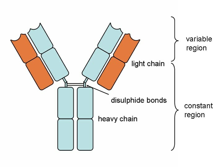
Above is a typical antibody. Notice that the structure is actually made of 4 different protein chains. There are two heavy chains and two light chains. The two heavy chains are connected by a disulfide bond, which exists between two sulfide atoms present in the amino acids of each chain. The light chains attach to the sides of the heavy chain, through a series of non-covalent bonds and weak interactions.


Each chain is broken into two regions, the constant region and the variable region. The constant region is produced directly from the DNA, and is the same on all antibody molecules of the same type. The variable region is the part of the antibody which changes according to the antigen present. The B lymphocytes are in charge of a complex process which matches the variable region to the antigen, and then mass produces the correct antibody.


It is the variable region that has a binding site, capable of binding to the antigen. The binding site is specific in that it is designed to attach only to the intended antigen. It does this by being as compatible to the antigen as possible. If the antigen is hydrophobic, so is the binding site. If the antigen is negatively charged, the binding site will optimally be positively charged to help bind the antigen. Further, the entire shape of the antibody head is specifically formed to the shape of the antigen. This ensures that the antibody will be specific to the antigen. The constant region of the antibody can come in a number of forms, and can be assembled into larger complexes with different shapes.


Antibody Action in Autoimmune Disease


In some cases, the antigen is so close to a molecule produced the organism that the immune system ends up attacking itself. This is known as an autoimmune disease. The immune system, upon being presented with an antigen, forms a defense. In these cases, the antigen is usually a protein. The protein is similar to a protein produced by the organism. While the antibody forming system can be very specific, it cannot accurately identify two molecules which have the same shape. Thus, even if the molecules are actually “self” it can end up attacking them.


Autoimmune diseases can be caused by a number of conditions. Some include viruses, such as HIV, which make the immune system target itself. Still other autoimmune diseases, such as certain forms of diabetes, can be caused by the immune system attacking the pancreas, an insulin-secreting organ. Some research has been done which may link this autoimmune reaction to the proteins found in animal products. While plant proteins have evolved on a completely different trajectory, humans and farm animals share many of the same genes. This means that they produce many of the same proteins. If these proteins leak into the body without being broken down, they could be identified as an antigen.


Upon seeing this antigen, the immune system will create an antibody, to contain it. These antibodies will be mass produced, and sent all over the body to attack any protein of the same shape. This can cause a serious problem for your body. Let’s say you just had a hot dog. All parts of a pig and cow are used to create hot dogs. Needless to say, you will likely get proteins which originated in the animal’s pancreas, cartilage, or other organs. Because their proteins are so similar to yours, your body will begin to have an immune reaction in places these proteins are present. This could be a major cause of diseases like diabetes, arthritis, and possibly even conditions like Multiple Sclerosis.


Antibody Use in Analytical Techniques


An antibody can be a very useful tool in the laboratory, as well. Because an antibody is so specific, and binds tightly in certain conditions, antibodies are used in a number of applications used to filter a solute from a solution. In column chromatography, they are used to bind to solute molecules passing by. As the solution is drained off, the antibody retains the solute. A different solution, with a different pH, can be washed over the antibody media, and the antibody will change shape and release the solute.


Another common use of an antibody in the laboratory is to detect certain substances. An antibody is attached to another protein, used to create a visible molecule. When the antibody is in the presence of the antigen, the antibody changes shape and activates the enzyme. This action creates visible molecules, and can be detected either visually or through a computer. This allows scientist to detect very small samples for a substance, relatively cheaply. This can be used to diagnose disease, test products, and test the safety of consumer products.


Quiz


1. A scientist has an antibody specific to guanine, an amino acid. He puts the antibody in a column, and pours in a mixture of guanine, taurine, and adenine. The solution is drained off into Beaker 1. A new acidic solution is placed in the column. The acid changes the shape of the antibody. The solution is drained off into Beaker 2. Where is the guanine?
A. Beaker 1
B. Beaker 2
C. In the Column

Answer to Question #1

2. In some autoimmune diseases, sometimes a treatment includes destroying parts of the immune system. How can this help?
A. With the cells destroyed, no antibodies can be produced
B. This cannot be helpful
C. With no immune system, the body cannot be overwhelmed by bacteria

Answer to Question #2

3. When scientists first learned about bacteria and germs, sterilization was the resounding call. Why is this method being rethought?
A. The immune system needs practice
B. An antibody cannot form without severe disease
C. Producing less antibodies makes you healthier

Answer to Question #3

References


  • Campbell, T. C., & Campbell, T. M. (2006). The China Study. Dallas: Benbella Books.

  • Nelson, D. L., & Cox, M. M. (2008). Principles of Biochemistry. New York: W.H. Freeman and Company.

  • Pough, F. H., Janis, C. M., & Heiser, J. B. (2009). Vertebrate Life. Boston: Pearson Benjamin Cummings.

  • Widmaier, E. P., Raff, H., & Strang, K. T. (2008). Vander’s Human Physiology: The Mechanisms of Body Function (11th ed.). Boston: McGraw-Hill Higher Education.


Antibody

Sunday, April 22, 2018

Ligand

Ligand Definition


In biochemistry, a ligand is any molecule or atom which binds reversibly to a protein. A ligand can be an individual atom or ion. It can also be a larger and more complex molecule made from many atoms. A ligand can be natural, as an organic or inorganic molecule. A ligand can also be made synthetically, in the laboratory. This is because the key properties of a ligand are found in its chemical structure. If that structure can be recreated in the laboratory, the synthetic ligand will be able to interact in the same ways a natural ligand acts.


How a Ligand Works


The ligand travels through the watery fluids of an organism, within the blood, tissues, or within a cell itself. The ligand travels at random, but once the concentration is high enough, a ligand will eventually reach a protein. Proteins receiving ligands can be receptors, channels, and can even be the start of a complex series of intertwined proteins. When the ligand binds to the protein, it undergoes a conformational change. This means that while no chemical bonds have been formed or broken, the physical action of the ligand fitting into the protein changes the overall shape of the entire structure. This can trigger many actions. In most cases, the movement of the protein itself activates another chemical pathway, or triggers the release of another messenger ligand, to carry the message to other receptors.


The reversibility of the bond between ligand and protein is a crucial aspect of all forms of life. If ligands bound irreversibly, they could not serve as messengers, and most biological processes would fall apart. If ligands were changed, the way an enzyme changes a substrate, the ligand would become something else after the interaction, and could not be as easily recycled as a messenger. Biologically active proteins are active because of their shape. This shape interacts with the chemistry of the ligand to create a stable connection between the two molecules, which will eventually reverse, leaving both molecules the same. In a substrate and enzyme reaction, the substrate is permanently changed.


It is this ability of the ligand, to activate a protein for a short amount of time and then be recycled, which allows for the biological control of many interactions. The amount of time a ligand spends attached to its receptor or specific protein is a function of the affinity between the ligand and the protein. If there is a high affinity, the ligand will tend to stick to the protein and modify its function for longer. If the ligand has a low affinity for the protein, it will be less likely to bond in the first place and will release from the receptor faster.


The affinity of a particular ligand for a particular protein is determined entirely by its chemical makeup and that of the binding site of the protein. At the binding site, amino acids will be exposed which tend to complement the desired ligand. The amino acids will match the ligand in certain aspects. For instance, both will be hydrophilic or hydrophobic. This increases the attraction between the substances. The amino acids tend to differ from the ligand in terms of electrical activity. If the ligand is positively charged, the binding site should be negatively charged. This creates the strongest interaction. In this way, proteins can obtain a certain degree of specificity for a ligand.


While this is the basis for how cells can begin to tell different molecules apart, it is also at the heart of one of an organism’s biggest problems. Many poisons and toxic substances are so toxic because of their ability to interfere with the protein-ligand binding process. Either the toxin directly binds to the protein itself, because it has a higher affinity, or the toxin otherwise prevents the normal bonding of a ligand to its target protein. Examples of ligands and some competitive toxins can be seen below.


Examples of a Ligand


Oxygen


One ligand that people often overlook is oxygen. In the bloodstream and body tissues, oxygen must reach all the mitochondria in the body if the organism is to survive. But, it is not an easy task to get oxygen everywhere. If oxygen were left to diffuse through the tissue to the cells, it can only pass a few cell layers thick. That is why all organisms of a certain size must contain some sort of circulatory system. Even still, it is hard to move the oxygen ligand where it is needed. Many organisms use specialized proteins for this.


In humans and other mammals, hemoglobin is the major blood protein responsible for transporting oxygen. The hemoglobin protein first attaches to a ligand called heme, which has an iron atom and can help bind oxygen. Thus, hemoglobin picks up oxygen in the lungs. As it travels to the body, the carbon dioxide content in the blood rises. As this happens, the pH lowers, and the conformation of hemoglobin changes. This forces the release of the ligand, oxygen, which can then be absorbed by the cells which need it.


A main competitor of oxygen is carbon monoxide. This is because carbon monoxide has a higher affinity for hemoglobin than oxygen has. In other words, once carbon monoxide is bound to the hemoglobin, it won’t come off. This means that someone exposed to large amounts of carbon monoxide will soon have all their hemoglobin saturated by the wrong ligand. Their body will have no ability to transfer oxygen to the brains and tissues. Even if the person gets oxygen after this, they can still suffocate because of their inability to transport the oxygen.


Dopamine


Dopamine is a ligand used heavily in the brain. When the brain releases dopamine, it is as a signal of a pleasure coming from success. In other words, dopamine is tied to the sensation of motivation. The dopamine receptors in your brain are activated when the ligand dopamine is released by the brain. When the receptors are full of dopamine, your brain feels as if you’ve done something good. This common reward center can be easily thrown off by drugs such as cocaine and methamphetamine.


These drugs, instead of being in direct competition with the ligand, actually increase its effectiveness. They do this by limiting the amount of dopamine which can be recycled. Thus, the brain stays in a constant state of feeling “rewarded”. This is the dangerous feeling which can easily lead to drug addiction. Even though logic tells you drugs are bad, the feelings produced by your brain and the extra dopamine feel real, and tell you to use the drug more.


Other Ligand Uses


Ligands are used in many other applications by cells. The proteins they control can range widely in type and function. Some ligands, like insulin, are used to signal various things to the metabolism of each cell. Another ligand, such as acetylcholine, is used by the brain to transfer nerve impulses between nerves. In this case, it opens an ligand-gated channel, which allows the electrical impulse to flow into the cell and down the length of it. This cell will then transmit acetylcholine to the next cell, and the signal will continue.


Some enzymes are controlled by regulatory ligands, which effectively turn the enzyme on. Without it, they do not have the proper shape to transform the molecules they operate on. When the ligand is present, however, these enzymes spring to life and function properly. Many ligands are needed for controlling the metabolism and other complex processes. Each ligand has a certain affinity, which is important, and also a point at which the receptors become saturated. Above this limit, no higher concentration of ligand will bring a greater reaction.


In general chemistry, a ligand may refer to any molecule bound to a transition metal. This is not the case in biology. In biology, a ligand is any molecule which attaches reversibly to a protein. These are typically used in cellular signaling and cellular regulation, but have many other uses.


Quiz


1. Which of the following is NOT a ligand?
A. Air
B. Insulin
C. Bacterial protein

Answer to Question #1

2. Select the true statement.
A. All ligands evolved for their specific protein
B. A ligand can only be used once
C. A ligand changes the conformation of the protein it affects

Answer to Question #2

3. Which organism below DOES NOT use ligands?
A. Tree
B. Fish
C. Worm
D. All organisms use ligands

Answer to Question #3

References


  • Bruice, P. Y. (2011). Organic Chemistry (6th ed.). Boston: Prentice Hall.

  • Nelson, D. L., & Cox, M. M. (2008). Principles of Biochemistry. New York: W.H. Freeman and Company.

  • Widmaier, E. P., Raff, H., & Strang, K. T. (2008). Vander’s Human Physiology: The Mechanisms of Body Function (11th ed.). Boston: McGraw-Hill Higher Education.


Ligand

Sexual Dimorphism

Sexual Dimorphism Definition


Sexual dimorphism is when the genders of a particular species have different characteristics, not related to their sexual organs. On the other hand, a sexually monomorphic species would look nearly identical, except for their sexual organs. Sexual dimorphism can be expressed in a number of different traits. Typically these include size, coloration, bone shape, and even bodies that hardly resemble one another. Sexual dimorphism is seen in birds, reptiles, mammals, fishes, humans, and many other sexually reproducing species. Sexual dimorphism is thought to be caused by sexual selection, a type of selection driven by the desire of every organism to find an evolutionarily fit mate.


Causes of Sexual Dimorphism


Sexual dimorphism is a product of evolution. There are many benefits and detriments to sexual dimorphism, and each species has evolved to further this trait or reduce it. In all sexually reproducing animals with distinct genders, there is a fundamental difference in the sexual organs. In hermaphroditic species, both sets of organs are present and these species are monomorphic. Sexual dimorphism then encompasses a huge spectrum. It goes from creatures which are only slightly different between the genders to animals which hardly look like they belong to the same species.


One force which usually drives sexual dimorphism in evolution is sexual selection. In this form or selection, the interaction between the genders seeking and choosing mates causes differences in the male and female populations. For instance, females seeking a fit partner might look for a large male, as confirmation of his success. Over time, males would tend to become larger, as a sign to females they were the most successful mate. This is not an active process on the part of the males, but simply the result of the largest males having the most offspring. Smaller males have less offspring, and thus have less of a contribution to the next generation. This type of size-based sexual selection can be seen in many animals.


Size is a commonly sexually selected trait for many reasons. Not only does it suggest to females that the male is capable of surviving, but also of fighting. Many males which compete for mates will develop high levels of sexual dimorphism. If not seen directly in their size, it can often be seen in other appendages, such as horns or enlarged appendages used for battles. Both Big Horn Sheep and Crabs share these enlargements caused by evolution, and both use them to battle for mates. However, size is far from the only trait selected for.


Other traits include color and other visual indications of success. A lion’s mane does not make the lion any bigger, or protect him in battle against other lions. However, a large mane simply looks more formidable. Manes may have developed as a type of sexually selected sexual dimorphism. Birds are often a clear example of sexual dimorphism. Males and females have starkly different feathers and markings, while their bodies are usually of similar size. Males are often brightly colored, with extravagant and cumbersome ornamental feathers. Think of the peacock. The extra weight of the tail feathers should be a detriment to his success. However, in attracting females, it is extremely useful and the benefits outweigh the consequences.


There are many other examples, from simple things like eye color all the way to very complex rearrangements of each organism. Remember too that each species is different. Not all birds are sexually dimorphic. There are many species which have evolved monomorphic patterns. The following examples will provide insight into several different species, and how evolution may have formed the sexual dimorphism seen.


Sexual Dimorphism Examples


Birds of Paradise


The Bird-of-Paradise is actually an entire family of bird species found from Indonesia to Australia. There are many different species, but almost all of them have evolved some of the most extreme forms of sexual dimorphism of any animals. Seen below is a male of one of the species.


Wilson


The females are typically the same size, but contain no special coloration. Each species has a unique and complex pattern. In addition, many species have complex courtship rituals in which the male tries to seduce the female with his pattern. The extremely bright coloration is a dead give-away to predators, but it is also the only way to attract females.


This form of sexual selection displays one theory of sexual selection, the so-called sexy-sons hypothesis. In the theory, females try to select males which will produce offspring which will be most attract to the next generation of females. In other words, the mother wants her son to be able to attract a mate. Therefore, she selects the brightest and most flashy male. In doing so, the males who are the brightest and have the best patterns are selected and contribute more offspring to the next generation. The power of this form of sexual selection can transform similar species in a very short time. The family of the Birds-of-Paradise has around 42 different species, which have emerged over only 24 million years from a common ancestor. Each species has a unique and different pattern, which further drives their separation form the other groups.


Ornate Box Turtle


A box turtle is any turtle which has a hinge on its shell, allowing it to completely enclose itself within the shell. Ornate box turtles live on the plains of the United States, across several states. The turtle is a sexually dimorphic species, but only to the trained eye. Both genders have similar markings on their shell, and from afar would appear to be the same.


However, the males have several traits which are sexually dimorphic from the females. Males have red eyes, where females have brown. Like the Bird-of-Paradise, this may be a trait which has no real benefit, but females find attractive. It may also have benefits which scientists are not yet aware of. While the two genders are roughly the same size, they are not the same shape. This is for a very good reason: turtle sex.


Turtle sex is much more difficult than most forms of mating. The shell, which does so much to protect the turtle from predators, is a huge hindrance for male turtles. That is, until sexual dimorphism. Male turtle have a unique bend on the underside of their shell. Instead of being flat, like the female, the male has a large inward bend. This allows him to slide on top of the female. The curve of her shell fits into his curve, and they can balance. He also has a much longer tail, in which his penis is housed. This allows him to tuck it under the female once he is in position, and successfully mate. The sexual dimorphism in turtles, therefore, is caused less by sexual selection and more out of necessity for evolutionary success. This is a good example of the difference between sexual dimorphism and sexual selection.


Humans


Humans have clearly sexual dimorphic traits. Males are slightly larger than females, as in many primate species. However, in humans it is much reduced. A male orangutan is much larger than a female, where a human male is only slightly larger than a human female. Human breasts are different between males and females. Besides Bonobos, this is not necessarily common. A dog, for instance, will only develop visible breasts when it is breastfeeding or has in the past. Humans and bonobos develop larger breasts on females. This could be a symbol to males that the female will be a supportive mother for their offspring. This would give these females more opportunities to reproduce, and with the best males.


There are a few other areas which differ between men and women, particularly body hair and bone structure. Men tend to have more body hair, and narrower hips. While these traits aren’t related, both are sexually-distinct traits which were likely caused by sexual selection. While humans do have some sexual dimorphism, we are near the monomorphic side of the scale.


Quiz


1. Which of the following is an example of sexual dimorphism?
A. A female flamingo picks the pinkest male, a sign of success
B. The male Black Widow spider is about a tenth of the size of the female
C. A male and female Rattlesnake look identical, except their reproductive organs

Answer to Question #1

2. Many species of spider, upon breeding with a male, will eat him. Scientist speculate that this process has driven the size difference in the spiders. What is this process called?
A. Sexual Dimorphism
B. Sexual Selection
C. Mating

Answer to Question #2

3. Why do some scientists argue that “sexual monomorphism” is an oxymoron (a phrase which contradicts itself)?
A. Sexual describes two forms already
B. That is a bad argument
C. Only hermaphrodites can be monomorphic

Answer to Question #3

References


  • Brusca, R. C., & Brusca, G. J. (2003). Invertebrates. Sunderland, MA: Sinauer Associates, Inc.

  • De luliis, G., & Pulera, D. (2007). The Dissection of Vertebrates. Amsterdam: Academic Press.

  • Feldhamer, G. A., Drickamer, L. C., Vessey, S. H., Merritt, J. F., & Krajewski, C. (2007). Mammology: Adaptation, Diversity, Ecology (3rd ed.). Baltimore: The Johns Hopkins University Press.

  • Pough, F. H., Andrews, R. M., Cadle, J. E., Crump, M. L., Savitzky, A. H., & Wells, K. D. (2004). Herpetology. Upper Saddle River, NJ: Pearson Prentice Hall.


Sexual Dimorphism

Nocebo Effect

Nocebo Effect Definition


The nocebo effect, the opposite of the placebo effect, is a negative reaction caused by a non-medicated control substance, such as a sugar pill. In the placebo effect, a patient experiences the benefits of a medicine under trial, without receiving the medicine. This is thought to be caused by the underlying belief of the patient that the treatment they are receiving will bring beneficial effects. The nocebo effect is similar, in that the patient’s expected outcomes can manifest real symptoms. Often, the nocebo effect is observed when patients are told potential side-effects of a drug, but are only given the placebo, or non-medicated treatment. If patients manifest the side-effects they were told about and never took the drug, this is a clear example of the nocebo effect.


The placebo and nocebo effect have similar causes. In both cases, the body is reacting to what the mind expects. While doctors and scientists first denied the mind’s ability to manifest symptoms or cures, the effect is well documented, as is its counterpart. Apparently, a patient’s expectations can be just as damaging as real chemicals which are introduced to the body. This has been documented in a number of clinical trials, and the nocebo effect itself can introduce a wide array of symptoms, or even increase the symptoms already present.


Meaning of the Nocebo Effect


The phrase nocebo effect was derived after the phrase placebo effect, which means in Latin, “I shall please”. Nocebo effect, as the opposite, means “I shall harm”. The differences in these phrases describes not what type of medicine or fake medicine administered, but is rather a reflection of the patient’s attitude toward the medicine. Those who experience the nocebo effect are consciously or subconsciously under the impression that the medicine they are taking will do them harm. This can be a sugar pill or actual medicine. The nocebo effect, while still present and active in those that took a real medicine, cannot be distinguished from real side effects. In the patients who took a placebo, or fake pill, any side effects seen were caused only by the patient’s mind, as they didn’t take any medicine.


Medical Implication of the Nocebo Effect


Researchers trying to study the effects of their medicine must take extra precautions to detect and exclude symptoms caused by the nocebo effect, as well as those caused by the placebo effect. The standard operation for controlling for these effects is to introduce a placebo into the study. The placebo treatment, which appears to be the same as the medicine but is inert, can show the nocebo effect. It must be remembered that this effect could also be affecting those who took the medicine. Therefore, any symptoms in that group could be any combination of the nocebo effect and the medicine. Scientists can use the baseline effect observed in the placebo patients to understand how much the nocebo effect is adding to the observed symptoms.


While this is a serious concern for medical practitioners, there is another reason the nocebo effect should be studied. Clinical trials have been conducted in which patients experiences symptoms not actually caused by a medicine, simply because they were told it would happen. In effect, the nocebo effect is the mind alone causing visible and quantifiable bodily reactions. The lesson to be learned is that a patient can do damage to themselves if they are not in the right mindset. On the other hand, the placebo effect has shown that patients can improve significantly only on the idea that the medicine or treatment will heal them. Therefore, doctors and researchers should see the nocebo effect as a way to judge the mindset of their patients. Further research is needed into how the mind can play a crucial role in affecting the healing process.


Another concern arising from the nocebo effect is the effect that advertising and negative announcements cause on medicated patients. Some studies have even suggested that telling someone on a certain medication that it can have negative effects will help manifest those symptoms. A serious concern includes inducing nocebo effects in a patient who normally had no side-effects.


The nocebo effect can manifest in a number of different ways. Everything from rashes to pain to changes in body chemistry have been observed as part of the nocebo effect. Other conditions which don’t have a clear cause, could also be related to the nocebo effect. For instance, electromagnetic hypersensitivity is a sensitivity to electronic devices. While there is no understood reason why someone would be affected by normal electronic devices, people suffering from the condition feel pain, headaches, and can even develop visible symptoms from electromagnetic fields. One theory on this condition is that these people are suffering from the nocebo effect, or simply the idea that these devices are harmful to them. More research needs to be done on if the nocebo effect can be reversed through therapy.


A final and significant concern the nocebo effect raises is that of patients giving up early, based solely on their expectations. If a patient expects a treatment to fail, the success rate of that procedure drops tremendously. Real symptoms and perceived symptoms can often not be distinguished from one another. Studies have shown that the effect also increases as the price and stress of a situation increases. Expensive treatments are must subject to both the placebo and nocebo effects because the patient has attached such a high material value to it. While the mind used to be considered separate from the actions and performance of the body, it is becoming increasingly clear that both the mind and body must be in a good place for treatments and medicine to work properly.


Quiz


1. A patient is in a trial of a new drug. The patient shows up at the testing center and is administered a pill. An hour later, the patient starts showing negative side-effects. Is this the nocebo effect?
A. Yes
B. No
C. Maybe

Answer to Question #1

2. Why is it important to test for the nocebo effect?
A. To determine which effects are created by the drug
B. To exclude patients who don’t have the right mentality
C. It is not important

Answer to Question #2

3. In the practice of Yoga, people try to improve their wellbeing by exercising their minds and bodies together. What can the placebo and nocebo effect tell us about the effectiveness of this practice?
A. Yoga is a joke
B. While it might work, it is just a trick of your mind
C. It really can work

Answer to Question #3

References


  • Colloca, L. (2017, Oct). Nocebo effects can make you feel pain. Science, 358(6359), 44. doi:10.1126/science.aap8488

  • Rothman, K. J., Greenland, S., & Lash, L. T. (2008). Modern Epidemiology. Philadelphia: Lippincott Williams & Wilkins.

  • Widmaier, E. P., Raff, H., & Strang, K. T. (2008). Vander’s Human Physiology: The Mechanisms of Body Function (11th ed.). Boston: McGraw-Hill Higher Education.


Nocebo Effect

Crystallization

Crystallization Definition


Crystallization is a natural process which occurs as materials solidify from a liquid, or as they precipitate out of a liquid or gas. This can be caused by a physical change, such as a temperature change, or a chemical change such as acidity. Crystallization is a process directed by the size and shapes of the molecules involved, and their chemical properties. Crystals can be formed out of a single species of atom, different species of ions, or even large molecules like proteins. Some large molecules have a harder time undergoing the crystallization process, because their internal chemistry is not very symmetrical or interacts with itself to avoid crystallization.


The smallest unit of a crystal is called the unit cell. This is the base formation of atoms or molecules upon which additional units can be attached. You can think of this as a children’s building block, to which other blocks can be attached. Crystallization proceeds as if you were attaching these blocks in all directions. Some materials form different shaped crystals, which accounts for the great variation in shape, size, and color of various crystals.


Crystallization Process


Nucleation


The first step in the crystallization process is nucleation. The first atoms in the mass to form a crystal structure become a center, and more atoms organize around this nucleus. As this happens, more unit cells assemble around the nucleus, a small seed crystal is formed. The process of nucleation is extremely important in crystallization, is the nucleus of a crystal will determine the structure of the entire crystal. Imperfections in the nucleus and seed crystal can lead to drastic rearrangements as the crystal continues to form. Nucleation happens in a supercooled liquid or a supersatured solvent.


A supercooled liquid is any liquid on the verge of becoming a solid. In order for that to happen, an initial nucleus must form. It is around this nucleus that the process of crystallization will continue. In a cooling liquid, the nucleus will form when atoms or molecules no longer have the kinetic energy to bounce off of each other. Instead, they begin to interact with each other and form stable crystal formations. Pure elements typically form a crystal structure, while larger molecules may be hard to crystalize at normal temperatures and pressures.


In a supersaturated solution, the solvent carrying the desired crystal is at capacity. As the temperature cools, or the acidity changes, the solubility of the atoms or molecules in the solution changes, and the solvent can hold less of them. As such, they “fall out” of the solution, colliding into each other. This too causes nucleation, and subsequent crystallization.


Crystal Growth


As other molecules and atoms surround the nucleus, they branch of from the symmetry which has already been set up, adding to the seed crystal. This process can happen very quickly, or very slowly, depending on the conditions. Water can crystalize into ice in a matter of minutes, while it takes millennia to form “typical” geological crystals like quartz and diamonds. The basic formation set up around the nucleus determines the entirety of the crystal structure. This difference in formation accounts for the differences in crystals, from the uniqueness of a snowflake to the clarity of a diamond.


There are only a handful of geometric shapes that crystals can take. These are determined by the bonds and interactions of the molecules involved. The different shapes are caused by the different bond angles of atoms, based on the original nucleus. An impurities in the solution or material will lead to diversion from the typical pattern. As seen in snowflakes, even tiny impurities in the nucleus lead to completely new and unique designs.


Laboratory Uses of Crystallization


Crystallization is a common and useful laboratory technique. It can be used to purify substances, and can be combined with advanced imaging techniques to understand the nature of the substances crystallized. In laboratory crystallization, a substance can be dissolved into an appropriate solvent. Heat and changes in acidity can help the material dissolve. When these conditions are reversed, the materials within the solution precipitate out at different rates. If the conditions are controlled properly, pure crystals of a desired substance can be obtained.


An advanced imaging technique, called crystallography, x-rays or other high-energy beams and particles can be shot through the crystal structure of a pure substance. While this doesn’t create a visible image, the rays and particles are diffracted in specific patterns. These patterns can be detected by special developing paper or electronic detectors. The pattern can then be analyzed by mathematics and computers, and a model of the crystal can be formed. The diffraction patterns are created when particles or beams are redirected by dense electron-clouds within the crystal structure. These dense areas represent the atoms and bonds present in the crystal, formed during crystallization. Using this method, scientists can recognize almost any substance based on its crystal form.


Crystallization Examples


Human time-scale


Crystals can take an enormous amount of time to form, or they can form quickly. Scientists were able to study crystallization, because there are many events in nature in which crystallization takes place quickly. As already discussed, ice and snowflakes are great examples of the crystallization of water. Another interesting example is the crystallization of honey. When bees regurgitate honey into the honeycomb, it is a liquid. Over time, sugar molecules within the honey begin to form crystals, through the process of crystallization described above. If you have an old bottle of honey, look inside. There will likely be little crystals of sugar within the liquid. If you want to speed the process up, put the honey in the refrigerator. The cooling of the liquid decreases the solubility of the sugar within the liquid, and it will rapidly form crystals.


Geological time scale


While the process is similar, the time it takes to form things like quartz, ruby, and granite are much longer. These crystals are formed under extremely high pressures within the crust and magma of the Earth. While the process of crystallization is the same, it takes a long time for the conditions and atoms to unite in just the right way to crystallize. These processes can be replicated in the laboratory, in shorter times, by creating ideal conditions for the crystallization to occur. Laboratories can also grow seed crystals, which can be introduced to greatly speed the production of large batches of crystal at once.


On a slightly shorter timescale, mineral buildups like stalactites and stalagmites are also formed through the crystallization process. As small drops of water are dropped onto these crystals, the minerals within are integrated into the crystal structure already present, and the water drains off.


Quiz


1. Some scientists argue that crystals are a form of life. Which of the following statements supports this idea?
A. Crystals can move about freely
B. Through crystallization, crystals assembly and grow naturally
C. Crystals are sentient beings, with nervous systems

Answer to Question #1

2. Which of the following is NOT a crystal?
A. Ruby gemstone
B. Gold bar
C. Helium Gas

Answer to Question #2

3. You take some sea water from the ocean. You pour it into a flat pan, and leave it in the sunlight. As the water evaporates, you begin to see small crystals forming in the bottom of the pan. What is happening?
A. Nothing, they were there before
B. As the water evaporates, the crystals present are simply more visible
C. As the water evaporates, the salts crystalize out of the solution

Answer to Question #3

References


  • Bruice, P. Y. (2011). Organic Chemistry (6th ed.). Boston: Prentice Hall.

  • Moore, J. T. (2010). Chemistry Essentials for Dummies. Indianapolis: Wiley Publishing, Inc.

  • Silberberg, M. S. (2009). Chemistry: The Molecular Nature of Matter and Change (5th ed.). Boston: McGraw-Hill Higher Education.


Crystallization

Bioavailability

Bioavailability Definition


When a substance such as a medicine or supplement enters your system, the portion of the total substance introduces which can effectively create a response determines that substance’s bioavailability. The bioavailability of a substance can fluctuate, depending on the route of administration. Intravenous administration, or a direct line into the bloodstream, is typically considered 100% bioavailability, as all of the substance will reach target cells. In oral administration routes, AKA when you take a pill, the amount of medicine or supplement you receive depends on many factors, including your diet and your personal metabolism.


Bioavailability has become a new and upcoming science in recent decades. Many researchers have been concerned with marketing for food and supplements. Many producers make claims that their foods or nutritional supplements carry certain nutrients. However, the science behind how these nutrients are absorbed into our system is very different. For example, milk claims to have huge amounts of calcium. Calcium is known to be a constituent of bone. Therefore, milk producers have claimed the enormous benefits of milk. However, the bioavailability of calcium in milk has never been shown. In fact, researchers are finding that milk and dairy products tend to pull calcium from the bones, to correct for the acidity they caused in the bloodstream. Countries that drink larger amounts of milk are shown to have higher incidences of hip fractures and poor bone health.


Clearly, the bioavailability of the calcium in milk is very low. On the other side of the spectrum, spinach also has a lot of calcium. Scientists have found that when you eat spinach, calcium is not depleted from your bones, and is able to be extracted from the spinach. In part, this is due to the high amount of fiber spinach has, which changes the way it moves through the intestines. This allows more calcium, and other nutrients, to be extracted. The bioavailability of nutrients in plants is typically higher than that of nutrients in animal products. In part, this is because the human body has evolved to be a frugivorous, not necessarily an omnivore or carnivore.


Factors Influencing Bioavailability


Route of Administration


Every medicine and nutrient must be taken into the body in some way. One of the largest hurdles to pass when creating a medicine is to understand how the medicine will reach the cells it needs to target. While it was mentioned before that the intravenous route is often considered 100% bioavailable, this is not always the case. A medicine which has hydrophilic (water-loving) tendencies will find it hard to make it through the cell membrane, which is very hydrophobic. To increase their bioavailability, they must often be coupled with another substance which is hydrophobic, so they can slip into cells.


Oral supplements must also conform to this rule. Further, they must make it through the digestive system and into the bloodstream. To do this, they often need to be designed to endure acid pH balances and high temperatures. Once they make it to the intestines, they can be absorbed into the bloodstream. But, like all nutrients and the food we eat, not all of it will make it into the bloodstream before we defecate and remove the substance from our system. These limitations severely lower the bioavailability of most orally administered drugs.


Metabolism


Every person’s biochemistry is slightly different, based on their DNA and how it has interacted with the environment over the course of their life. Therefore, their body will react differently with every substance. This will also affect a drug’s ability to enter the body, absorb through the tissues, and a drug’s overall ability to affect target cells. Thus, the bioavailability of any substance is also affected by individual and unique metabolisms.


Further, beyond your individual metabolism, all bodies go through different phases. When you are full of food, your body is actively working on digesting it. Your membranes become more active, your stomach and intestines actively work to move food around, and your cells are ready to receive materials. In this state, the bioavailability of supplements and medicines increases. In a fasted state, your body is not ready to move materials quickly from the intestines to blood stream, which may significantly lower the bioavailability of many substances.


Type of Substance


As discussed in the definition of bioavailability, the type, size, shape, and chemical properties of any given substance are of utmost importance. These properties determine if the molecule will even be able to make it into the body, and will determine how it interacts with the cell. Some substances are less bioavailable than others. This has become markedly clear in the use of supplements. While nutritional supplements do have some bioavailability, it is often found that the same nutrients found in natural foods have a much higher bioavailability. This is often because the supplements do not have any of the fiber or sugars, which are needed to help move the nutrients into the body.


Quiz


1. Which of the following substances would have the highest bioavailability?
A. Pain-reliever delivered orally
B. Pain-reliever delivered intravenously
C. Pain-reliever as a topical gel

Answer to Question #1

2. Two people are given the same dose of medicine, in a pill form. One person took the pill with food, the other person took the pill on an empty stomach. For which person will the medicine have a higher bioavailability?
A. With food
B. Without food
C. They will be the same

Answer to Question #2

3. Birds are often seen eating rocks. These rocks are used to grind up food into more digestible bits. Do the rocks themselves have a bioavailability to birds?
A. Yes
B. No
C. Maybe

Answer to Question #3

References


  • Bruice, P. Y. (2011). Organic Chemistry (6th ed.). Boston: Prentice Hall.

  • Campbell, T. C., & Campbell, T. M. (2006). The China Study. Dallas: Benbella Books.

  • Nelson, D. L., & Cox, M. M. (2008). Principles of Biochemistry. New York: W.H. Freeman and Company.


Bioavailability

Coenocytic Hyphae

Hyphae Basics


Hyphae are masses of tubular thread-like filaments filled with cytoplasm and have a cell wall. Some hyphae are one long tube while others are divided into compartments by walls called septa. The septa are perforated to allow cytoplasm and molecules to move between cells, but small enough so that organelles cannot.


Structure and Growth of Hyphae


Hyphae are a structural component of fungi used for penetrating the soil and other surfaces, secreting enzymes, breaking down organic material, and absorbing nutrients. Another part of fungi, the mycelium, is made from networks of hyphae that cluster together and comprise the vegetative portion of the organism, like what is seen with mushrooms, truffles, etc. The mycelium develops spores which give rise to new hyphae. Hyphae grow longer at the tips using an organelle called the spitzenkörper (a German word which means pointed body).


Coenocytic Hyphae


Coenocytic hyphae are nonseptate, also called aseptate, meaning they are one long cell that is not divided into compartments. The word coenocytic (coenocyte) comes from the Greek words koinós meaning ‘common’ and kýtos which means ‘box’ (cell). Coenocytic hyphae result from nuclear divisions within a cell without an accompanying division of the cytoplasm (cytokinesis). Coenocytic hyphae have several nuclei scattered around in the cytoplasm along with ribosomes, Golgi apparatus, and endoplasmic reticulum.


The lack of septa is generally seen in more primitive fungi such as those in the class Zygomycetes (now called Mucormycetes) which are the distant relatives of fungi with septate hyphae like Basidiomycetes and Ascomycetes. Coenocytic hyphae have septa, but they are only at the branching points. This prevents the entire tubular mass from being compromised if one hypha is damaged.


A 2013 study examined the DNA of a fungus with coenocytic hyphae, Rhizopus irregularis, to help understand how it and others in its phylum Glomeromycota, evolved to have a symbiotic relationship with the roots of crop plants and why it was an essential phylum for the evolution of terrestrial plants. The researchers found that Rhizopus irregularis has a low level of gene polymorphism, meaning there is a lack of genetic variation one would expect to see in a species that gives rise to highly diverged genomes. A hypothesis to explain this comes from their finding of extra genes related to mating. This could mean there were important, yet not understood reproductive differences in the evolutionary past of the organism. In addition, the study found no genes that encode for enzymes that degrade cell walls, or that are involved in toxin and thymine synthesis. Also, the researchers detected an abundance of DNA coding for secreted proteins in the symbiotic tissues that interact with the roots of crops.


Zygomycosis, mucormycosis

The image above shows coenocytic hyphae in a fungus of the class Mucormycetes.


References


  • Ahmadjian, V., Alexcopoulos, C.J. & Moore, D. (2018). Fungus. In Encyclopedia Britannica online. Retrieved from https://www.britannica.com/science/fungus

  • Coenocyte. (n.d.). In Wikipedia. Retrieved April 18, 2018 from https://en.wikipedia.org/wiki/Coenocyte

  • Tisserant, E., Malbriel, M., Kuo, A., Kohler, A., Symeonidi, A., Balestrini, R.,…Martin, F. (2013). Genome of an arbuscular mycorrhizal fungus provides insight into the oldest plant symbiosis. Proceedings of the National Academy of Sciences. 110, 20117-20122. DOI: 10.1073/pnas.1313452110


Coenocytic Hyphae

Hyphae vs. Mycelium

The fungi kingdom contains eukaryotic organisms such as mushrooms, molds, mildews, and yeasts. Fungi differentiate themselves from other eukaryotes like plant and animals by having chitin in their cell walls. It is estimated that there are 2.2 million to 3.8 million species of fungi but only about 120,000 have been identified and described. Fungi play an important role in nature as the principal decomposers in ecosystems. Structurally, the hyphae and mycelium are the two main components of fungi.


The Hyphae


Hyphae are the masses of branched, tubular, thread-like filaments about 4-6 micrometers in diameter that penetrate into substrates and absorb nutrients. They secrete enzymes that break down nutrients into smaller molecules before being absorbed. Masses of hyphae are sometimes called a shiro. When hyphae come together and fuse, they form a mycelium. Hyphae grow at the tip with the help of an organelle called the spitzenkörper (German, meaning pointed body). Sometimes hyphae create individual cells by forming walls called septa. New hyphae come from spores on the mycelia.


Fungal hyphae mass

The image above shows the masses of thread-like filaments that make up hyphae.


The Mycelium


Hyphae branch into a complicated and expanding patchwork called a mycelium which forms the thallus, or vegetative part of the fungus. This part can be microscopic or visible as mushrooms, toadstools, puffballs, and truffles. Spores are formed on the mycelium which develop and grow into hyphae. Only dikaryotic (those with 2 nuclei) mycelium are capable of sexual reproduction while homokaryotic mycelium reproduce asexually. In addition, multi-cellular fungi are classified based on the structure of their mycelium.


Mycelium growing on coffee grounds

The image above shows the patchwork appearance of the mycelium in the oyster mushroom Pleurotus ostreatus.


Comparison Table


HyphaeMycelium
The vegetative part of a fungusNoYes
The filamentous part of a fungusYesNo
Produces sporesNoYes
Main area of growth on a fungusYes, at the tipsNo
Present in yeast?YesNo, individual cells
Also called shiroYesNo
Are thread-like in appearanceYesNo
Has an organelle called the spitzenkörperYesNo

References


  • Fungus. (n.d.). In Wikipedia. Retrieved April 18, 2018 from https://en.wikipedia.org/wiki/Fungus

  • Alexopoulos, C.J., Moore, D. & Ahmadjian, V. (2018). Fungus. In Encyclopedia Britannica. Retrieved from https://www.britannica.com/science/fungus


Hyphae vs. Mycelium

Isotonic vs. Hypotonic vs. Hypertonic Solution

The effects of isotonic, hypotonic, and hypertonic extracellular environments on plant and animal cells is the same. However, due to the cell walls of plants, the visible effects differ. Although some effects can be seen, the rigid cell wall can hide the magnitude of what is going on inside.


Osmosis and Diffusion


Osmosis has different meanings in biology and chemistry. For biologists, it refers to the movement of water across a semipermeable membrane. Chemists use the term to describe the movement of water, other solvents, and gases across a semipermeable membrane. Both biologists and chemists define diffusion as the movement of solute particles (dissolved materials) from an area of higher concentration to lower concentration until equilibrium is reached.


How Osmosis Works


Osmosis is a passive transport system, meaning it requires no energy. It causes water to move in and out of cells depending on the solute concentration of the surrounding environment. This movement is caused by a concentration gradient created when there are different solute concentrations inside and outside the cell. It doesn’t matter what dissolved materials make up the solute, only the overall concentration. It is important to note that cells do not regulate the movement of water molecules in and out of their intracellular fluid. They rely on other systems in the body (such as the kidneys) to provide an isotonic external environment (see below).


Isotonic Solution


A cell in an isotonic solution is in equilibrium with its surroundings, meaning the solute concentrations inside and outside are the same (iso means equal in Latin). In this state there is no concentration gradient and therefore, no large movement of water in or out. Water molecules do freely move in and out of the cell, however, and the rate of movement is the same in both directions.


Hypotonic Solution


A hypotonic solution has a lower solute concentration than inside the cell (the prefix hypo is Latin for under or below). The difference in concentration between the compartments causes water to enter the cell. Plant cells can tolerate this situation better than animal cells. In plants, the large central vacuole fills with water and water also flows into the intercellular space. The combination of these two effects causes turgor pressure which presses against the cell wall causing it to bulge out. The cell wall helps keep the cell from bursting. However, if left in a highly hypertonic solution, an animal cell will swell until it bursts and dies.


Hypertonic Solution


In Latin, the prefix hyper means over or above. Hypertonic solutions have a higher solute concentration than inside the cell. This causes water to rush out making the cell wrinkle or shrivel. This is clearly seen in red blood cells undergoing a process called crenation. Plant cells in a hypertonic solution can look like a pincushion because of what’s going on inside. The cell membrane pulls away from the cell wall but remains attached at points called plasmodesmata. Plasmodesmata are tiny channels between plant cells that are used for transport and communication. When the inner membrane shrinks, it constricts the plasmodesmata resulting in a condition called plasmolysis.


Comparison Chart


Isotonic SolutionHypotonic SolutionHypertonic Solution
High level of solutes outside of the cellNoNoYes
Low level of solutes outside of the cellNoYesNo
Water movement depends on the type of soluteNoNoNo
If uncontrolled, may lead to cell deathNoYesYes
Can cause the cell to wrinkle/shrivelNoNoYes
Can cause the cell to swell/burstNoYesNo
In plants, results in plasmolysisNoNoYes
In plants, results in turgor pressure inside the cellNoYesNo
Causes water movement via osmosisNoYesYes
Represents a homeostatic stateYesNoNo

Tonicity

The image above shows what happens to a cell in isotonic, hypertonic, and hypotonic solutions.


References


  • OpenStax College. (2018). Anatomy & Physiology. Houston, TX. OpenStax CNX. Retrieved from http://cnx.org/contents/14fb4ad7-39a1-4eee-ab6e-3ef2482e3e22@8.119

  • Tonicity. (n.d.). In Wikipedia. Retrieved April 17, 2018 from https://en.wikipedia.org/wiki/Tonicity


Isotonic vs. Hypotonic vs. Hypertonic Solution

What Happens to a Cell in a Hypertonic Solution

Understanding Cell Pressure Gradients


In animals, cells are always striving to maintain an equilibrium between their internal (intracellular) environment and the surrounding (extracellular) environment. The barrier between the cell and the outside world is a semipermeable membrane called the cell membrane. Besides water, the extracellular environment for cells in the human body includes plasma, proteins, fats, glucose, waste products, ions, and other substances. These dissolved materials are called solutes. Similar solutes are also present inside cells.


Osmosis is a spontaneous homeostatic process where water moves from an area of low solute concentration to high solute concentration through a semipermeable membrane. This is a natural process reflecting the preference of systems to achieve and maintain equilibrium. The amount of water outside a cell compared to the inside creates an osmotic pressure gradient which causes water to move. In other words, if there are more solutes outside the cell than inside, water will move out of the cell to equalize the solute level inside. Conversely, more solutes inside the cell compared to the outside environment causes water to enter the cell. The process by which organisms maintain water balance is called osmoregulation.


Hypertonic Solutions


For a discussion about what happens to a cell in a hypertonic solution, ‘solution’ refers to the extracellular environment. Hyper is a Latin prefix meaning over or above. Therefore, a hypertonic solution has more solutes than the intracellular environment, so water will leave the cell to try to achieve equilibrium. If enough water is lost, the cell will take on a wrinkled or shriveled appearance. In red blood cells this is called crenation and the surface of the cells take on a scalloped appearance. A high amount of water loss can be damaging or even fatal for a cell.


How Some Organisms Overcome Hypertonic Solutions


Marine organisms often live in hypertonic environments compared to their internal body chemistry. Species can live in such environments because they have evolved adaptive mechanisms. Fish, for example, use the large surface area of their gills for gas exchange with the saltwater. However, due to osmosis, the cells in the gills continually lose water to the sea. The fish overcome this by drinking large amounts of saltwater and excreting the excess salt. This process allows them to maintain fluid homeostasis while living in a hypertonic environment.


Isotonic and Hypotonic Solutions


An isotonic solution has a solute concentration equal to that inside of the cell. This is a state of equilibrium and no water moves in or out through the semipermeable membrane. In contrast, a hypotonic solution has less solute than inside the cell, like putting a cell in distilled water. In this situation, water enters the cell, and if left uncontrolled it can cause the cell to burst (lyse) and die.


Osmotic pressure on blood cells diagram

The image above shows what happens to red blood cells in hypertonic, isotonic, and hypotonic solutions. Note the movement of water based on the solute concentration of the extracellular fluid.


References


  • OpenStax College. (2018). Anatomy & Physiology. Houston, TX. OpenStax CNX. Retrieved from http://cnx.org/contents/14fb4ad7-39a1-4eee-ab6e-3ef2482e3e22@8.119

  • Tonicity. (n.d.). In Wikipedia. Retrieved April 16, 2018 from https://en.wikipedia.org/wiki/Tonicity


What Happens to a Cell in a Hypertonic Solution

How Does the Excretory System Maintain Homeostasis

The buildup of waste and excess materials in the human body can quickly throw it out of homeostasis, or even be fatal, without an efficient system of elimination. Wastes result from normal metabolic processes, the natural breakdown of other materials, and the death of cells. The excretory system is passive, meaning it requires no conscious thought or effort to function.


The Urinary System


The kidneys, ureter, urethra, and bladder make up the urinary system. Blood is filtered by the kidneys, removing nitrogenous wastes like urea, salts, and excess water. The leftover liquid is urine which passes through the ureter, into the bladder, and is excreted through the urethra.


Kidney stones can have a detrimental effect on the urinary system because they can lodge themselves in the urinary tract, blocking the flow of urine and causing pain. The stones come from a breakdown in the homeostatic mechanisms of the urinary system caused by dehydration, diet, obesity, and other medical conditions. The result is urine having high concentrations of minerals and salts which stick together and form stones.


Urinary tract

The image above shows the main components of the urinary system.


The Respiratory System


The lungs expire gaseous wastes like carbon dioxide from the body. These gases arrive at the lungs from the tissues via the bloodstream. Specifically, gas exchange from the blood to the lungs (and vice versa) occurs by diffusion through capillary walls at the alveolar sacs.


Any damage to the lung tissue from smoking, asthma, disease, cancer, or other causes disrupts the homeostatic balance maintained by this part of the excretory system. Too much carbon dioxide building up in the blood causes fatigue, shortness of breath, confusion, headache, and metabolic acidosis. Very high levels can lead to coma and death.


Capillary beds

The image above shows the capillary beds that surround alveoli (singular alveolus) in the lungs.


The Gastrointestinal Tract


By the time food reaches the large intestine (also known as the bowel or colon), most of the nutrients have been absorbed and what remains is primarily waste material. Wastes concentrate here and move through the sections of the colon—ascending, transverse, descending, and sigmoid and then to the rectum. Elimination occurs through the anus.


There are various diseases and conditions of the gastrointestinal tract that can disrupt the homeostasis. These include constipation, hemorrhoids, colitis, celiac disease, Crohn’s disease, diverticulitis, and irritable bowel syndrome.


Lower digestive system

The image above shows the components of the lower digestive system.


References


  • Excretory System. (n.d.). In Wikipedia. Retrieved April 16, 2018 from https://en.wikipedia.org/wiki/Excretory_system


How Does the Excretory System Maintain Homeostasis

How Does the Skeletal System Maintain Homeostasis

The 206 bones in the human body have several functions that maintain homeostasis.


Mineral and Fat Storage


Bones serve as reservoirs for calcium and phosphorous. About 99% of the body’s calcium and 85% of the phosphorus are stored in the bones of the skeleton. Calcium is needed for muscle contraction and nerve impulse conduction. The amount in circulation must be kept tightly controlled inside a narrow range. If the concentration is too high or too low, these cells cannot function.


While the red marrow is where red and white blood cells and platelets are made, the yellow marrow stores fat in the form of triglycerides. This serves as a reservoir of quick energy that the body can use when needed.


Red and Yellow Bone Marrow

The image above is a cross section of bone showing the red and yellow marrow inside.


Blood Cell and Platelet Production


The spongy bones of the body contain bone marrow that produces red blood cells to replace those that have reached the end of their life span and what is lost during bleeding and hemorrhaging. In addition, marrow is a lymphoid organ that generates lymphocytes such as natural killer cells, T cells, and B cells that are essential to the immune system and maintain good health. The bone marrow of the average human produces about 500 billion blood cells every day.


Platelets are also formed in the bone marrow. These tiny pieces of specialized tissue (they’re actually fragments of cells) are essential for blood clotting. In addition, platelets send chemical signals into the blood stream that attract other platelets to the site of a wound. They also activate other coagulation mechanisms in the body.


Bone marrow WBC

The image above shows red bone marrow with red and white blood cells in various stages of development.


Protection, Support, and Movement


The ribs surround and protect the lungs and the skull encases and protects the brain. Also, the spinal cord is protected by the vertebrae that surround it. Bones make it possible for the body to move and also provides support by being the attachment points for tendons which, in turn, attach to the muscles. Support and movement of the body is needed to hunt for food and fight off predators, both essential functions for maintaining homeostasis.


References


  • OpenStax College. (2018). Anatomy & Physiology. Houston, TX. OpenStax CNX. Retrieved from http://cnx.org/contents/14fb4ad7-39a1-4eee-ab6e-3ef2482e3e22@8.119


How Does the Skeletal System Maintain Homeostasis

How Does the Muscular System Maintain Homeostasis

The muscular system of the human body is indispensable for homeostasis. Specialized muscle types evolved over millions of years form the foundation of systems that monitor, detect, communicate, and react to keep the body healthy and in equilibrium.


Skeletal Muscle


This type of muscle is under voluntary control and is attached to bones using tendons. When it contracts it moves individual bones or entire groups of bones to move the whole organism. This maintains homeostasis by allowing individual body parts to move (pulling away from a hot surface), or the entire body to move away from danger, hunt, capture food, or to mate.


Smooth Muscle


Smooth muscle is ubiquitous in the body. It lines the digestive system, the respiratory tract, the uterus, urinary bladder, and the walls of arteries and veins. All these systems are involved in maintaining homeostasis. Most smooth muscle is specialized to propel fluids, semi-solids, and solids by being a single-unit which means the entire muscle contracts or relaxes at one time. The digestive system moves food along its length using peristalsis, a wavelike movement of the intestinal wall that results from repeated contractions.


In addition, smooth muscles in the eye allow pupils dilate and contract and alters the shape of the lens for focusing on objects. In the skin, smooth muscle causes the hairs on the skin to stand up in response to fear and cold temperatures. Unlike skeletal muscle, smooth muscle movement is involuntary.


Cardiac Muscle


This type of muscle is so specialized that it is only found in the heart, one of the basic organs needed for homeostasis in large, complex organisms. Cardiac muscle is involuntary like smooth muscle. The muscle tissue of the heart is called the myocardium and is made of cardiac cells, intercalated disks, and collagen fibers.


The heart contributes to homeostasis of the body of many ways including pumping blood to the tissues which contains oxygen and nutrients and propelling waste products like carbon dioxide to the lungs to be exhaled. In addition, control centers in the vessels and the brain help to modulate heart rate which is essential to maintaining the proper balance of nutrients and gases in the blood and tissues during exertion. The heart is also responsible for working with the kidneys to maintain blood pressure.


Skeletal Smooth Cardiac

The image above shows the three types of muscle tissue as seen under the microscope: (a) skeletal, (b) smooth, and (c) cardiac.


References


  • OpenStax College. (2018). Anatomy & Physiology. Houston, TX. OpenStax CNX. Retrieved from http://cnx.org/contents/14fb4ad7-39a1-4eee-ab6e-3ef2482e3e22@8.119


How Does the Muscular System Maintain Homeostasis

Sunday, April 15, 2018

How Do the Kidneys Maintain Homeostasis

The kidneys are essential for cleansing the blood and eliminating urine waste from the body. They also have other important functions that maintain homeostasis in the body including regulating acid-base balance, the concentration of electrolytes, controlling blood pressure, and secreting hormones.


Kidney failure causes a very serious and possibly fatal disruption of homeostasis in the body. Complications include weakness, shortness of breath, widespread swelling (edema), metabolic acidosis, and heart arrhythmias.


Acid-Base Balance


Along with the lungs, the kidneys are the main organs for regulation of pH in the body. They do this by recovering and regenerating bicarbonate (HCO3) from urine and excreting hydrogen ions (H+) into the urine. The kidneys use the enzyme carbonic anhydrase to catalyze reactions involving bicarbonate. This is the same enzyme used in acid-base balance functions in the red blood cells, the stomach, and pancreas.


Electrolyte Concentrations


Some of the electrolytes the kidney helps to keep in homeostasis are sodium, potassium, chloride, bicarbonate, magnesium, copper, and phosphate. For example, the hormones aldosterone and angiotensin II regulate the reabsorption of sodium from the renal filtrate and the excretion of sodium into the renal collecting tubule, respectively.


Blood Pressure- Extracellular Fluid Volume


The kidneys don’t directly sense blood pressure, but they act to regulate blood pressure over the long term. They do this via the renin-angiotensin system that regulates the amount of extracellular fluid in the body, which, in turn, is regulated by the levels of sodium in the blood plasma. Over time, untreated high blood pressure can damage the arteries around the kidneys leading to kidney disease.


Hormone Secretion


The kidneys synthesize two important hormones that help support homeostasis—erythropoietin and renin. Erythropoietin stimulates the production of red blood cells in the bone marrow. This happens in response to the normal turnover rates (life spans) of these cells, and in response to cellular hypoxia when the tissues aren’t getting enough oxygen.


Renin is both a hormone and an enzyme, also known as angiotensinogenase. It is used to help synthesize angiotensin II that has several effects on the body, ultimately leading to increased blood pressure.


References


  • OpenStax College. (2018). Anatomy & Physiology. Houston, TX. OpenStax CNX. Retrieved from http://cnx.org/contents/14fb4ad7-39a1-4eee-ab6e-3ef2482e3e22@8.119

  • Kidney. (n.d.). In Wikipedia. Retrieved April 14, 2018 from https://en.wikipedia.org/wiki/Kidney


How Do the Kidneys Maintain Homeostasis

How Does the Respiratory System Maintain Homeostasis

Gas exchange in the lungs is one obvious way that the respiratory system helps maintain homeostasis. However, the respiratory system has several other strategies that keep the body in equilibrium.


Mechanical Functions to Maintain Homeostasis


The mouth and nose are the first lines of defense against invaders trying to enter via the respiratory system. Coughing and sneezing are important for expelling mucus and clearing the airways. Mucus traps bacteria, viruses, and dust before they progress further into the body. Also, tiny hairs in the nose and trachea have a thin coating of mucus to catch and hold particulates until they are expelled. The lungs also have vessels containing a fibrinolytic system that dissolves clots that try to pass through.


Gas Exchange


One of the main homeostatic functions of the respiratory system is the gas exchange that occurs in the alveoli in the lungs. As blood passes through the tiny capillaries in the alveolar sacs, changing pressure gradients allow oxygen and carbon dioxide to diffuse in and out of the blood.


Gas exchange in the lungs also helps maintain acid-base balance in the body. If the pH of the blood becomes too acidic, the breathing rate increases. This reduces the amount of carbon dioxide in the blood so the pH increases toward normal. Blood that is too alkaline will trigger slowing of the breathing to increase the amount of carbon dioxide (and thus, carbonic acid) and lower the pH.


Ventilation, or breathing, is controlled by the sympathetic and parasympathetic portions of the autonomic nervous system. The sympathetic nervous system causes bronchodilation like what happens during exercise. Conversely, parasympathetic stimulation results in bronchoconstriction. Coughing and sneezing are also under the control of the autonomic nervous system.


Temperature Regulation


In humans, one way the body dissipates excess heat to maintain homeostasis is through exhalation. Air that enters the lungs is warmed by body heat and then exhaled. This coupled with the evaporation of sweat from sweat glands cools the body. Animals like cats and dogs do not have sweat glands, so their method for cooling is rapid in and out breathing called panting.


Immunity


The lungs secrete an antibody known as IgA and cytokines like interleukin 25 (IL-25) and interleukin 33 (IL-33) to destroy invaders. Lymphoid tissue lines the respiratory system and it produces white blood cells such as lymphocytes that are ready to recognize and deactivate microbes entering the lungs. Cells called alveolar macrophages make up the largest population of immune cells in the lungs.


Respiratory System

The image above shows the major features of the human respiratory system.


References


  • OpenStax College. (2018). Anatomy & Physiology. Houston, TX. OpenStax CNX. Retrieved from http://cnx.org/contents/14fb4ad7-39a1-4eee-ab6e-3ef2482e3e22@8.119

  • Respiratory System. (n.d.). In Wikipedia. Retrieved April 13, 2018 from https://en.wikipedia.org/wiki/Respiratory_system


How Does the Respiratory System Maintain Homeostasis