Paraphyletic Definition
Paraphyletic is a term used in evolutionary biology to describe a group of animals which contains a common ancestor and some, but not all, of the descendants. Describing a group of organisms as a paraphyletic group implies that for some reason, some members of the natural group have been placed into another group. There are many reasons this could happen.
Reasons a Group is Paraphyletic
New Understanding
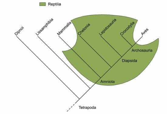
Sometimes, organisms which look incredibly different are actually closely related. Look at the graph below, describing the phylogenetic relationships between different groups of animals.
Traditional Reptilia
This cladogram shows different groups of animals, with the green shaded area representing reptiles, or the taxonomic group “Reptilia”. As is made clear by the picture, reptiles include a group of animals which is paraphyletic. This is a paraphyletic group because it excludes the mammals (“Mammalia”) and the birds (“Aves”). Both of these groups are descendants of the first animals with amniotic development, the “Amniota”. The Amniota, as a group, would include both the birds and the mammals, and would be monophyletic.
Many groups which we consider natural groups, like the reptiles, are actually paraphyletic. While they include many related animals and their ancestors, these paraphyletic groups fail to take into account the whole picture and the diversity of life. While many of these classifications were made in the days when animals were judged solely by their looks. When modern techniques like DNA analysis were able to inform the relationships between animals, new patterns were observed.
Undiscovered Species
Often, we don’t even know that a group we are discussing is paraphyletic. Many species in the world remain undiscovered, which makes them unable to be placed in a phylogeny. If a group doesn’t include all of the existent species, it is a paraphyletic group. A paraphyletic group is not necessarily wrong, as it does show the relationship between organisms and their descendants. However, in analyzing paraphyletic groups, scientists cannot get a full view of the relationships between animals.
Language
Often, the common language for animals outweighs any scientific names ascribed to them. While this is extremely beneficial for average people trying to communicate about various animals, it is often a pain for scientists trying to describe the complex relationships between animals. Consider the word “wasp”. What does that mean to you? It probably means an insect, with wings, a thin abdominal section, and a sharp stinger. While that definition is enough to understand what someone means when they say the word “wasp”, it doesn’t tell you nearly enough if you are an entomologist. Consider the paraphyletic phylogenetic tree below.
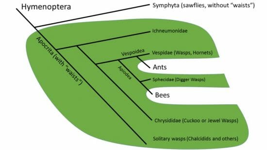
Wasps are Paraphyletic
Here, you can clearly see that what laypeople call “wasps” are actually a paraphyletic group which excludes the ants and the bees. When you think about it, it is easy to see how these insects are related to wasps, but it is still wrong to call an ant or a bee a wasp. Yet, according to genetic relationships between the animals, they should all be a part of the same phylogenetic grouping.
Language is a common barrier for evolutionary biologists, and creates many paraphyletic groupings. This is often done unconsciously, as we simply inherit our language from our parents and have to learn how to use it best. For example, while it is now recognized that ants and bees are actually a subset of the paraphyletic wasp grouping, we will always call them ants and bees. Ants will not be called “wingless wasps”, nor will bees become “hairy wasps”. Language has a tendency to stick, making paraphyletic groups more or less a requirement when discussing evolution and the relationships between species.
Quiz
1. A researcher studying the evolution of flying animals groups a bat and a butterfly, and labels them “Things which fly”. What kind of group is this?
A. Paraphyletic
B. Polyphyletic
C. Monophyletic
2. The advent of modern DNA analysis techniques revealed many paraphyletic groups within the classification schemes backed by science. Why is DNA more revealing than other traits animals have?
A. DNA is unlikely to have a homoplasy.
B. DNA cannot produce mistaken phylogenies.
C. DNA is not more revealing than other methods.
3. In the lab, a scientist goes through pain-staking efforts to create an entirely synthetic species of bacteria. Although the synthetic bacteria looks and functions like a regular bacteria, it is composed of entirely synthetic, non-natural parts. Even the DNA is composed of unique nucleotides, not found in the animal kingdom. The scientist claims that the new bacteria is in a monophyletic group of its own. Is the scientist correct?
A. No, the group is paraphyletic
B. Yes
C. No, there could be other species
References
- Brusca, R. C., & Brusca, G. J. (2003). Invertebrates. Sunderland, MA: Sinauer Associates, Inc.
- Feldhamer, G. A., Drickamer, L. C., Vessey, S. H., Merritt, J. F., & Krajewski, C. (2007). Mammology: Adaptation, Diversity, Ecology (3rd ed.). Baltimore: The Johns Hopkins University Press.
- Hartwell, L. H., Hood, L., Goldberg, M. L., Reynolds, A. E., & Silver, L. M. (2011). Genetics: From Genes to Genomes. Boston: McGraw Hill.
Paraphyletic