What is a Moss
Moss is a type of non-vascular plant, classified in the division Bryophyta in the kingdom Plantae. Moss, while typically associated with dark, damp environments, has actually adapted to occupy many drier, sunny regions. There are over 12,000 species of moss recognized, which span 8 classes and 23 different genera.
Examples of Moss
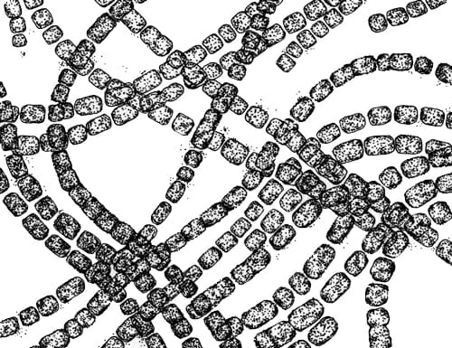
Bryopsida
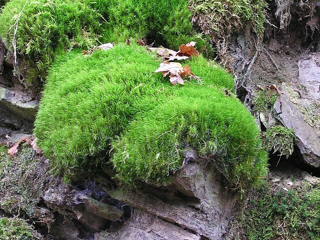
The Bryopsida, the largest class of mosses, contains most of the recognized species. A typical species can be seen above. In this image the gametophyte form is seen, as the sporophytes have not developed. Moss in the class Bryopsida can be found all over the world and grows on nearly any available surface, from concrete to bare fields, given the right conditions. In all, there are over 11,500 species of moss in the class. Before genetic and anatomical evidence suggested the division of more classes, all species of moss were found within this class.
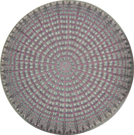
Andreaeobryopsida
The moss found in the class Andreaeobryopsida represents only a couple species. These moss species are endemic to only a few parts of Alaska and Western Canada. These moss plants have developed a unique tolerance to the climate in this region. This, plus differences in their genetics and the development of their spore capsules, led scientists to remove them from the Bryopsida and into their own unique class. Many of the other types of mosses have been divided into their own classes, eight in total. However, the large majority are still classified as Bryopsida.
Types of Moss
While there are not necessarily different types of moss, there are currently 8 recognized classes, which are distinguished by their genetics, anatomy, and physiology. Importantly, scientists look at their reproduction habits and structures to help identify and categorize the various moss groups. The eight different classes are listed below:
- Takakiopsida
- Sphagnopsida
- Andreaeopsida
- Andreaeobryopsida
- Oedipodiopsida
- Polytrichopsida
- Tetraphidopsida
- Bryopsida
As an example, the Sphagnopsida class holds the genus Sphagnum, which has important industrial uses. This moss, known for creating thick sheets of moss over large areas, can be commercially harvested as peat. The moss can be identified by the way it grows, which is in large flat sheets. Further, Sphagnum moss species have a unique way of spreading their spores. Instead of mildly cracking open the case surrounding the spores and letting them fall out, the moss in this class use a more explosive strategy. By compressing air in the chamber, pressure builds. The cells of the sporophyte continue this process until the operculum holding the spores back ruptures. This shoots the spores into the air, like a “party-popper” or overfilled balloon. This greatly increases the area the spores can reach and is unique to the class.
Life Cycle of Mosses
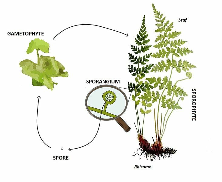
Like all plants, moss species show an alternation of generations, in which two different classes of individuals carry out separate parts of the reproductive process. In a system like this, one organism, the sporophyte, is a diploid organism which creates haploid spores through the process of meiosis. In the picture below, the tall stems with small structures at the top are the sporophyte.
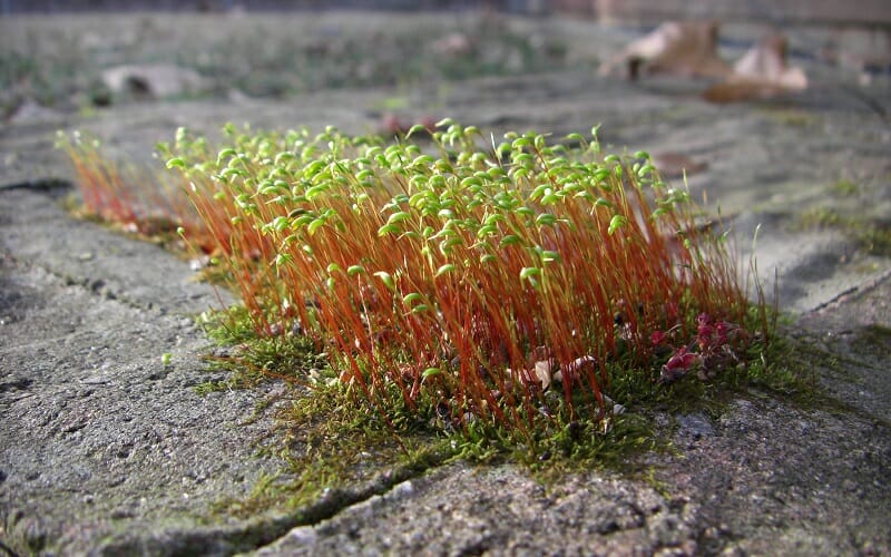
However, after the sporophyte generation has released the spores, it dies off. The spores find a place to settle, and develop into a haploid organism, the gametophyte. This is the dominant structure of moss, what you typically see if the moss is not reproducing. This can be seen in the image at the base of the sporophyte, much shorter and seemingly a different species. The gametophyte is responsible for producing gametes, which are capable of fusing together. Look at the image below, of moss reproduction.
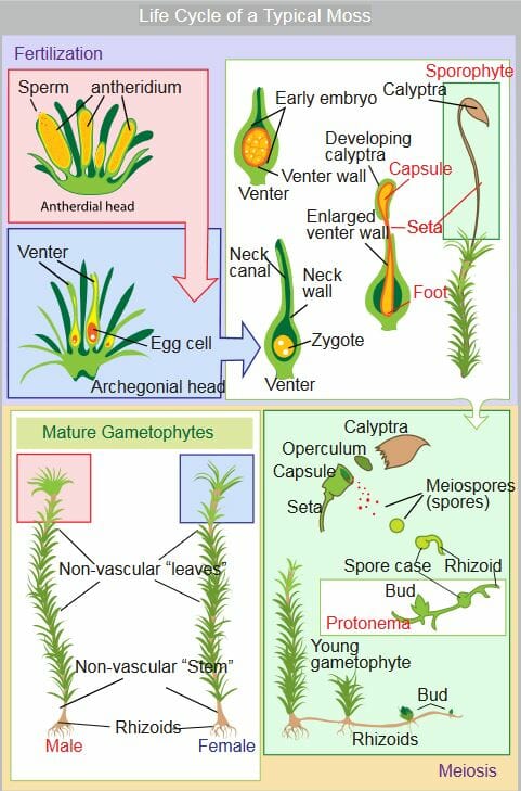
In the top left of the image, fertilization is occurring. Sperm and eggs, gametes, are produced in special organs of the gametophyte. The sperm are released into the environment, and travel to the archegonial head, which houses the egg. Once the sperm fertilizes the egg, the zygote is formed. The zygote will develop into the sporophyte, which actually grows out of the gametophyte. The sporophyte, again a diploid organism after the fusion of two haploid gametes, is responsible for undergoing meiosis, and starting the process over again.
Further, many moss species have the ability to reproduce asexually using bundles of cells called gemmae. These cells, produced on the gametophyte, fall off when exposed to running water. This allows them to be carried to a new location, where an entire new plant can be established. If you have ever seen moss growing below a drip of water, this is likely the route in which it got there. Sexual reproduction takes a lot of energy, and is generally good for diversifying the genetic pool. Asexual reproduction is much faster, and can happen every time it rains.
Within this life cycle, some moss species have the same sex represented on one gametophyte, while others have different gametophytes for different sexes. This is another way in which moss species can be distinguished and identified against each other.
Commercial Uses of Moss
The main commercial use of moss is as peat, a renewable fuel source. As the moss grows, it pushes down old moss and creates dense mats of biofuel. The peat can be burnt in a fire or stove, as it has been for centuries in many different countries. Peat moss can also be used as a fertilizer and growing medium for various commercially important plants and mushrooms. Even Scottish whiskey famously uses peat fires to smoke the malt, giving the whiskey a distinct flavor.
Moss is also becoming a more important and widespread landscaping plant. Several cultures, like the Japanese, have used moss for centuries as a way to decorate an outdoor space. Like a grass turf lawn, it is comfortable, pleasingly green, and easy to maintain. In more extreme uses, it can even be used as a base for a green roof, a new conservation technique aimed at reducing the urban heat effect.
In the past, moss has even had uses in the medical and consumer fields. Moss, when dried, is extremely absorbent. Even more absorbent than cotton. This lead to the use of moss in bandages for wounded soldiers. Some even claimed that the moss had antibacterial properties, which helped wounds heal. Further, moss has been used as a diaper alternative product in several countries. Moss, which is completely biodegradable, is said to outperform many plastic and cotton products used today.
Quiz
1. If moss can reproduce asexually, what is the benefit of reproducing sexually?
A. It uses less energy
B. It takes less time
C. It recombines and diversifies the gene’s an organism can use
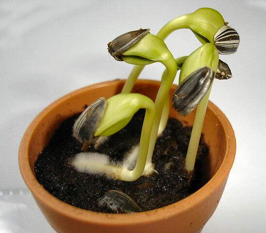
2. You identify a new form of plant. It is small, with tiny leaves that resemble moss. You take a closer look at the stem under the microscope. There are small bundles of vascular tissue, clearly distinguished from the rest. You determine that this new species is:
A. A moss
B. Not a moss
C. Impossible to tell
3. A small insect, the springtail, is attracted to moss, and may be responsible for pollinating the moss plants. If an insecticide is developed which targets theses insects, how could the energy industry be affected?
A. It cannot be affected by an insect
B. The moss making peat could die, affecting energy consumers
C. The moss would reproduce more, making energy cheaper
References
- Hartwell, L. H., Hood, L., Goldberg, M. L., Reynolds, A. E., & Silver, L. M. (2011). Genetics: From Genes to Genomes. Boston: McGraw Hill.
- McMahon, M. J., Kofranek, A. M., & Rubatzky, V. E. (2011). Plant Science: Growth, Development, and Utilization of Cultivated Plants (5th ed.). Boston: Prentince Hall.
- Rubinstein, C. V., Gerrienne, P., de la Puente, G., Astini, R. A., & Steemans, P. (2010). Early Middle Ordovician evidence for land plants in Argentina (eastern Gondwana). New Phytologist, 188(2).
Moss