Showing posts with label 4k. Show all posts

Monday, February 12, 2018

Supine Position

Supine Position Definition


The supine position is a term used in anatomy and medicine to describe an organism on its back. This position can be used to describe any organism with clear dorsal and ventral sides. In the supine position, the dorsal side, or back, goes towards the ground. The ventral side then points toward the sky. The supine position is opposed to the prone position, where an animal or person is lying on their stomach. This can be seen in the image below.


Supine and prone


Uses of Supine Position


The supine position is used in a number of fields to orient a specimen, subject, or person into the correct orientation. For instance, many surgeries are conducted in the supine position to allow access to the majority of the internal organs. Other surgeries, like certain back surgeries, must be conducted with the patient in the prone position to allow access to the dorsal side of the spine. Other doctors may study how a patient sleeps using the supine position. A polysomnograph is a test done on a sleeping person which records how they sleep in various positions, supine included. The supine position is also used in veterinary medicine to position animals on their backs for various procedures.


Other, non-medical professionals also use the position. In comparative anatomy scientists may place two different specimens in the supine position to compare their ventral anatomical features. In rehabilitation, sports training, weight-lifting, and yoga, the supine position is an important starting position. From this position you can begin a wide variety of exercises and stretches. It should be noted that while the supine position is in general any position with the subject on its back, many professions and fields may have specific adjustments or nuances that accompany their field.


References


  • De luliis, G., & Pulera, D. (2007). The Dissection of Vertebrates. Amsterdam: Academic Press.

  • Nakos, G., Batistatou, A., Galiatsou, E., Konstanti, E., Koulouras, V., Kanavarous, P., . . . Bai, M. (2006). Lung and ‘end organ’ injury due to mechanical ventilation in animals: comparison between the prone and supine positions. Critical Care, 10(1), R38.


Supine Position

Biomechanics

Biomechanics Definition


Biomechanics is an interdisciplinary field that applies the principles of physics to biological systems to understand how organism move and interact with their surroundings. Biomechanics is concerned with everything from microscopic systems like muscle contraction in cells, all the way to large-scale, whole-body motions like a jumping cat. Biomechanics applies the laws of physics with regards to levers, pulleys and other known functions to define and understand the complicated forces involved in biological systems. A career in biomechanics means studying biological systems and learning from them or creating devices based on a combination of biological and physical principles.


History of Biomechanics


Biomechanics has a long and intricate history, reaching back to the days of Aristotle and the first philosophers. These men sought to understand the driving forces behind life, and as such, they studied how animals moved and what caused those actions. Building on their successes, the thinkers of the Renaissance added to these notions. Leonard DaVinci is still known for his works of anatomy and physiology, which incorporated some of the first math-driven biomechanics on record. Modern biomechanical engineers have followed in their footsteps.


The ideas behind biomechanics gained footing in the 1500s, with the writings of Descartes, and others that saw the world in a mechanic way. Thus was born the science of automatons, or the idea that all creatures were simply biological machines that reacted to stimuli in the same way a machine would. This idea has captivated scientists for hundreds of years, as it would give the ultimate ability to control and manipulate these machines. However, as science progressed, the complexities of the living machines became infinitely intricate. The field branched into many subdivisions.


Modern biomechanics has innumerable advantages over the early pioneers of the science. Modern technology can provide insights and measurements that science has never before been able to obtain. For instance, a greater understanding of nerve impulses came after the invention of the EEG, a test in which a computer monitors the electrical signals passed between cells. Further advances into microbiology and chemistry have revealed the internal microscopic structure of muscles. This allows biomechanical engineers to have a full and clear view of the body and how it works. These advancements have not been left to waste. New advancements in biomechanics and biotechnology are allowing for novel treatments like artificial limb and organ replacement. Not only can science produce new joints for old bodies, but the science has advanced far enough that we can now grow organs using specialized stem cells sprayed onto 3D printed models. The possibilities of biomechanics are expanding rapidly.


Careers in Biomechanics


Pathways to a career in biomechanics can vary, but almost always include engineering and biology courses. General Bachelor’s degrees are offered which provide introduction to biomechanics, but most professionals have a Master’s degree or higher. Further coursework is often needed beyond a Bachelor’s degree to understand the complex math and biology underlying the field. It is not rare for a biomechanical engineer to receive a degree in engineering and a medical doctorate. However, once the field is understood the possibilities are limitless.


Medicinal biomechanics deals with the human body, and is involved in everything from replacing limbs and organs to understanding the complex forces athletes deal with while playing sports. Orthotics and Prosthesis are the fields that deal with replacing lost or missing limbs. These scientist integrate invention with biology as they seek to reverse debilitating conditions. Others, like those who study sports biomechanics, focus on physics involved during complex sports. These professionals provide estimates for the damage being done, and ways to avoid that damage, like wearing a helmet. Still others focus on the strain of repetitive tasks. The field of ergonomics studies the body’s natural position and how stresses are created. Still others, like rehabilitation specialists, practice biomechanics to give bedridden patients zero-strain exercise.


Other scientists use biomechanics for different reasons. Understanding the physics of an animal can often lead to understandings about its role in the environment. Ecologists and other field-scientists use biomechanics to understand the different stresses on an organism. They could measure the strain a tree faces in the wind or measure the amount of friction a dolphin experiences while it is swimming. These observations can lead to understanding about the animal or ecosystem or even lead to novel devices to help humans. Modern biomechanical engineers often employ computers in their modeling. This is known as computational biomechanics and will lead to greater understanding of all biological systems. Experts in this area also have training in computer information systems to exploit the power of modern computers in their studies.


References


  • De luliis, G., & Pulera, D. (2007). The Dissection of Vertebrates. Amsterdam: Academic Press.

  • Nelson, D. L., & Cox, M. M. (2008). Principles of Biochemistry. New York: W.H. Freeman and Company.

  • Rollin, B. E. (2006). Animal Rights and Human Morality (3rd ed.). New York: Prometheus Books.


Biomechanics

Friday, September 8, 2017

Autopsy

Autopsy Definition


An autopsy is a surgical procedure performed on a corpse after death (a period called, “post-mortem.”). It is typically conducted in an attempt to understand the person’s cause of death. The autopsy will be conducted by a trained physician who has specialized in pathology, as determining the cause of death will require a vast understanding of disease and injury.


A brief history of autopsies will tell us that humans have been performing autopsies since the dawn of time, it seems. There is historical data to support that autopsies were performed in ancient Egyptian times. Ancient Egypt was notably known for performing elaborate death rituals and valuing the afterlife, so it makes sense that an autopsy would be part of that ritual. However, a distinction between those early times and now is that whereas ancient Egyptian examiners were removing organs for preservation, autopsies today are done with the intention to explain disease and death.


Autopsy of a Japanese victim killed in the Jinan Incident

The image depicts an old photograph of a Japanese victim of the Jinan Incident who is undergoing an autopsy


Autopsy Method


An autopsy, at its base, is a surgical dissection. There are different correct ways to perform it, but the Letulle method has become the principle protocol for training pathologists. This particular method finds the pathologist or medical student commencing the dissection at the abdomen. After piercing the abdominal area, the abdominal organs will be removed in one block per this method – understandably called the “en bloc” method. The direction the Letulle method will take is called a retroperitoneal approach. This essentially means that the organs will be removed starting with the organs situated directly behind the peritoneum, and moving backward. The peritoneum, in turn, is a serous lining located in the abdominal cavity. It appears like a light colored sheet that will cover and thus protect our abdominal organs. The retroperitoneal organs will include the adrenal glands, the pancreas, the lower segments of the small intestine’s duodenum, and the ascending and descending parts of the colon. The examiner will take out organs by layer, all while using the vasculature (or veins and arteries) as reference points to orient themselves. This method is considered to be best for the pathologist in –training, as it follows anatomical relationships learned in class. However, there are faster ways to perform an autopsy, as well.


The Virchow technique will see that the organs are removed one by one, starting at the cranium and moving down to the thoracic, abdominal, and cervical organs. In other words, it may follow a top-down approach, if you will.


The Rokitansky technique is an in situ (or local) dissection that starts at the neck and trails down, and the organ is removed as a bloc as well. The first cut pierces the larynx to separate the esophagus and pharynx, then the larynx and trachea, followed by the chest organs that are cut to expose those in the abdomen.


Finally, the Ghon technique is similar to the Rokitansky in that the thoracic, cervical, and abdominal organs are removed using the bloc method, but the Ghon will not employ an in situ dissection instead opting for “en bloc” removal.


Types of Autopsy


An autopsy is performed for three main reasons that we will discuss shortly. A clinical autopsy will be done on a patient that has died while under the care of a hospital or clinical staff and in cases where the physicians have failed to identify the cause of a sudden death. This type of autopsy will be useful for attaining the time and cause of death, as well as for giving doctors a cause of death to appropriately file a death certificate.


A forensic autopsy, on the other hand, will be a type of autopsy performed when a corpse has been retrieved from a crime or murder site. This autopsy will reveal any trace of bullets, blows or injuries, and poison in the system. A medical examiner must be present, and will decide if the cause of death was an accident, murder, or suicide. This autopsy will guide police through their investigation.


Lastly, an academic autopsy is one that is performed by medical students to teach them about human anatomy. Likewise, some may be used for research purposes, as well. The source of the bodies will be patients who have willingly donated their bodies to science, or unclaimed bodies after filing the needed legal paperwork.


Importance of Autopsies


While any family, or next of kin, can request an autopsy of their deceased loved one, autopsies are most certainly the golden standard when the cause of death is uncertain. This may take place if a person was found deceased from a possibly accidental circumstance, or if a person was murdered. This clearly has crucial significance in the realm of criminal law, but also within the human experience of grieving and finding closure. For this reason, the autopsy has been performed for legal and medical reasons for a long time.


Quiz


1. Which of the following characterizes the Rokitansky method of autopsy?
A. Start at the abdominal cavity and start removing organs by layer
B. Remove organs employing in situ removal
C. Begin at the cranium and move downward
D. Remove organs employing en bloc removal

Answer to Question #1

2. Which type of autopsy is one that requires the presence of a medical examiner?
A. Clinical
B. Forensic
C. Academic
D. None of the above

Answer to Question #2

References


  • Newsmax Health (2017). “What happens during an Autopsy?” Newsmax. Retrieved on 2017-8-19 from http://www.newsmax.com/Health/Health-Wire/autopsy-death-organs-forensic/2014/04/29/id/568274/

  • Culora GA, Roche WR (1996). “Simple method for necropsy dissection of the abdominal organs after abdominal surgery.” J Clini Pathol 49(9):776-9.

  • Forensic Pathology (2017). “Methods/Technique.” NCSSM Forensic Pathology. Retrieved on 2017-08-19 from http://ncssmforensicpathology.weebly.com/methodstechniques.html


Autopsy

Sunday, August 20, 2017

Leaf

Leaf Definition


The term leaf refers to the organ that forms the main lateral appendage on the stem of vascular plants. In general, leaves are thin, flat organs responsible for the photosynthesis of the plant. Although photosynthesis typically only occurs on the upper surface of the leaf, it can occur on both sides in some plant species. Leaves are typically comprised of a distinct upper and lower surface, stomata for gas exchange, waxy coating, hairs, and venation. Each side of the leaf differs in regarding the level to which these features are expressed. Although leaves are typically located above ground, some species have leaves which reside underground (e.g., bulb scales) or underwater (e.g., aquatic plant species). Moreover, the leaves of some plants may not be associated with photosynthesis (e.g., cataphylls). Leaves are typically oriented on a plant to avoid blocking the sunlight of the leaves situated underneath.


Function of the Leaf


As one of the most important constituents of plants, leaves have several essential functions:


Photosynthesis


The primary function of the leaf is the conversion of carbon dioxide, water, and UV light into sugar (e.g., glucose) via photosynthesis (shown below). The simple sugars formed via photosynthesis are later processed into various macromolecules (e.g., cellulose) required for the formation of the plant cell wall and other structures. Therefore, the leaf must be highly specialized to combine the carbon dioxide, water, and UV light for this process. Carbon dioxide is diffused from the atmosphere through specialized pores, termed stomata, in the outer layer of the leaf. Water is directed to the leaves via the plant’s vascular conducting system, termed the xylem. Leaves are orientated to ensure maximal exposure to sunlight, and are typically thin and flat in shape to allow sunlight to penetrate the leaf to reach the chloroplasts, which are specialized organelles that perform photosynthesis. Once sugar is formed from photosynthesis, the leaves function to transport it down the plant via specialized structures called the phloem, which run in parallel to the xylem. The sugar is typically transported to the roots and shoots of the plant, to support growth.


Plants


Transpiration


Transpiration refers to the movement of water through the plant, and subsequent evaporation via the leaves. When the stomata open to accommodate the diffusion of carbon dioxide into the plant for photosynthesis, water flows out. This process also serves to cool the plant via evaporation of the water from the leaf, as well as regulate the plant’s osmotic pressure.


Guttation


Guttation refers to the excretion of xylem from the edges of leaves and other vascular plants due to increased levels of water in the soil at night, when the stomata are closed. The pressure caused at the roots results in the leakage of water from the xylem out of specialized water glands at the edges of leaves.


Storage


Leaves are a primary site of water and energy storage since they provide the site of photosynthesis. Succulents are particularly adept at water storage, as evidenced by the thick leaves. Due to the high levels of nutrients and water, many animal species ingest the leaves of plants as a source of food.


Defense


Some leaves have also evolved defense mechanisms to avoid being eaten or damaged. Some examples include the spines of cacti, cones of gymnosperms, respectively. In addition, hairs found on leaves prevent water loss in dry climates and sting animals that detour herbivores (e.g., Urticaceae). Moreover, the waxy coatings found on leaves serve to protect against water loss, rain, and forms of contamination. Oils and other secreted substances also detract from being consumed by herbivores.


Types of Leaf


In general, the types of leaf can be divided into six major types, although there are also plants with highly specialized leaves:


Conifer Leaf


Conifer leaves are needle-shaped or in the form of scales. Conifer leaves are typically heavily waxed and highly adapted to colder climates, arranged to dispel snow and resist freezing temperatures. Some examples include Douglas firs and spruce trees. The images below illustrate this type of leaf.


Conifer needle


Microphyll Leaf


Microphyll leaves are characterized by a single vein that is unbranched. Although this type of leaf is abundant in the fossil record, few plants exhibit this type of leaf today. Some examples include horsetails and clubmosses. The image below illustrates this type of leaf.


Baragwanathia fossil land plant


Megaphyll Leaf


Megaphyll leaves are characterized by multiple veins that can be highly branched. Megaphyll leaves are broad and flat, and generally comprise the foliage of most plant species. The image below illustrates this type of leaf.


Setaria megaphylla leaves


Angiosperm Leaf


Angiosperm leaves are those found on flowering plants. These leaves are characterized by stipules, a lamina, and a petiole. The illustration below shows an example of an angiosperm leaves.


Medicago prostrata


Fronds


Fronds are large, divided leaves characteristic of ferns and palms. The blades can be singular or divided into branches. The image below presents an example of a frond.


Fern


Sheath Leaf


Sheath leaves are typical of grass species and monocots. Thus, the leaves are long and narrow, with a sheathing surrounding the stem at the base. Moreover, the vein structure is striated and each node contains only one leaf. The image below presents an example of a sheath leaf.


Juncus bufonius


Quiz


1. The primary function of a leaf is:
A. Water evaporation for cooling
B. Photosynthesis
C. Provide shade to the shoot and root structures of the plant
D. Transpiration

Answer to Question #1

2. Which of the following statements is TRUE regarding guttation:
A. It typically occurs at night.
B. It occurs when the stomata are closed.
C. It results from increased water pressure in the soil.
D. All of the above

Answer to Question #2

References


  • Brodersen C and McElrone A. (2013). Maintenance of xylem Network Transport Capacity: A Review of Embolism Repair in Vascular Plants. Front Plant Sci.4:108.

  • El-Sharkawy, M. and Hesketh, J. (1965) Photosynthesis among species in relation to characteristics of leaf anatomy and CO2 diffusion resistances. Crop Science. 5(6):pp. 517-521.

  • Roth-Nebelsick A, Uhl D, and Kerp H. (2001). Evolution and Function of Leaf Venation Architecture: A Review. Ann Bot. 87(5): 553-566.

  • Sadras and Milroy. (1996). Soil-water thresholds for the responses of leaf expansion and gas exchange: A review. Field Crops Research. 47(2): 253-266.


Leaf

Wednesday, July 5, 2017

Ovary

Ovary Definition


An ovary refers to the female reproductive organ in vertebrates that produces the female gametes (eggs/oocytes) and functions as an exocrine gland. The ovaries are typically found in pairs; however, both are not functional in all animals (e.g., birds and some fish species). The number of eggs produced by each species differs, with fish and amphibian species capable of producing millions of eggs throughout their lifetime. In addition, depending on the species, the eggs produced by the ovary may be small, and invisible to the naked eye (e.g., humans), or they may be large, encompassing the entire follicle (e.g., birds). In humans, one of the two ovaries (or both) will release an egg during ovulation each menstrual cycle.


Where are the Ovaries Located?


In humans, the ovaries are situated along the lateral wall of the uterus, under the external iliac artery and in front of the internal iliac artery. This region that houses the ovaries is termed the ovarian fossa. The ovaries attach to both sides of the uterus via specialized fibrous tissue termed the ovarian ligament. Since the ovaries lie uncovered within the peritoneal cavity, they are considered intraperitoneal organs and attach to the fallopian tubes via the suspensory ligament of the ovary (shown below).


Ovaries, Uterine Tubes, and Uterus


Ovary Function


The ovaries function as an endocrine gland, as well as a location for the production and maturation of gametes.


Endocrine Functionality


One of the most important functions of the ovary is the secretion of the hormones estrogen, progesterone, and testosterone, each of which serves a distinct purpose. Estrogen is required for the development of the secondary sexual characteristics, such as the breasts, the maturation of the reproductive organs, and pubic hair. Progesterone is required for the thickening of the uterine lining in preparation for the potential implantation of a fertilized egg. Progesterone is responsible for the menstrual cycle as levels surge following ovulation via secretion from the corpus luteum. As the levels of progesterone and estrogen fluctuate throughout the menstrual cycle, the processes of ovulation, thickening of the uterine lining, and shedding of the uterine lining (menstruation) occur (shown below). Testosterone is also produced by the ovaries, and is secreted directly into the blood stream. In females, testosterone is required for bone and muscle growth, as well as libido (sex drive).


Menstrual Cycle


Production of the Female Gametes


The female gametes are the oocytes, which develop within the ovaries within a specialized location termed the follicle. As the oocyte develops, the size and shape of the follicle also changes. When maturation of the oocyte is complete, it is released from the follicle following the secretion of luteinizing hormone from the pituitary gland. The release of the oocyte involves the rupture of the follicle, the process of which is termed ovulation. Following ovulation, the follicle forms the corpus luteum, which prepares the uterine lining for implantation of a fertilized egg via the secretion of progesterone. A detailed diagram of the menstrual cycle is shown above. In females, testosterone is required for proper bone and muscle development, as well as libido (sex drive). It has been found that female testosterone levels are associated with the degree of sexual arousal.


Age-related Changes Exhibited by the Ovaries


As the number of ovarian follicles declines with age, the reproductive capacity of females wanes until menopause, typically occurring around the age of 50. Despite the presence of over one million oocytes in the human ovary (this number differs by species), only approximately 5% mature and ovulate. As fertility wanes, there is an increased likelihood of chromosome abnormalities and risk of miscarriage associated with pregnancy. Menopause involves the reduction in estrogen and progesterone production by the ovaries as a result of the dwindling number of available oocytes. It has been found that the ability to repair DNA breaks in the ooctyes becomes increasingly compromised with age, resulting in the depletion of viable oocytes in the ovaries. The decreased levels of estrogen and progesterone have substantial consequences for menopausal/post-menopausal women, who without the aid of exogenous hormones, experience vaginal tissue atrophy, increased risk of osteoporosis, and the ceasation of menstruation.


Quiz


1. Which of the following statements is false:
A. Testosterone is secreted directly into the bloodstream from the ovary.
B. Estrogen levels decrease during menopause.
C. Progesterone is produced by the ovarian fossa.
D. An oocyte is a female gamete.

Answer to Question #1

2. The primary function(s) of the ovary is/are:
A. Secretion of estrogen
B. Site of oocyte maturation
C. Secretion of progesterone
D. All of the above

Answer to Question #2

3. Which of the following statements is true regarding menopause?
A. The onset of menopause corresponds with increased secretion of progesterone.
B. Menopause is characterized by follicle maturation without ovulation.
C. During menopause, the ovaries secrete increasing amounts of luteinizing hormone, which inhibits oocyte maturation.
D. None of the above.

Answer to Question #3

References


  • Channing CP, Hillensjo T, and Schaerf FW. (1978). Hormonal control of oocyte meiosis, ovulation and luteinization in mammals. Clin Endocrinol Metab. 7(3):601-24.

  • Channing CP, Schaerf FW, Anderson LD, and Tsafriri A. (1980). Ovarian follicular and luteal physiology. Int Rev Physiol. 22:117-201.

  • Linton et al. (2016). Associations of Serum Levels of Sex Hormones in Follicular and Luteal Phases of the Menstrual Cycle with Breast Tissue Characteristics in Young Women. pLoS One. 11(10):e0163865.

  • Oktem O and Oktay K. (2008). The ovary: anatomy and function throughout human life. Ann N Y Acad Sci. 1127:1-9.

  • Rimon-Dahari N, Yerushalmi-Heinemann L, Alyagor L, and Dekel N. (2016). Ovarian Folliculogenesis. Results Probl Cell Differ. 58:167-90.


Ovary

Fetus

Fetus Definition


Fetal development occurs between the embryonic stage of development and birth in humans. This stage begins after 11 weeks of gestation, when the embryo begins to exhibit human characteristics, and lasts until birth. Typically, all the major organs and tissues can be observed; however, they are not yet fully developed or appropriately situated within the body.


Fetus Stages


Throughout the process of fetal development, the growing fetus goes through three distinct stages, each characterized by specific events.


2 ¼ to 4 months


As the embryo enters the fetal stage of development, the placenta becomes functional. The fetus typically measures 30 mm from the crown to the rump and weighs approximately 8 g (shown below). By the end of this stage, the fetus is approximately 15 cm. During this time, several organs can be observed, including the hands, feet, heart, and brain. The pancreas and liver begin to secrete fluids. In addition, the genitals begin form and the head is prominent, comprising almost half of the fetal body. The fetus also exhibits unregulated movements required for the lung, muscle, and neurological development that is occurring.


Abortus


4 ¼ to 6 ¼ months


During this period of fetal development, the mother begins to feel the movements of the growing fetus, which grows from approximately 15 cm to 38 cm and weighs about 500 g by the end of this stage (shown below). During this stage, the eyebrows and eyelashes form, muscle development increases and the fetus becomes more active, the lung continues to develop with the formation of the alveoli. In addition, the nervous system rapidly develops, exhibited by the development of the inner ear, control over the opening and closing of the eyelids, as well as other bodily processes. The genitals are fully formed and the sex can be reliably discerned.


20 weeks pregnant


6 ½ to 9 ½ months


At this stage, the fetus begins to gain weight as the body fat increases. The lungs continue to mature and become capable of gas exchange. In addition, hair begins to thicken on the head and breast buds appear. The fetus is considered to be full-term at approximately 38 weeks (between 36 and 40 weeks) of pregnancy (shown below). While fetuses born prior to 36 weeks can survive outside the uterus, medical intervention is required to promote survival, particularly due to the underdevelopment of the lungs in premature infants.


40 weeks pregnant


Quiz


1. Which of the following statements is TRUE:
A. The lungs are one of the first organs to fully mature in a developing fetus.
B. The heart is one of the first organs to form in the developing fetus.
C. The sex of the fetus can be observed as early as 13 weeks.
D. A fetus born at 37 weeks has a low probability of survival.

Answer to Question #1

2. The fetus becomes increasingly active due to:
A. Increased neurological development
B. Increased muscle development
C. Increased bone and limb development
D. All of the above

Answer to Question #2

References


  • Del Valle et al. (2017). A genomic atlas of human adrenal and gonad development. Wellcome Open Res.2:25.

  • Gilbert SF. (2000). Developmental Biology. 6th edition. Sinauer Associates; Sunderland (MA).

  • Lubchenco L, Hansman C, Dressler M, and Edith Boyd. (1963). Intrauterine growth as estimated from liveborn birth-weight data at 24 to 42 weeks of gestation. Pediatrics. 32(5).

  • Meyeer G, Schaaps, JP, Moreau L, and Goffinet AM. (2000). Embryonic and Early Fetal Development of the Human Neocortex. Journal of Neuroscience. 20 (5): 1858-1868.

  • Sandikcioglu M, Mølsted K, and Kjaer I. (1994). The prenatal development of the human nasal and vomeral bones. Journal of Craniofacial Genetics and Developmental Biology. 14(2):124-134.


Fetus

Hyperplasia

Hyperplasia Definition


Hyperplasia or “hypergenesis” refers to an increase in the number of cells within a given tissue as a result of cellular proliferation. Since hyperplasia refers to an increased number of cells, the cell appears normal in size, but can lead to an enlargement of an organ or tissue. Such proliferation occurs in response to a particular stimulus and remains under mechanisms of regulation by the cell. In some cases, hyperplasia can be a pathological response to abnormal levels of growth factors or hormones, resulting in a number of disorders. Moreover, since the cells are rapidly dividing, it increases the risk of cancer, due to unregulated cellular proliferation in the absence of physiological stimuli.


Causes of Hyperplasia


There are numerous causes of hyperplasia, including the demand for increased tissue to compensate for a loss of cells (e.g., the skin or wound healing), chronic inflammation, hormones, growth factors, and diseased tissue within the body. Some forms of hyperplasia are required on an ongoing basis, such as the replacement of skin cells as they are sloughed off from the epidermal layer. In addition, hyperplasia is also required in the breasts of pregnant women for the growth of the milk glands in order to breastfeed the newborn. The process of hyperplasia is also used (and abused) in various sports for the purpose of increasing the number of skeletal muscle cells to improve athletic performance.


Types of Hyperplasia


There are many manifestations and disorders of hyperplasia, depending on the affected tissues or organs. Some of the most common types of hyperplasia are listed below:


Benign Prostatic Hyperplasia


Benign prostatic hyperplasia is an enlargement of the prostate due to hyperplasia of the epithelial and stromal cells comprising the prostate (shown below). Such hyperplasia causes the formation of discrete nodules on the prostate which can eventually obstruct the bladder, causing complications such as bladder stones, kidney disease, and urinary tract infections. It is thought that testosterone and its metabolites play a key role in the induction of hyperplasia of the prostate. One reason for increased levels of testosterone in this tissue is hypothesized to be the result of the insufficient drainage of the spermatic venous system, which increases the hydrostatic pressure and testosterone in the prostate, thereby inducing hyperplasia.


Nodular hyperplasia of the prostate


Cushing’s Disease


Cushing’s disease results from hyperplasia of the adrenal cortex in response to the enhanced secretion of adrenocorticotropic hormone from the anterior pituitary. Such enhanced secretion is often caused by the overproduction of hypothalamus corticotrophin releasing hormone or a pituitary adenoma.


Sebaceous Hyperplasia


Sebaceous hyperplasia involves hyperplasia of the sebaceous glands located in the skin. This condition is commonly observed in newborn infants and older adults and is characterized by the increased secretion of sebum (the oily substance secreted by the sebaceous glands) and formation of yellow-colored papules on the face. This condition is typically self-resolving, but laser treatments are also available.


Hemihyperplasia


Hemihyperplasia occurs when the growth of one side of the body is greater than that of the other. This condition can result in the generation of limbs that are longer on one side than the other, with differing levels of severity. Since this condition can be debilitating, there are several treatment options ranging from bone lengthening, bone resection, and the insertion of a growth plate in order to correct the condition. An example of an individual with this condition is presented below:


Hemihyper


Intimal Hyperplasia


Intimal hyperplasia refers to hyperplasia of the tunica intima (endothelial lining) of blood vessels in response to injury. Since this process is a normal physiological response, it often causes graft failure following any form of vascular reconstructive or bypass surgery.


Compensatory Liver Hyperplasia


Compensatory liver hyperplasia involves hyperplasia of the liver hepatocytes in response to damage or injury. This gives the liver its unique regenerative ability and allows for substantial liver resections (e.g., for the purpose of liver transplantation).


Endometrial Hyperplasia


Endometrial hyperplasia refers to hyperplasia of the uterine inner lining in response to elevated levels of estrogen. Estrogen can be overproduced in conditions, such as obesity, various cancers, polycystic ovary syndrome, and other exogenous sources of estrogen (e.g., hormone therapy). This condition is associated with an increased risk of cancer.


Quiz


1. Which of the follow terms correctly describes “hyperplasia”? (Multichoice)
A. The enlargement of skeletal muscle cells.
B. The proliferation of skeletal muscle cells.
C. The proliferation of ductal cells in the breasts during pregnancy.
D. The enlargement of ductal cells in the breasts during pregnancy.

Answer to Question #1

2. Which of the following is NOT a characteristic of hyperplasia: (Multichoice)
A. Regulated cellular proliferation in response to environmental stimuli.
B. Tissue regeneration.
C. Tissue repair in response to injury.
D. Cellular proliferation in the absence of environmental stimuli.

Answer to Question #2

References


  • Charalampakis V, Tahrani AA, Helmy A, Gupta JK, and Singhal R. (2016). Polycystic ovary syndrome and endometrial hyperplasia: an overview of the role of bariatric surgery in female fertility. Eur J Obstet Gynecol Reprod Biol. 207:220-226.

  • Chaudhary MA, Guo LW, Shi X, Chen G, Gong S, Liu B, and Kent KC. (2016). Periadventitial drug delivery for the prevention of intimal hyperplasia following open surgery. J Control Release. 233:174-80.

  • Craiglow BG, Ko CJ, and Antaya RJ. (2014). Two cases of hemihyperplasia-multiple lipomatosis syndrome and review of asymmetric hemihyperplasia syndromes. Pediatr Dermatol. 31(4):507-10.

  • Eleazu C, Eleazu K, and Kalu W. (2017). Management of Benign Prostatic Hyperplasia: Could Dietary Polyphenols Be an Alternative to Existing Therapies? Front Pharmacol. 8:234.

  • Kholodenko IV and Yarygin KN. (2017). Cellular Mechanisms of Liver Regeneration and Cell-Based Therapies of Liver Diseases. Biomed Res Int.2017:8910821.

  • Loriaux DL. (2017). Diagnosis and Differential Diagnosis of Cushing’s Syndrome. N Engl J Med. 376(15):1451-1459.

  • Nussey S and Whitehead S. (2001). Endocrinology: An Integrated Approach. BIOS Scientific Publishers; Oxford, UK.

  • Simmons BJ, Griffith RD, Falto-Aizpurua LA, Bray FN, and Nouri K. (2015). Light and laser therapies for the treatment of sebaceous gland hyperplasia a review of the literature. J Eur Acad Dermatol Venereol. 29(11):2080-7.


Hyperplasia

Tuesday, July 4, 2017

Thymus Gland

Thymus Gland Definition


The thymus is an organ that is secretory in pre-pubescence, which earns its status as a gland. The thymus gland has an important role in immune function. One of its main secretions is the hormone thymosin. Thymosin stimulates the maturation of T cells, which are derivatives of the white blood cells that circulate our system. T cells “kill” or are cytotoxic to damaged cells. The damaged cells may be cancerous cells that have lost the ability to stop proliferating, or even cells infected with viruses. T cells will be able to bind the T receptor on the target cell’s surface that will initiate its eventual death. The T cell’s cytotoxicity comes from the cytokines it produces.


Despite the thymus’ essential role in immune health, the thymus gland is not active during our entire lifetime. In fact, it is only active until puberty and becomes non-functional in adulthood. But its actions are instrumental in preventing the body from having an autoimmune response, which is when the immune system cannot distinguish between itself and foreign agents. Chronic periods of fever, fatigue, and malaise mark the lives of patients with autoimmune diseases. Therefore, the thymus gland is closely tied to the lymphatic system as it is the body’s natural defense network. The network of vessels and tissues that make up the lymphatic system make it possible for the body to expel or “drain” toxins and waste from the body.


Thymus Gland Location


The thymus is a soft organ located behind the breastbone and between the lungs. In relation to the organs in the human body, the thymus is a two-lobed structure that lies almost on top of the heart and traces up along the trachea. The thymus gland is more or less triangular in shape and has two lobes that are encased in a fibrous exterior. Its thymic lobes are an opaque pink, and the most superficial layer is named the cortex. When the thymus is sliced for a histology study, it will reveal a deeper layer called the medulla. If the human chest were divided into four regions, the thymus would be located right in the center of the upper quadrants with both clavicles beside it.


Thymus gland position diagram

The image depicts an anatomical illustration of the thymus gland, in relation to other important organs.


Thymus Gland Anatomy


The thymus gland is made up of a patchwork of epithelial tissue and lymphatic tissue. Likewise, the tissues will contain dendritic, or “antigen presenting,” cells that will signal off the killer T cells. Meanwhile macrophages will be present as well to eat away any cellular debris or to directly ingest foreign pathogens. The macrophage and dendritic cells really populate the thymus, which of course is crucial as they assist the thymus gland in performing its immune functions of discarding harmful waste and identifying diseased cells to destroy.


The thymus gland is active in childhood and reaches it maximum weight of around one ounce during puberty. However, after reaching this pinnacle, the thymus will become less and less active. This decline in activity will correspond to a decrease in size as well, until the thymus tissue is almost completely replaced by fat. This shrinking will take place after puberty and into adulthood.


Function of Thymus Gland


As previously alluded to, the main function of the thymus gland is to release thymosin hormone that will stimulate the maturation of T cells. All of our childhood, white blood cells or lymphocytes will come in contact with the thymus gland. This contact will transform them into T cells. Once the T cells have matured, they will migrate to the lymph nodes which are the stores of immune cells in the body. So, the thymus gland can be understood to be a recipient of immature T cells that were created in the bone marrow but have yet to reach full maturation. Once the thymus receives the cells, they will be trained to only attack foreign agents. The way this happens is through positive selection. Only the T cells that have properly responded to foreign antigens will be selected to survive and eventually migrate to the medulla. The T cells that do not make the cut will die by apoptosis in a healthy patient.


Once the surviving T cells have reached the medulla, the T cells will proceed to mature. The remaining T cells will go on to kill pathogens, will activate helper B cells that make antibodies against specific antigens, and will store the memories of previous infections and viruses so that the body can be better prepared to fight them off if they ever return.


Quiz


1. Which of the following best describes the location of the thymus gland?
A. In front of the breastbone
B. Between the lungs
C. Behind the trachea
D. Below the heart

Answer to Question #1

2. The maturation of functional T cells requires which of the following natural processes?
A. Evolution
B. Bottleneck selection
C. Positive selection
D. Natural selection

Answer to Question #2

References


  • Inner Body (2017). “Thymus Gland.” InnerBody. Retrieved on 2017-07-01 from http://www.innerbody.com/image_endoov/lymp04-new.html

  • Sargins, Robert MD (2017). “An Overview of the Thymus.” Endocrine Web. Retrieved on 2017-07-01 from https://www.endocrineweb.com/endocrinology/overview-thymus

  • New Health Advisor (2017). “Functions and Disorders of Thymus Gland.” New Health Advisor. Retrieved on 2017-06-30 from http://www.newhealthadvisor.com/Thymus-Gland-Function.html


Thymus Gland

Saturday, June 24, 2017

Frenulum

Frenulum Definition


A frenulum is a small ridge or fold of skin that helps to anchor a semi-mobile body part. In the human body, frenulums are found on the penis, under the tongue, inside the lips, as part of the female genitalia, and internally in the brain and digestive tract.


An oral frenulum, the , is pictured below. This person has gotten their frenulum labii superioris pierced.
Lip frenulum piercing closeup


Because the two most important locations of frenulums outside the body are in the mouth and on the genitalia, “frenulums” are most often discussed either in dentistry and oral health, or in reproductive health and sexual advice.


While these structures often go unnoticed in the healthy body, frenulums which are too thick or too short can lead to health problems. Unusually short or thick frenulums can also be symptoms indicating more serious illnesses, such as genetic syndromes that may effect both the development of the frenulum and the internal organs.


Internal frenulums such as those found in the gut and brain are rarely discussed or operated on except when they are subject to internal injury. As a result, most people do not even know they exist!


Function of Frenulum


The term “frenulum” comes from the Latin term meaning “little bridle.” As the name suggests, frenulums help to “rein in” organs and structures that are not completely connected to each other.


In the mouth, the function of frenulums is obvious: the lips, cheeks, and tongue are largely separate from the jaw. The presence of intermittent thick skin folds connecting these structures helps them to remain anchored and properly aligned with the jaw.


A similar purpose is served by genital frenulums, which similarly help to connect external sheaths of skin and tissue to the reproductive organs they are supposed to be protecting.


Frenulums may sometimes be surgically cut in cases where they are unusually strong and thick, resulting in unusual restraint of the mouth or genitalia. They may also be cut as part of circumcision.


In rare cases, “frenulum” may refer to a part of the anatomy of moths. We will not discuss the moth anatomical characteristic here.


Examples of Frenulum


Oral Frenulums


In the mouth, one frenulum can be found under your tongue. The thick band of skin that runs along the under side of your tongue, anchoring it to the bottom of your mouth, is the “frenulum linguae.” The smaller bands of skin many people have running between their lips and front teeth are “frenulum labii.


Some people also have bands of tissue connecting the jaw to the cheek at various points, which are called “buccal frena.” In some cases, dentists might elect to cut one or both of the frenulum labii, since especially thick frenulums can pull teeth together and effect their alignment.


Dentists may sometimes cut oral frenulums, if they are unusually thick and cause problems for the mobility or alignment of the jaw or teeth. This can often be done painlessly with local anesthetic.


Doctors also sometimes use oral frenulums to look for signs of violence or abuse in a patient. Because oral frenulums are easily torn by impacts or jerking movements, torn or scarred oral frenulums can be a clue that a patient has experienced violence.


Genital Frenulums


In the penis, the frenulum of prepuce of penis anchors the foreskin to the glans. In uncircumcised people, this helps the foreskin to contract over the penis, protecting it. Circumcision may involve removal of the frenulum of the penis as well as the foreskin.


The frenulum is considered be one of the most sensitive parts of the penis to touch. Stimulation of the frenulum alone has been found to produce orgasm in some men, and some men retain feeling in their frenulum and surrounding issues even after spinal cord injury.


The frenulum of the penis can also cause significant bleeding if injured due to the presence of a small artery in the frenulum.


Frenulums which, like those of the penis, contain small blood vessels, can also be found helping to anchor structures near the clitoris and the bottom opening of the vagina.


Circumcision of men, which involves removal of the foreskin and sometimes the frenulum, is presently a controversial practice in the medical community. Some doctors believe that circumcision prevents certain diseases and disorders of the penis, while others feel that it removes the penis’ natural protection and is mostly performed for religious and cosmetic reasons that may ultimately be harmful to the health of men and boys.


“Female circumcision,” which often involves cutting of other genital parts such as the clitoris and labia, is not the same procedure as male circumcision and is often referred to by doctors as “female genital mutilation,” because it is anatomically more comparable to cutting off most or all of the penis than to the removal of the male foreskin and frenulum.


There are no medical benefits to female circumcision, which typically results in severe damage to female sexual functioning and may sometimes result in dangerous bleeding or infections. Many governments in Africa and the Middle East have mounted campaigns to discourage female genital mutilation, which is traditional among some groups in those regions.


Internal Frenulums


Frenulums inside the body that don’t normally see the light of day include the frenulum veli in the brain and the frenulum valvae ilocaecalis in the digestive tract.


Quiz


1. In which of the following settings would you expect to hear frenulums being discussed?
A. In a dentist’s office
B. In a sexual health clinic
C. In an operating room
D. All of the above

Answer to Question #1

2. Which of the following body parts does NOT have a frenulum?
A. The mouth
B. The brain
C. The genitals
D. None of the above

Answer to Question #2

3. Why are structures in such different organs as the mouth, genitals, and brain all referred to as “frenulums?”
A. Because “frenulum” is the name for an anatomical structure that helps to anchor two body parts together.
B. Because scientists named these body parts independently, without consulting each other.
C. Because the structures all look a bit similar.
D. A and C.

Answer to Question #3

References


  • Priyanka, M., Emmadi, P., Ambalavanan, N., Sruthi, R., & Ramakrishnan, T. (2013). An overview of frenal attachments. Journal of Indian Society of Periodontology, 17(1), 12. doi:10.4103/0972 124x.107467

  • Preiser;, G., Herschel;, M., Bartman;, T., Andersson;, C., Bailis;, S. A., Shechet, R. J., . . . Lannon, C.M. (2000). Circumcision—The Debates Goes On. Pediatrics, 105(3), 681 684.doi:10.1542/peds.105.3.681

  • Frenulum. (n.d.). Retrieved from https://en.oxforddictionaries.com/definition/us/frenulum


Frenulum

Monday, June 12, 2017

Lesion

Lesion Definition


A lesion is an area of inflammation in a tissue that suffered trauma or the impact of chronic illness. Typically, a lesion involves an abnormal structural change to a tissue. So, it is often defined as a site of abnormal tissue change. These changes can take place in the form of wounds or bruising, but is also a term used by oncologists to describe wounds caused by malignant or benign (innocuous) cancers.


Lesions, of course, vary in severity depending on their size and location. While some are completely harmless, others are serious or even life threatening. Luckily, inspection during a doctor’s visit can identify a lesion and predict its harmful impact. Tools clinicians have at their disposal for diagnosis are x rays, biopsies, MRI, blood analysis tests, and neurologic or physical exams. These latter exams are important in evaluating the effect the lesion has had on the patient’s reflexes, coordination, balance, vision, and language centers. A lesion can certainly compromise these abilities given that the body is wired so that any severed connection can have a lasting impact on coordinated processes like movement or hormone production (if an organ or gland is severed).


Lesions can be classified by the visual patterns they create. An x ray scan of the gastrointestinal tract, for instance, can reveal ulcer craters. The sickle cell “s” shape of red blood cells with anemia is an example of a molecular lesion that can be seen histologically. Likewise, a tumor can present an either target or bull’s-eye pattern. Lesions left behind by illness, like the Ghon lesion scar in the lungs of adults with childhood tuberculosis, is another example of the insight that lesion analysis can give.


Lesion Examples


Lesions can arise in varied places and for varied reasons ranging from the impact of an injury to a viral infection. Below we will discuss a few groups of commonly acquired lesions.


Skin Lesions


While lesions can occur on any site inside our bodies, one of the more visible places to incur lesions is on our skin. Skin lesions are patches of skin that have undergone abnormal structural changes. They can be separated into either primary or secondary groups. Primary lesions refer to those that vary in color or texture and may have been present at birth. These lesions are quite common since our epidermis has a large surface area, and therefore has the potential to acquire many types of lesions over the course of our lifetimes. Blisters, for example, are commonly attained and can arise from sun exposure or friction against our skin. Acne sufferers will be familiar with papules and pustules, which are really small skin lesions filled with pus. Cysts are similar pockets of pus but are buried deeper in the skin. Freckles and flat moles are examples of macule skin lesions. Nodules refer to “knot” like growths of abnormal tissue that develop under the skin. For instance, lymph nodes can develop nodules that are visible from the surface of the skin upon inspection. Skin tags and moles are also common skin growths that often call into question whether they should be left alone, or removed if malign.


Secondary skin lesions are a bit more nuanced. Some examples of secondary lesions include ulcers, which perforate the epidermis such that the dermis layer is exposed. The scales or dead skin flakes that form from psoriasis or dermatitis are another example. Scabs and scars that form in the healing (and post-healing) stages after an injury are also classified as secondary tissue lesions. Erosion occurs when the surface-most dermal layers are lost, and excoriation creates a hollow area from picking at a primary lesion. The underlying factors that give rise to skin lesions vary. Eczema or birth marks are inherited at birth. Scarring and scabs result after incurring skin infections by viruses, bacteria or fungi, or even acne – which affects not only teens but adults, also.


Detecting these outer skin lesions is helpful in identifying (and treating) skin disease. But perhaps one of the best detection guidelines in examining moles and skin irregularities lie in the ABCs of skin cancer:


  • Asymmetry: an abnormal mole will not be round or even

  • Border: an abnormal mole will not have a defined or regular border

  • Color: an abnormal mole will likely not be uniformly tan but instead a mixture of colors.


Tularemia lesion

Pictured is a tularemia skin lesion on a patient’s right hand, caused by a bacterium Francisella tularensis that causes skin ulcers


Brain Lesions


Brain lesions are a site of injury to the brain. Understanding cerebral lesions is quite complex, since any severed connection can have permanent consequences on personality or thinking and action processes. Brain lesions come in different sizes and can be varied in number. Depending on these factors, they can be determined to be harmless or a threat to life. There are numerous ways of acquiring a brain lesion, such as injury, immune problems, or infection, but the truth is many are acquired by unknown means. Similarly, brain lesions may vary by size, location, and type.


Brain abscesses, or cysts, are sites of inflamed tissue that develop puss. They are rare, but life threatening. They are known to appear after a nearby infection (i.e. ear infection) but also from a blow to the skull. Cerebral infarctions (or “strokes”) are another type of lesion caused by a cluster of cell dying as a result of not receiving adequate blood. Arteriovenous malformations, or AVMs, are tangled knots of arteries and veins that prevent brain tissue from getting a healthy supply of blood. Multiple sclerosis is another disease that is auto immune and severs the myelin that coat nerves in the brain and spinal cord. These lesions muddle the information sent between the brain and the body. In infants, cerebral palsy emerges from brain lesions received in the womb. Brain tumors, likewise, can lead to blockage or worse, metastasize into cancer. Removal may involve surgical resection, but also chemotherapy or radiation therapy for cancerous lesions.


Common clinical signs of a brain lesion include:


  • Nausea

  • Vision changes

  • Memory loss

  • Seizures

  • Mood changes


Inner Organ Lesions


As previously alluded to, our inner organs are also subject to lesions that often require surgical resection. Liver lesions, for instance, may either be asymptomatic or symptomatic. Benign (or harmless) masses of cells form in the liver and are either solid, or cysts (with fluid). Of the most common solid masses to form, hemangiomas are the most common and are lesions of less than 6 cm that form from compression injuries. The most common liver cyst forms in the bile duct and causes vomiting, jaundice, and right rib cage pain. In lungs, a single pulmonary nodule (SPN) growth can form. It is typically asymptomatic but can be spotted by a chest x ray or CT scan, followed by a biopsy to test if the nodule is cancerous or benign. Lesions in the spinal cord also form, secondary to trauma or infections like HIV. These can result in motor or sensory issues, if not worse in death.


Quiz


1. Define primary skin lesions:
A. They are raised sections of skin
B. They vary in size and texture
C. They are embedded deep, under the epidermis
D. They vary in color and texture

Answer to Question #1

2. Correctly name what the ABCs of skin lesions stand for:
A. Abnormality, base, color
B. Asymmetry, border, color
C. Asymmetry, base, color
D. Application, border, color

Answer to Question #2

References


  • Web MD (2017). “Brain Lesions: Causes, Symptoms, Treatments.” Web MD: Brain & Nervous System. Retrieved on 2017-06-09 from http://www.webmd.com/brain/brain-lesions-causes-symptoms-treatments#1

  • Page, Elizabeth MD (2017). “Diagnostic Tests for Skin Disorders.” Merck Manuals. Retrieved on 2017-06-10 from http://www.merckmanuals.com/professional/dermatologic-disorders/approach-to-the-dermatologic-patient/diagnostic-tests-for-skin-disorders

  • National Multiple Sclerosis Society (2017). “Diagnosing Tools.” NMS. Retrieved on 2017-06-09 from http://www.nationalmssociety.org/Symptoms-Diagnosis/Diagnosing-Tools

  • Medical Dictionary (2017). “Skin Lesions.” The Free Dictionary. Retrieved on 2017-06-08 from http://medical-dictionary.thefreedictionary.com/Skin+Lesions

  • Sutter Health CPMC (2017). “Non-Cancerous Liver Lesions Diagnosis and Treatment.” CPMC. Retrieved on 2017-06-08 from http://www.cpmc.org/advanced/liver/patients/topics/noncancerous-lesions-profile.html


Lesion

Friday, June 9, 2017

Glycogen

Glycogen Definition


Glycogen is a large, branched polysaccharide that is the main storage form of glucose in animals and humans. Glycogen is as an important energy reservoir; when energy is required by the body, glycogen in broken down to glucose, which then enters the glycolytic or pentose phosphate pathway or is released into the bloodstream. Glycogen is also an important form of glucose storage in fungi and bacteria.


Glycogen Structure


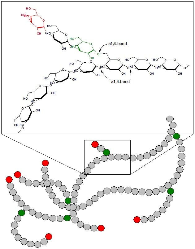
Glycogen is a branched polymer of glucose. Glucose residues are linked linearly by α-1,4 glycosidic bonds, and approximately every ten residues a chain of glucose residues branches off via α-1,6 glycosidic linkages. The α-glycosidic bonds give rise to a helical polymer structure. Glycogen is hydrated with three to four parts water and forms granules in the cytoplasm that are 10-40nm in diameter. The protein glycogenin, which is involved in glycogen synthesis, is located at the core of each glycogen granule. Glycogen is an analogue of starch, which is the main form of glucose storage in most plants, but starch has fewer branches and is less compact than glycogen.


Glycogen

This figures shows the structure of glycogen. Green circles represent α-1,6 linkages at branch points, and red circles represent the nonreducing ends of the chain.


Glycogen Function


In animals and humans, glycogen is found mainly in muscle and liver cells. Glycogen is synthesized from glucose when blood glucose levels are high, and serves as a ready source of glucose for tissues throughout the body when blood glucose levels decline.


Liver Cells


Glycogen makes up 6-10% of the liver by weight. When food is ingested, blood glucose levels rise, and insulin released from the pancreas promotes the uptake of glucose into liver cells. Insulin also activates enzymes involved in glycogen synthesis, such as glycogen synthase. While glucose and insulin levels are sufficiently high, glycogen chains are elongated by the addition of glucose molecules, a process termed glyconeogenesis. As glucose and insulin levels decrease, glycogen synthesis ceases. When blood glucose levels fall below a certain level, glucagon released from the pancreas signals to liver cells to break down glycogen. Glycogen is broken down via glycogenolysis into glucose-1-phosphate, which is converted to glucose and released into the bloodstream. Thus, glycogen serves as the main buffer of blood glucose levels by storing glucose when it levels are high and releasing glucose when levels are low. Glycogen breakdown in the liver is critical for supplying glucose to meet the body’s energetic needs. In addition to glucagon, cortisol, epinephrine, and norepinephrine also stimulate glycogen breakdown.


Muscle Cells


In contrast to liver cells, glycogen only accounts for 1-2% of muscle by weight. However, given the greater mass of muscle in the body, the total amount of glycogen stored in muscle is greater than that stored in liver. Muscle also differs from liver in that the glycogen in muscle only provides glucose to the muscle cell itself. Muscle cells do not express the enzyme glucose-6-phosphatase, which is required to release glucose into the bloodstream. The glucose-1-phosphate produced from glycogen breakdown in muscle fibers is converted to glucose-6-phosphate and provides energy to the muscle during a bout of exercise or in response to stress, as in the fight-or-flight response.


Other Tissues


In addition to liver and muscle, glycogen in found in smaller amounts in other tissues, including red blood cells, white blood cells, kidney cells, and some glial cells. Additionally, glycogen is used to store glucose in the uterus to provide for the energetic needs of the embryo.


Fungi and Bacteria


Microorganisms possess mechanisms for storing energy to cope in the event of limited environmental resources, and glycogen represents a main energy storage form. Nutrient limitation (low levels of carbon, phosphorus, nitrogen, or sulfur) can stimulate glycogen formation in yeast, while bacteria synthesize glycogen in response to readily available carbon energy sources with limitation of other nutrients. Bacterial growth and yeast sporulation have also been associated with glycogen accumulation.


Glycogen Metabolism


Glycogen homeostasis is a highly regulated process that allows the body to store or release glucose depending on its energetic needs. The basic steps in glucose metabolism are glycogenesis, or glycogen synthesis, and glycogenolysis, or glycogen breakdown.


Glycogenesis


Glycogen synthesis requires energy, which is supplied by uridine triphosphate (UTP). Hexokinases or glucokinase first phosphorylate free glucose to form glucose-6-phosphate, which is converted to glucose-1-phosphate by phosphoglucomutase. UTP-glucose-1-phosphate uridylyltransferase then catalyzes the activation of glucose, in which UTP and glucose-1-phosphate react to form UDP-glucose. In de novo glycogen synthesis, the protein glycogenin catalyzes the attachment of UDP-glucose to itself. Glycogenin is a homodimer containing a tyrosine residue in each subunit that serves as an anchor or attachment point for glucose. Additional glucose molecules are subsequently added to the reducing end of the previous glucose molecule to form a chain of approximately eight glucose molecules. Glycogen synthase then extends the chain by adding glucose via α-1,4 glycosidic linkages.


Branching is catalyzed by amylo-(1,4 to 1,6)-transglucosidase, also called the glycogen branching enzyme. The glycogen branching enzyme transfers a fragment of six to seven glucose molecules from the end of a chain to the C6 of a glucose molecule located further inside the glycogen molecule, forming α-1,6 glycosidic linkages.


Glycogenolysis


Glucose is removed from glycogen by glycogen phosphorylase, which phosphorolytically removes one molecule of glucose from the nonreducing end, yielding glucose-1-phosphate. The glucose-1-phosphate generated by glycogen breakdown is converted to glucose-6-phosphate, a process that requires the enzyme phosphoglucomutase. Phosphoglucomutase transfers a phosphate group from a phosphorylated serine residue within the active site to C6 of glucose-1-phosphate, producing glucose-1,6-bisphosphate. The glucose C1 phosphate is then attached to the active site serine within phosphoglucomutase, and glucose-6-phosphate is released.


Glycogen phosphorylase is not able to cleave glucose from branch points; debranching requires amylo-1,6-glucosidase, 4-α-glucanotransferase, or glycogen debranching enzyme (GDE), which has glucotransferase and glucosidase activities. About four residues from a branch point, glycogen phosphorylase is unable to remove glucose residues. GDE cleaves the final three residues of a branch and attaches them to C4 of a glucose molecule at the end of a different branch, then removes the final α-1,6-linked glucose residue from the branch point. GDE does not remove the α-1,6-linked glucose from the branch point phosphorylytically, meaning that free glucose is released. This free glucose could in theory be released from muscle into the bloodstream without the action of glucose-6-phosphatase; however this free glucose is rapidly phosphorylated by hexokinase, preventing it from entering the bloodstream.


The glucose-6-phosphate resulting from glycogen breakdown may be converted to glucose by the action of glucose-6-phosphatase and released into the bloodstream. This occurs in liver, intestine, and kidney, but not in muscle, where this enzyme is absent. In muscle, glucose-6-phosphate enters the glycolytic pathway and provides energy to the cell. Glucose-6-phosphate may also enter the pentose phosphate pathway, resulting in the production of NADPH and five carbon sugars.


Exercise and Glycogen Depletion


In endurance exercise, athletes may undergo glycogen depletion, in which most of the glycogen is depleted from the muscle. This can result in severe fatigue and difficulty moving. Glycogen depletion can be mitigated by continuously consuming carbohydrates with a high glycemic index (high rate of conversion to blood glucose) during exercise, which will replace some of the glucose using during exercise. Specialized exercise regimens may also be employed that condition muscle to utilize fatty acids as an energy source at a greater rate, thus breaking down less glycogen. Athletes may also use carbohydrate loading, the consumption of large amounts of carbohydrates, to increase the capacity for glycogen storage.


Examples of Glycogen Storage Diseases


There are two main categories of glycogen storage diseases: those resulting from defective glycogen homeostasis in the liver and those resulting from defective glycogen homeostasis in the muscle. Diseases resulting from defective liver glycogen storage generally cause hepatomegaly (enlarged liver), hypoglycemia, and cirrhosis (liver scarring). Diseases resulting from defective muscle glycogen storage generally cause myopathies and metabolic impairment. Examples of glycogen storage diseases include Pompe Disease, McArdle Disease, and Andersen Disease.


Pompe Disease


Pompe disease is caused by mutations in the GAA gene, which encodes lysosomal acid α-glucosidase, also referred to as acid maltase, and affects skeletal and cardiac muscle. Acid maltase is involved in glycogen breakdown, and disease-causing mutations result in the detrimental buildup of glycogen in the cell. There are three types of Pompe Disease: the adult form, juvenile form, and infantile form, which are progressively more severe. The infantile form leads to death by one to two years of age if left untreated.


McArdle Disease


McArdle Disease is caused by mutations in the PYGM gene, which encodes myophosphorylase, the glycogen phosphorylase isoform present in muscle. Symptoms are often observed in children, but the disease may not be diagnosed until adulthood. Symptoms include muscle pain and fatigue, and the disease may be life-threatening if not properly treated.


Andersen Disease


Andersen Disease is caused by a mutation in the GBE1 gene, which encodes glycogen branching enzyme, and affects muscle and liver. Symptoms are usually observed at a few months of age, and include stunted growth, liver enlargement, and cirrhosis. Complications of the disease can be life-threatening.


Quiz


1. Which best describes the function of glycogen?
A. Provides structural support to muscle cells
B. A transcription factor that regulates cell differentiation
C. Stores glucose in plants
D. Buffers blood glucose levels and serves as a readily mobilized energy source

Answer to Question #1

2. What is the main hormone that stimulates glycogen breakdown?
A. Glucagon
B. Thyroid
C. Insulin
D. Estrogen

Answer to Question #2

3. What are the possible fates of the glucose-1-phosphate produced by glycogenolysis?
A. Conversion to glucose-6-phosphate followed by entrance into the glycolytic pathway
B. Conversion to glucose-6-phosphate followed by entrance into the pentose phosphate pathway
C. Conversion to glucose followed by release into the bloodstream
D. All of the above

Answer to Question #3

References


  • Cohen, P., Nimmo, H.G., and Proud, C.G. (1978) “How does insulin stimulate glycogen synthesis?” Biochemical Society Symposium. (43):69-95.

  • Eicke, S., Seung, D., Egli, B., Devers, E.A., and Streb, S. (2017) “Increasing the carbohydrate storage capacity of plants by engineering a glycogen-like polymer pool in the cytosol.” Metabolic Engineering. 40:23-32.

  • Hargreaves, M. and Richter, E.A. (1988) “Regulation of skeletal muscle glycogenolysis during exercise.” Canadian Journal of Sport Sciences. 13(4):197-203.

  • Ivy, J.L. (1991). “Muscle glycogen synthesis before and after exercise.” Sports Medicine. 11(1):6-19.

  • Wilson, W.A., Roach, P.J., Montero, M., Baroja-Fernández, E., Muñoz, F.J., Eydallin, G., Viale A.M., and Pozueta-Romero J. (2010) “Regulation of glycogen metabolism in yeast and bacteria.” FEMS Microbiology Reviews. 34(6):952-85.


Glycogen