Showing posts with label 5k. Show all posts

Saturday, August 25, 2018

Sucrose

Sucrose Definition


Sucrose, commonly known as “table sugar” or “cane sugar”, is a carbohydrate formed from the combination of glucose and fructose. Glucose is the simple carbohydrate formed as a result of photosynthesis. Fructose is nearly identical, except for the location of a double-bonded oxygen. They are both six-carbon molecules, but fructose has a slightly different configuration. When the two combine, they become sucrose.


Plants use sucrose as a storage molecule. For quick energy, cells may store the sugar for later use. If far too much is accumulated, plants may begin to combine the complex sugars like sucrose into even large and denser molecules, like starches. These molecules, and oily lipids, are the main storage chemicals used by plants. In turn, animals eat these sugars and starches, break them back down into glucose, and use the energy within the bonds of glucose to power our cells.


Sucrose has been an important sugar for humans because it is easy extracted from plants such as sugar cane and sugar beets. These plants tend to store an excess of sugar, and from this we produce the majority of the sugar that we use. Even most “natural” sweeteners, which claim to be healthier than sucrose, are simply a different version of glucose combined in a different manner by plants.


Sucrose Structure


As mentioned above, sucrose is disaccharide, or a molecule made of two monosaccharides. Glucose and fructose are both monosaccharides, but together they make the disaccharide sucrose. This is an important process for the storage and compression of energy. Plants do this to make it easier to transport large amounts of energy, via sucrose. This process can be seen in the following image.


Sucrose condensation

Sucrose condensation


Glucose is seen on the left. Glucose is known as an aldose, meaning the carbonyl group (carbon double bonded to an oxygen) is found at the end of the chain of carbons. When the molecule creates a ring back on itself, it forms a 6-sided ring. Fructose, on the other hand, is a ketose. This means that the carbonyl group is found in the middle of the middle of the molecule. In this case, it forces fructose into a five-sided ring.


In a plant creating sucrose, an enzyme comes along to smash these two rings together, and extract a molecule of water. This process is called a condensation reaction, and forms a glycosidic bond between the two molecules. As you can see in the image, the reaction can also go the other way. To dissolve sucrose into fructose and glucose, a molecule of water can be added back in. This is what happens to sucrose as you digest it.


Sucrose Uses


Sucrose is the most common form of carbohydrate used to transport carbon within a plant. Sucrose is able to be dissolved into water, while maintaining a stable structure. Sucrose can then be exported by plant cells into the phloem, the special vascular tissue designed to transport sugars. From the cells in which it was produces, the sucrose travels through the intercellular spaces within the leaf. It arrives at the vascular bundle, where specialized cells pump it into the phloem. The xylem, or vascular tube which carries water, adds small amounts of water to the phloem to keep the sugar mixture from solidifying. The sucrose mixture then makes its way down the phloem, arriving at cells in the stem and roots which have no chloroplasts and rely on the leaves for energy.


The sucrose is absorbed into these cells, and enzymes begin breaking the sucrose back into its constituent parts. The six-carbon glucose and fructose can be broken down into 3-carbon molecules, which are imported into the mitochondria, where they go through the citric acid cycle (AKA the Krebs Cycle). This process reduces coenzymes, which are then used in oxidative phosphorylation to create ATP. The energy within the bonds of ATP can power many of the reactions these cells need to complete in order to maintain the stem and roots.


Likewise, all other life on Earth is dependent upon sucrose and other carbs produced by plants. Sucrose was one of the first substances to be extracted from plants on a mass-scale, creating the white table sugar we know today. These sugars are extracted and purified from large crops, including sugar cane and sugar beets. To extract the sugar, the plants are usually boiled or heated, releasing the sugar. “Sugar in the Raw” is sugar which has not been treated further, while white table sugar undergoes more purification.


Quiz


1. Which of the following terms describes sucrose?
A. Disaccharide
B. Monosaccharide
C. Ketose

Answer to Question #1
A is correct. Sucrose is a disaccharide, meaning it is made of two monosaccharides, or sugars. You can also think of it as a “complex sugar” or “complex carbohydrate”.

2. Which of the following is NOT a use of sucrose?
A. Sweetener
B. Information storage
C. Carbon-transport

Answer to Question #2
B is correct. Humans use sucrose as a sweetener, while plants use it to transport carbon they have acquired and imbued with energy. Ribose, a five-carbon sugar, is used in the construction of DNA, which does store information. The uses of carbohydrates are truly incredible.

3. The items listed below all store energy within their bonds. Which of the following molecules stores the MOST energy?
A. Glucose
B. Sucrose
C. Starch

Answer to Question #3
C is correct. Remember that sucrose is made of two smaller molecules. Each of these holds a similar number of bonds, meaning sucrose stores twice as much energy as glucose, approximately. Starch is made up of many, many glucose molecules. Therefore, starch stores the most energy of these substances. However, starch is hard to transport throughout the plant, making sucrose the preferred method of transporting energy through a carbon source.

References


  • Jones Jr., J. B. (2005). Hydroponics: A Practical Guide for the Soilless Grower (2nd ed.). Boca Raton: CRC Press.

  • McMahon, M. J., Kofranek, A. M., & Rubatzky, V. E. (2011). Plant Science: Growth, Development, and Utilization of Cultivated Plants (5th ed.). Boston: Prentince Hall.

  • Nelson, D. L., & Cox, M. M. (2008). Principles of Biochemistry. New York: W.H. Freeman and Company.


Sucrose

Wednesday, June 20, 2018

Extinction

Extinction Definition


Extinction is a term applied to a known species, of which there are no known living individuals. Some species which have suffered extinction are known only from their fossilized remains. Others were at one point known to humans, but are gone now. Still others suffered directly at the hands of humans, driven to extinction. An extinct species, or one that has suffered extinction, no longer contributes to the evolution of organisms, but can help us understand the relationship between extant, or living animals.


Extinction has many causes, some of which are caused directly by humans and others which are parts of natural cycles or apocalyptic events. An extinction event is when many species are driven to extinction by a particular species, natural disaster, or other phenomenon. While these mass extinctions sometimes wipe out a large majority of life, extinction itself is a continual part of evolution. Extinction happens on some scale all the time, as organisms adapt and outcompete others. It has been estimated that extinction has claimed at least 99 percent of all species that have ever lived. However, new species are also being generated through the process of speciation. As they spread, diversify, and recover the niches lost to extinction, the tree of life flourishes. However, it might flourish in a new direction.


Examples of Extinction


Thylacine


Welcome to Tasmania, mate! The year is 1800, and the island of Tasmania is overflowing with a variety of interesting marsupials. Among these is the thylacine, an apex predator resembling a mixture of a tiger and a wolf. Like other marsupials, the thylacine had an external pouch. Its newborn young, underdeveloped and tiny, would make their way to the pouch to continue developing in safety. Unfortunately for the thylacine, human expansion in Australia and Tasmania would lead to their extinction.


Bagged thylacine

As seen above, the thylacine was often hunted. Thylacine were top predators, and the sheep and livestock of the new human population seemed no exception. As the human population spread on Tasmania, the competition grew fiercer, and bounties were put on the thylacine by the 1830s. Less than 100 years later, the thylacine would go extinct in the wild in 1930. While there were populations in zoos these too would die off by 1933. Thus, extinction for the thylacine was complete.


Passenger Pidgeon


Once a species that formed the vastest flocks known to man, the Passenger pigeon went extinct almost entirely at the hands of man. Before 1800, the Passenger pigeon enjoyed a range from New York to Denver, across most of the Continental United States. First described by Carl Linnaeus, the bird had been known to mankind for a long time. As the Europeans arrived in the New World, the saw the pigeon as useful and plentiful food source. At the time, hunting technology and population size would not allow for the mass harvesting of the birds, and they sustainably provided food.


Fast forward several hundred years, and man had multiplied across the North American continent. Where Native American populations were small, and more sustainable, the new colonizers needed vast resources to maintain their way of life. As such, the passenger pigeon started to see steady declines into the late 1800. By the end of the 1800s, there was a massive drop off. While bills were drafted and passed to protect the Passenger pigeon, it was too late. The biology of the Passenger pigeon made it an animal prone to gathering and flocking, driven by millions of years of evolving to escape solitary predators. This social feature of the bird which had protected it for so long, made it easy prey for human hunters. Extinction quickly ensued. By the early 1900s, the last Passenger pigeon had died in a zoo.


Megalodon


The largest known shark to ever live suffered extinction. Carcharocles megalodon, or simply Megalodon, has been identified from fossilized remains of its jaw and teeth. Possibly related to the Great White Shark, its teeth suggest it was much bigger. One of these teeth can be seen below, next to two Great White teeth.
Megalodon tooth with great white sharks teeth


Comparing measurements from these teeth and the jaw, scientists have estimated Megalodon to be somewhere around 60 feet long. The largest living shark currently, the whale shark, is only around 30 feet long, and even the Great White tops out at around 21 feet long. Scientific data suggests that extinction for Megalodon occurred around 2.6 million years ago. At this time, humans did not exist. It is suggested that extinction occurred because of a shift in the food supply for Megalodon as well as increased competition from other megapredators, such as early killer whales.


Interestingly, like other extinctions, there is always an air of doubt. Just because humans have not witnessed an animal thought to be extinct does not mean it is actually extinct. Extinction, in this regard, is simply a category used by the International Union for the Conservation of Nature (IUCN) and other agencies to categorize an animal thought to be extinct. For example, the black-footed ferret was thought to be extinct for several decades, until a population was found in Wyoming. Due to the unknown and vast nature of the ocean, even Megalodon is outliving its extinction. Often, claims of large sharks and attacks on boats are still attributed to Megalodon. However, no actual evidence has ever been found to refute that Megalodon suffered extinction.


Causes of Extinction


Ultimate Causes


Ultimately, every species has three “choices”. They can adapt to a situation, somehow evolving a novel or more efficient way to live. They can migrate, in the hopes that other areas will provide the resources they need with less competition. Or, as is the case for many animals, they can die. Extinction, as has been demonstrated in the fossil record, far surpasses survival for most species. While this may be seen as a negative thing, remember that extinction not only leaves new niches open to colonize, but can also be caused by a species becoming more successful. While one species may take over for a while, they usually undergo speciation into a variety of forms.


Proximate Causes


There are many more proximate causes of extinction. In mathematical terms, extinction happens any time the rate of reproduction is lower than the rate individuals are dying. This situation inevitably leads to extinction, but there are a number of factors which can drive these rates.


Predation, for example, is a major cause of extinction for many animals. Many species of fish in the Caribbean are currently threatened by the emergence of a new species, the Lionfish. Lionfish are not native to the Caribbean, and have no natural predators of their own. As such, they have pretty much free reign on the fish of the Caribbean. Many of these endemic species are being wiped out by the lionfish, and extinction is the likely result. In a similar story, extinction is plaguing many species of birds and lizards which have been exposed the brown tree snake. The snake, transported on cargo ships during WWII, has no natural predators on the islands to which is was transported. As such, the snake population has exploded and driven its prey items towards extinction, if not into it.


Other causes, which are directly the result of human action, involve habitat destruction and fragmentation. As we destroy the resources animals need to survive, we decrease the capacity an area can hold. As we further divide these areas with roads, fences, and other boundaries, we decrease the ability of species to migrate and successfully reproduce. This phenomena, as well as hunting and exploitation of animals for meat and game, has cause the extinction of a massive amount of animals. Scientists now speculate that, due to human interactions with the rest of nature, the world is entering another mass extinction event.


Quiz


1. How do we know an animal is really extinct?
A. We have no documented and confirmed sightings of the animal in recent times
B. We can never know
C. We find its fossils

Answer to Question #1
A is correct. While it may be believed by some that the thylacine and Megalodon are still out there, there would be so few individuals that they could not survive anyway. Due to the effects of genetic drift and bottlenecks in a small population, it is unlikely that a few individuals will survive an extinction. Remember that all animals leave fossil evidence, even animals which are still extant.

2. When considering extinct organisms which do not leave good fossils, how can scientists claim to pinpoint their extinctions?
A. Voodoo Magic
B. Only organisms with fossils can be determined
C. Chemical evidence points to many extinction events

Answer to Question #2
C is correct. While small microorganisms like bacteria and algae rarely leave reliable fossils, scientists have other ways of determining which organisms were most prominent. For example, by examining the composition of the air in modern times and comparing it to depositions in the soil, scientists can estimate the gas content of ancient times by looking at the composition of the soil and rocks.

3. Scientists want to revive the Woolly Mammoth. To do so, they supposed that they could use the DNA found in a frozen male mammoth to impregnate a female elephant. Would this “reverse” extinction?
A. Yes
B. No
C. Only if the baby comes out a Mammoth

Answer to Question #3
B is correct. First off, it is unlikely that the embryo would be viable, simply because the animals are separated by millions of years of evolution. Further, mixing two species is not actually recreating a mammoth, it is creating a hybrid. Lastly, a real way to bring the mammoth out of extinction would be to clone the mammoth DNA, and grow a new organism.

References


  • Feldhamer, G. A., Drickamer, L. C., Vessey, S. H., Merritt, J. F., & Krajewski, C. (2007). Mammology: Adaptation, Diversity, Ecology (3rd ed.). Baltimore: The Johns Hopkins University Press.

  • Pimiento, C., & Clements, C. (2014, October 22). When did Carcharocles megalodon become extinct? A new analysis of the fossil record. PLOS One. Retrieved from http://journals.plos.org/plosone/article?id=10.1371/journal.pone.0111086

  • Pough, F. H., Janis, C. M., & Heiser, J. B. (2009). Vertebrate Life. Boston: Pearson Benjamin Cummings.


Extinction

Thursday, May 17, 2018

Endemism

Endemism Definition


Endemism is the condition of being endemic, or restricted in geographical distribution to an area or region. The area or region can vary in size, and is defined or identified in different ways. Endemism is an ecological classification in that it describes the range or distribution of a species, or group of species. For instance, entire families of different species of birds are endemic to the island of Madagascar. The term endemism can applied to many things, including diseases and natural phenomenon. Endemism in these cases refers to the “normal” or standard level of some measured observation within a specific geographic region or area.


Endemism is not to be confused with indigenous, a term which refers to the origins of a species. Indigenous refers to where a group originated. A species can be both endemic and indigenous to an area. However, some species thrive and exceed the bounds of their original indigenous location. This means that the species is no longer endemic, but is still indigenous to the original area. Once a species has reached a wide-spread, global distribution it is said to be cosmopolitan. Animals like whales, once indigenous to a specific mainland in the form of their 4-legged ancestors, are now cosmopolitan in distribution.


Endemic Species


An endemic species is a species which is restricted geographically to a particular area. Endemism in a species can arise through a species going extinct in other regions. This is called paleoendemism. Alternatively, new species are always endemic to the region in which they first appear. This is called neoendemism. Both forms of endemism are discussed in more detail under the heading “Types of Endemism”, below.


Endemic species, regardless of how they came to be restricted to a particular area, experience the same threats to their existence. The smaller the region, the more dire the threat toward the survival of the species. Any action that reduces the size of the land, or divides it in any way can significantly affect the normal patterns of the endemic species. While endemism and being endangered or threatened are different things, being endemic to a small area is often a warning sign that a species may become threatened or endangered.


This is not always the case, as many globally distributed species are also considered threatened or endangered. In recent years, many sharks have joined the list. While they are distributed throughout many of the ocean’s waters, the harvesting of shark fins for soup has decimated their populations globally. Endemism sometimes protects species from being exploited globally, simply because of the fact that the species only exists in a small area. This can even make the species easier to protect, because the land can be placed under a conservation easement to restrict the construction and human impact on the land.


Endemic Disease


Scientists studying epidemiology, or disease outbreaks, have a similar definition of endemism. An endemic disease is a disease seen at consistent levels in specific location. For instance, endemic relapsing fever is a disease seen in Europe and in North America. The disease is not seen in any sort of observable amounts in other parts of the world. Other diseases, which are new to an area or are spiking in their prevalence, are known as epidemic diseases.


There are many endemic diseases, and their endemism has roots in the species and vectors which promote these diseases. In the case of relapsing fever, a vector carries the bacterium of the Borrelia species. There are several vectors which can carry these bacteria, mostly including ticks and lice. The species of ticks and lice which carry these bacteria are endemic to the Northern Hemisphere. Borrelia bacteria are also responsible for Lyme disease, a disease endemic to the Northern Hemisphere. A map of Lyme disease is shown below, and corresponds to the endemism seen in tick and lice species.


Geographical distribution of reported Lyme Disease cases


While Lyme disease and relapsing fever are endemic to these areas, they are not endemic to say, Australia. If there were even a few cases of Lyme disease in Australia, the disease would be considered epidemic, because the normal level of Lyme disease in Australia is zero.


Types of Endemism


Paleoendemism


There are two basic ways for a species to show endemism to a certain region. Basically, the difference between the two is whether the species is newly emerging, or historic and declining. Paleoendemism describes the later. In this form of endemism, a species which was once widespread has been reduced to a much smaller range. This is the case for many large predators today.


Before humans, large predators were widely distributed across the globe. As human society became more organized, large predators were driven away from society, and out of their historic ranges. Those which have not gone extinct are now restricted to limited ranges. Conservation efforts for these animal focus on protecting the current range and expanding it to encompass the historic range. This is hard however, as humans often oppose the re-introduction of large predators. Without protections from hunters, the species will easily be pushed back to their endemic range.


Neoendemism


On the opposite hand, new species are branching off the evolutionary tree every day. These species are both endemic and indigenous to the location in which they first appeared. They are restricted to a geographical location simply because that is where they started. This is known as neoendemism. There are many species, found on islands, which show this form of endemism.


Islands provide an interesting and isolated grounds for the development of new species. While the species on the island are now endemic, their ancestors were likely not. Take the Galapagos finches, as an example. The Galapagos archipelago contains many islands. Many thousands of years ago, a single finch species arrived on the islands. At first, it spread across the island as one species. However, evolution has now separated the birds so much that they represent different species. The differences in the vegetation on the islands divided the ancestor into many smaller species, which show endemism to the island they are found on.


Quiz


1. The Greenback Cutthroat Trout is a fish which is indigenous to many waters in Colorado. After heavy over-fishing, the species is now endemic to only a small handful of streams in Colorado. Which of the following statements says the same thing as the above sentences?
A. These trout are native to many waters, but have been reduced to only a handful
B. These trout both come from and reside in Colorado
C. Overfishing has not decreased the range of these trout

Answer to Question #1

2. Endemism is often mistaken with being endangered. How are the two terms different?
A. They are essentially the same.
B. Endemism simply describes the distribution, while endangered describes the threats to a population
C. Endemic species often become endangered

Answer to Question #2

3. Which of the following is NOT an endemic disease?
A. The common flu
B. Zika virus, found in Minnesota
C. Malaria, in Africa

Answer to Question #3

References


  • Blumstein, D. T., & Fernandez-Juricic, E. (2010). A Primer of Conservation Behavior. Sunderland: Sinauer Associates, Inc. Publishers.

  • Heymann, MD, D. L. (Ed.). (2015). Control of Communicable Diseases Manual. Washington: American Public Health Association.

  • Rogers, PhD, K. (2015). Colorado Outdoors – Piecing together the past (Cutthroat Trout). Retrieved from Colorado Parks and Wildlife: http://cpw.state.co.us/Documents/Research/Aquatic/CutthroatTrout/Rogers2012CoOutdoors.pdf


Endemism

Monday, May 14, 2018

Mesentery

Mesentery Definition


The mesentery is an organ which surrounds the organs of the gut, and suspends them from the abdominal wall. The mesentery is made of mesoderm cells, the middle of the three embryonic layers. This layer ends up surrounding all internal organs, as the peritoneum. In the gut, this layer folds over on itself and provides points of attachment for the other internal organs. The mesentery used to be known as a variety of different tissues related to the mesocolon. However, recent studies have revealed the mesentery to be a single organ, which suspends the internal organs in the abdominal cavity, and allows the various vessels of the body to reach these organs.


Mesentery Anatomy


The mesentery, for many decades, was often not considered an organ because of its thin and convoluted nature. The mesentery surrounds all of the organs in the abdomen. Because it is formed during embryogenesis, the mesentery ends up getting twisted and turned as the gut develops. Therefore, the mesentery is a complex shape which surrounds the organs. The organ consists of sheet of tissue, which surrounds organs and folds back on itself. This can be seen in the image below. The mesentery is red.


Horizontal disposition of the peritoneum in the lower part of the abdomen


Notice how the mesentery surrounds the small intestine. The mesentery is very thin. Not shown in this picture are the many blood and lymph vessels which traverse the mesentery on their way to the intestines. On a microscopic level, the mesentery is similar to other connective tissues. It is comprised of several layers of cells, derived from mesoderm, attached to a matrix of connective fibers. The extracellular matrix of the cells allows for the creation of a very strong cell and fiber network, which can heal itself if damaged. Within this structure, blood and lymph vessels can carry their respective fluids to the intestines.


Function of Mesentery


Recent studies have shown that within these folds of tissue are complex arrays of lymph vessels, blood vessels, and immune cells. This suggests that the mesentery functions as a complex organ which carries nutrients away from the intestines while at the same time protecting from infection. The intestines are busy digesting food. As it travels through the intestine, nutrients are released and get absorbed by the cells of the intestine. These nutrients are transported to the blood, where they can be distributed to the body. The mesentery provides a stable and secure route for these vessels to pass. Without the mesentery, the fragile vessels would be subject to the pulling and stretching the body goes through.


The mesentery also allows lymph vessels to reach the intestines. This is important because nutrients are not the only thing that makes it through the intestines. Often, bacteria and viruses manage to squeeze their way through the intestines. The second line of defense is the immune system. White blood cells can defend the body and create antibodies to target the invaders. However, they must be able to reach the invaders. The mesentery gives the immune system access to this area of the body, even though it is contained within a cavity.


Besides these functions of directing and protecting vessels, the mesentery has an important role in development and support of the gut. During development, the expansions and contractions of the mesentery direct the shape of the gut. The colon, for instance, is pulled into place against the abdominal wall when the mesentery connecting the two shrinks in size. In fact, without the mesentery your organs would fall into a puddle in the bottom of your gut. The complex folds and attachment points hold your organs in place. They will even work when you are hanging upside-down or doing a backflip!


Still further research into the mesentery must be done. It has also been found to be a holder or excess fat, and is riddled with nerves stretching to the intestines and other organs. The mesentery is likely to have other, undiscovered functions which will be uncovered with time. Among these are likely to be immune defenses for the body and roles in development.


Mesentery and Disease


Along those lines, research has begun to show the prominent role the mesentery plays in several diseases. Because it is a line of defense from the digestive system, it is not surprising to find out that it is heavily involved in the defense against food-borne illnesses. More interesting however, is its function in the spread of other diseases, such as cancer.


Some studies coming out in recent years have shown that the mesentery may by an important pathway for metastasizing cancer cells to travel. These cells often travel through the lymph or blood vessels. Because the mesentery is lined so prolifically with them, it can become a central highway for distributing the cells. This may feature prominently in future cancer treatments.


Other diseases, such as Crohn’s disease, are caused by a malfunctioning mesentery. People with Crohn’s disease often have a swollen or hardened mesentery. This makes it difficult for the vessels contained in the mesentery to function properly. People with this condition often have a hard time digesting food, and can have compromised immune systems.


Quiz


1. Which of the following is NOT part of the mesentery?
A. The peritoneum around the heart
B. The peritoneum around the appendix
C. The peritoneum around the colon

Answer to Question #1

2. Which of the following is NOT a function of the mesentery?
A. Digestion
B. Support
C. Access for vessels

Answer to Question #2

3. A lancelet is a very small fish-like creature. It is one of the smallest organism with a notochord. The lancelet does not have a mesentery. Why could this be?
A. The mesentery is only in complex organisms
B. The mesentery is only needed to distribute nutrients
C. No structural support or access to the intestines are needed in lancelets

Answer to Question #3

References


  • De luliis, G., & Pulera, D. (2007). The Dissection of Vertebrates. Amsterdam: Academic Press.

  • Feldhamer, G. A., Drickamer, L. C., Vessey, S. H., Merritt, J. F., & Krajewski, C. (2007). Mammology: Adaptation, Diversity, Ecology (3rd ed.). Baltimore: The Johns Hopkins University Press.

  • Widmaier, E. P., Raff, H., & Strang, K. T. (2008). Vander’s Human Physiology: The Mechanisms of Body Function (11th ed.). Boston: McGraw-Hill Higher Education.


Mesentery

Monday, April 2, 2018

Fluoride

Fluoride Definition


Fluoride is a negatively charged fluorine atom (F), also known as a fluorine anion. Fluoride is a naturally occurring ion, found in certain mineral and salty deposits. Certain levels of fluoride have been proven to be beneficial in fighting cavities and strengthening teeth. As a public health measure, fluoride has been maintained at low levels in municipal drinking water in many countries, including Australia, the United States, and several European countries. While there remains some controversy over the dangers of fluoride in water, the level is maintained well below the documented level at which negative symptoms are seen.


Biochemistry of Fluoride


Fluoride, as a negative ion, or anion, is capable of binding ionically to a cation, or positive ion. In the blood, there are many cations, but of utmost importance are H+ (hydrogen) and Ca2+ (calcium). These cations must be maintained in strict balance. If they are not in balance, the system will become more acid, and more calcium will be drawn from the bones and teeth, making them weaker and brittle. If a person has two little fluoride or too much fluoride in their system, these cations will also become unbalanced.


On the short hand, too little fluoride creates weak teeth, and allows bacteria to infiltrate and infect. This leads to tooth decay and cavities. Very low levels of fluoride prevent calcium from leaving the teeth, thereby strengthening them and preventing bacterial invasions. It is for this reason that water supplies are fluorinated on purpose. Fluoride provides an efficient, cost-effective method of providing a basic preventative health service.


On the other hand, too much fluoride in the system can be a bad thing. Just as too little fluoride weakens the bones, too much fluoride causes the same condition. Instead of limiting the system which balances calcium in the bones and teeth, fluoride can become the negative anion which pushes it out of balance. With too much fluoride in the system, the pH begins to lower. This causes the release of calcium, which can soak up acids in the blood. The calcium is then lost in the urine, and the bones and teeth become weaker.


Your body has this natural ability to process and remove excess fluoride, and can expel up to 50% of the fluoride intake per day. However, this means that long exposures to medium doses or single exposures to very high doses will take time to be removed from the system. At a certain point, they can become deadly, but this point is far above the threshold we keep our fluoride levels at.


Uses for Fluoride


Fluoride in Drinking Water


Health professionals have been maintaining low levels of fluoride in drinking water for many decades, as there is much evidence that it decreases tooth decay by killing bacteria in the mouth. The low levels of fluoride added to drinking water are far below the maximum limits of 7-10 mg per day that a human can safely consume. While many millions of people drink fluorinated water, controversy remains around its effects and potential side-effects. Much of this is due to the harsh effects of fluoride at high levels, seen in some natural ground water sources. Municipal sources of water are specially treated and screened to prevent high levels of fluoride. See “Dangers of Fluoride” below, for more information.


Fluoride in Dental Products


Fluoride is a common component in many dental products, including toothpastes, varnishes, and other teeth cleaning products. Fluoride has a proven history in the fight against mouth disease. It is also incredibly cheap, and reliable to work with. However, in the early days of using fluoride in dentistry there were some unfortunate accidents. Several people got sick, and even died, when they misused concentrated fluoride solutions provided by their dentists. While the solutions were intended to be spit out, the patients swallowed the concentrated fluoride. The high dose of fluoride disrupted their pH and calcium content enough to cause death.


These situation have been easily avoided by the setting of guidelines to the use of fluoride and strict warning labels on products which contain fluoride. Fluoride toothpastes usually contain a fraction of the daily advised amount of fluoride, and most of the fluoride is not absorbed when properly used. Concerns over children and fluoride poisoning have led to children’s toothpastes, which have a reduced fluoride content.


Other Places Fluoride is found


Fluoride is found naturally nearly everywhere. There is fluoride present in seawater, at nearly the same levels government fluorine their water. There is also some present in rainwater, which has fallen from the atmosphere after being deposited there by fires, volcanoes, and industrial pollution. The majority of fluoride on the Earth is found as the salt crystal fluorite (CaF2). This crystal can be mined, and the fluoride can be extracted for industrial applications. Because fluoride is found in rain and water, it is also found in plants. Therefore, everything we eat has trace amounts of fluorine.


Dangers of Fluoride


While there are virtually no well documented dangers to the levels of fluoride we are exposed to in the water, there is a danger of having no or too much fluoride. Fluoride is a necessary part of several biological reactions, and most living organisms need at least trace amounts of it to function properly. Where this threshold is exactly is not known, but even as little as half a milligram per liter of water can drastically improve bone and tooth health.


On the flip side, too much fluoride can have drastic consequences. Beginning symptoms of excess fluoride include dental and skeletal fluorosis. This is a condition in which the calcium starts to leave the bones to deal with the excessively high blood pH. As the calcium leaves, it is replaced by fluorine. This can make the bones and teeth excessively hard and brittle, causing them to break or shatter more easily. These symptoms can be slowly reversed by cutting fluoride out of the diet. These diseases have been documented in cases where the fluoride in the drinking water was above 10 mg/L, which is almost 10 times the amount in North American drinking water.


Exposure to higher levels of fluoride can cause more severe symptoms. At the far end of the spectrum is hypocalcemia. Your body, in order to counterbalance the huge amount of negation ions you just ingested, rapidly pulls the calcium from all of your tissues, not just the bones and teeth. As this happens, critical junctions at nerve and muscle cells can no longer function. Your body shuts down quickly, and you die. This condition is known as hypocalcemia. Fluoride has caused several deaths this way, mostly in accidents involving concentrated fluoride chemicals for industrial purposes. While fluoride is not a problem for nations with sophisticated water treatment facilities, high levels of fluoride are consumed by over 300 million people a year from untreated groundwater sources.


In drinking water in the United States, the fluoride is maintained between 0.7 and 1.2 mg per liter. A human can drink around 3-4 liters of water per day, which brings their total fluoride intake to somewhere between 4-5 mg. A daily intake of less than 10 mg is recommended, allowing much room before significant levels of fluoride are reached. In groundwater sources which contain more fluoride than this, the fluoride can quickly accumulate and cause a wide variety of issues.


Quiz


1. Is fluoride harmful or beneficial to your health?
A. Harmful
B. Beneficial
C. It depends how much you take

Answer to Question #1

2. Which of the following is a serious concern about fluoride?
A. The Communists are using it to control our brains
B. Too much fluoride will cause mutations and deformations
C. Too little fluoride will leave us with brittle bones and teeth

Answer to Question #2

3. Fluorite (CaF2) is the salt crystal which is mined to extract fluoride. Approximately how much fluoride is obtained from two grams of fluorite? Hint: you might want to look at the periodic table.
A. 0.25 grams
B. 1 gram
C. 1.5 grams

Answer to Question #3

References


  • Bruice, P. Y. (2011). Organic Chemistry (6th ed.). Boston: Prentice Hall.

  • Nelson, D. L., & Cox, M. M. (2008). Principles of Biochemistry. New York: W.H. Freeman and Company.


Fluoride

Monday, February 12, 2018

Toxicology

Toxicology Definition


Toxicology is the study of how an organism reacts to various concentrations of chemicals. Toxicologist use model organisms to test various chemicals and estimate the concentration of chemical which can cause effects. Chemicals which will be used to produce goods used by people will undergo rigorous testing to ensure their safety. Toxicology also focuses on the structure of poisons, toxins, and venoms. Advanced methods, such as Nuclear Magnetic Resonance Spectroscopy, are used in toxicology to classify new toxins and understand their chemical structure. As we understand more about the world around us and seek to use new chemicals to our advantage, toxicologists are continually presented with a broader field of study.


History of Toxicology


Toxicology is an ancient science. Since the dawn of man, humans have sought to understand and manipulate the substances found in their environment. Ancient Roman and Middle-Eastern and Asian texts have descriptions of toxins along with their treatments. While ancient forms of the field relied on various belief in magic, the influence of stars, and other fantastical origins to explain and cure the effects of toxins, modern toxicology is a much more scientific pursuit.


Many fields have influenced and informed toxicology, and our understanding of chemicals and their interactions in general. A general understanding of chemistry and biology allowed the father of toxicology, Mathieu Orfila, to publish the first work on toxicology in 1813. Orfila went on to establish forensic toxicology by using his techniques to find evidence of arsenic in the body of a murder victim. Since the 1800’s toxicology has greatly expanded and advanced. Today, toxicology contains many branches of study and specialization.


Careers in Toxicology


While some schools have Bachelor’s and Master’s level toxicology programs, the majority of toxicologist go on to receive a PhD and specialize in a certain area of toxicology. Many area of toxicology exist, but in general toxicologists focus on a few themes and specialize from there. Medicinal toxicology is the branch most concerned with humans and what we are exposed to. Many toxicologists in this field work for government organizations or testing labs, and approve the products released to consumers. Others in this field understand toxins and poisoning and assist criminal investigators as forensic toxicologists. Everyone with a human focus on toxicology must understand human biology, and most are medical doctors that specialize in toxins.


Another area of toxicology focuses on the toxins themselves, and the species that produce the toxins. Because the chemicals in venom and animal toxins are so biologically active, they are extremely interesting for academics and pharmacists alike. For instance, a mycotoxicologist studies the toxins produced by fungi and their relatives. Many professionals studying toxicology study how the toxins form, act on their target, and eventually break down. This information is crucial to those on the defending edge of toxicology, like the doctors treating deadly snake-bites or accidental poisonings. Others search for ways to use the effects of the toxin in therapeutic ways. Venom, toxins in plants, and other biologically active substances could possibly be used to target specific unwanted cells or simply shut down nerves temporarily. A key convention in toxicology is that the dose makes the poison. In other words, the way we use chemicals determines their effect on us. Pharmaceutical companies are very interested in using toxins in beneficial ways, and hire toxicologist to research and analyze new chemicals.


The final focus in toxicology is of a broader scope. Ecotoxicologist, a combination of ecology and toxicology, study toxins in a larger context, and how they affect ecosystems and populations. Organizations like the Environmental Protection Agency employ toxicologists to study and monitor the health of the environment and discover potential disruptive toxins. It is this branch of toxicology that is responsible for the discovery of PCBs affecting the ozone and of the pesticide DDT reducing populations of eagles. Using the same tools as their peers, these scientists try to isolate and detect large-scale sources of potentially environmentally hazardous materials. Since the chemical revolution of the 1960’s, this branch of toxicology has expanded rapidly. Books like Silent Spring, by Rachel Carson, helped shed light on environmental destruction being caused by unregulated chemical usage. Since then, many organizations have been created to monitor and try to control the spread of dangerous substances.


Ethics and Technology in Toxicology


Toxicology has always required the use of model organisms in testing. To understand the effects of a new chemical, toxicologists first introduce the chemical to these model organisms. The effects of the chemical in different concentrations and doses are observed. This helps inform scientists of how the chemical reacts with living cells in general, and how it can specifically irritate or mutate cells. The most common of these tests involves exposing a test population to varied concentrations until half of the organisms are dead. Throughout history, different organism have been used as models, with different ethical implications.


Traditionally, some reprehensible methods have been employed in testing toxicology. From experiments on human prisoners to forced toxin inhalation tests in primates, toxicology is one of many branches of science with a dark history of morally questionable experiments. While these more questionable practices have been phased out and abolished, the need for experimentation still exists. The chemical revolution of the 1960’s created millions of new commercial and agricultural chemicals. Poor practices in toxicology led to epidemics like the use of the insecticide DDT, which ended up affecting the thickness of bird eggs and greatly reduced populations of birds of prey. Modern organizations, like the FDA, test chemicals and products for various industries before they are allowed to be sold or used commercially.


While there is still a need for experimentation, the methods of experimenting have changed drastically. New methods include testing chemicals on non-sentient organisms such as bacteria, yeast, and other single-celled organisms. The effects found in observing these smaller organisms can be extrapolated to other organisms. For products that must be tested on higher organisms, limits have been set on the number of subjects needed and animals are not allowed to suffer. These methods have created a more ethics-friendly toxicology. However, even more advanced methods of computational toxicology are emerging and will soon replace animal models completely. Computer simulations and understanding of chemistry and biology has advanced to a point that toxicology simulations can now be carried out by the computer. Advances in this field may lead to the elimination of model organisms all together, and the ethics of testing would be much more agreeable for scientists.


References


  • American Chemical Society. (2018, February 5). Toxicology. Retrieved from ACS.org: https://www.acs.org/content/acs/en/careers/college-to-career/chemistry-careers/toxicology.html

  • Nelson, D. L., & Cox, M. M. (2008). Principles of Biochemistry. New York: W.H. Freeman and Company.

  • Rollin, B. E. (2006). Animal Rights and Human Morality (3rd ed.). New York: Prometheus Books.

  • Sarkar, S. (2005). Biodiversity and Environmental Philosophy: an introduction. New York: Cambridge University Press.


Toxicology

Friday, September 8, 2017

Ear

Ear Definition


The ear is the organ found in animals which is designed to perceive sounds. Most animals have some sort of ear to perceive sounds, which are actually high-frequency vibrations caused by the movement of objects in the environment. The human ear picks up and interprets high-frequency vibrations of air, while the sound-sensing organs of aquatic animals are designed to pick up high-frequency vibrations in water. Most vertebrates have two ears: one on either side of the head.


In some animals, including most mammals, the ear is also used for balance. In humans, the inner ear contains parts called the semicircular canals, where otoliths – tiny stone-like structures – shift in response to gravity and the movement of our body. By sensing the movements of these stones, the ear can tell our brain where we are relative to the directions up and down, and how our body is moving or accelerating. It is these signals sent to our brain from the ear which allow our eye muscles and other muscles to compensate for the small movements our body makes.


In this article, we will focus on the structure and anatomy of the human ear.


Function of Ear


Hearing


Just as the eyes turn certain wavelengths of light into images, so the ear turns certain wavelengths of vibration into sounds.


It does this through a system of many parts, including:


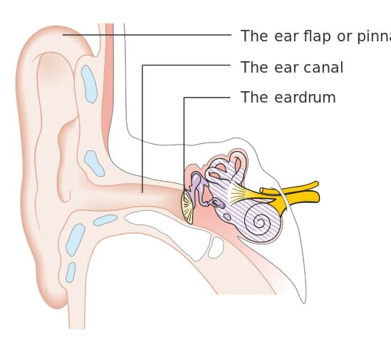
  • The outer ear, which includes the complex shell that is the visible ear we see on the outside of our heads. This outside structure, called the “pinna,” acts like a satellite dish or funnel, gathering and focusing sound so that we can hear better.

    The pinna is made mostly of cartilage. In some animals, this outer “shell” or “dish” can actually move, rotating enable it to collect sound from different directions. Some breeds of dogs and cats maintain this ability to move their ears to better focus on a sound without moving their whole head.

    Humans have largely lost this ability, our ears being firmly fixed to our heads and without much range of motion. But a few of us can still use vestigial muscles we inherited from our animal ancestors to wiggle our ears!

  • The middle ear consists of a series of bony tubes, which contain other bones that are designed to amplify vibrations they receive through the eardrum. This “eardrum” also called the “tympanic membrane,” vibrates in response to the sounds that enter through the ear canal.

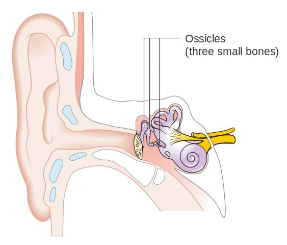
    Its vibrations are then transmitted through three tiny bones known as the “ossicles.” These are the malleus (also known as the “hammer”), the incus (also known as the “anvil”), and the stapes (also the “stirrup”).

    Unlike most bones which are used for structure and protection, the function of these three delicate bones is to vibrate as much as possible in response to sounds that enter the ear. They concentrate the vibrations from the ear canal and transmit them to the inner ear, where these vibrations ultimately reach the cells that send impulses to the auditory nerve.

  • The inner ear contains a series of fluid-filled chambers, which use hair cells to convert fine vibrations into neural impulses for purposes of both hearing and balance. The inner ear receives vibrations that have been amplified and transmitted from the ear canal and through the malleus, incus, and stapes.

    Located deep within the head, the hair cells of the inner ear are just what the name suggests: fine cells, shaped like hairs, which are extremely sensitive to vibration. When these hair cells are bent by vibrations, special proteins in the cell membrane cause the hair cells to create electrochemical impulses, very like nerve impulses, which are then carried to the auditory nerve in the brain.

    By determining which hair cells are bending in response to vibration, the brain can calculate with a high degree of detail and accuracy the “pitch” or frequency of the sound vibration; the volume; and the location of the sound.

Today, modern medicine allows many people with malformed or damaged cochleas to hear better using devices such as cochlear implants, which artificially produce electrochemical impulses that our auditory nerves can understand.


We will talk in more detail about these parts of the ear in the “Parts of the Ear” section below.


Balance


Within the parts of the ear known as the semicircular canals, hair cells just like those used for hearing have been adapted for a different purpose. This is called the “vestibular system,” and it assists with vision and balance.


In the semicircular canals, these hair cells respond to the movement of otoliths – tiny calcium carbonate crystals which can shift in response to gravity and motion, causing them to press on hair cells and release nerve impulses.


By using these nerve impulses to track the position of the otoliths, the brain can tell which way is up and down relative to the position of the body. It can also tell which way the head is moving relative to the outside world.


Most of us take this remarkable ability of the inner ear for granted, but anyone who has had an inner ear infection – in which viruses or bacteria can temporarily disrupt the balance signals going to our brain – knows how crucial these signals are.


When the activity of the inner ear is disrupted, our eye muscles are unable to instinctively adjust to our the movements of our heads. This results in the illusion that the world is unstable, and that it spins when we move! This happens because, without the input from our vestibular system, our eye muscles do not “know” that they have to follow objects in the environment when our heads move.


People with inner ear problems also have problems coordinating their muscle movements to keep their weight balanced. Many have problems walking without falling or running into walls, and may experience motion sickness-like symptoms such as nausea and vomiting.


Fortunately, most inner ear infections are only temporary. They may last a few days or a few weeks – just enough to help us appreciate the actions of these remarkable organs!


Parts of the Ear


The Pinna


The pinna is the outer, visible part of the human ear. Its curves and folds are specially designed to gather sound from the environment and funnel it into our ears. People with pinnas that have been damaged can still hear, but typically do not hear as well as people with intact pinnas.


The pinna, and the other parts of the outer ear, are labeled below:


Outer ear parts

Outer ear parts


The stiff, rigid parts of the pinna are made of cartilage, just like our noses. The soft, malleable “earlobe” is made of fatty tissue. Some people can still wiggle the external parts of their ears using muscles which our ancestors may have used to rotate our ears to better gather sound from different directions.


The opening in the center of the pinna is the opening to the ear canal, which will be discussed next.


The Ear Canal


The ear canal is the opening through which sound waves enter the middle ear. It serves to further focus and concentrate the vibrations collected by the pinna, ensuring that the vibrations will be clear and strong enough to be amplified and turned into nerve impulses.


The ear canal is only 2-3 centimeters deep – a little bit less than one inch. About an inch inside ear canal, the tympanic membrane, or the “eardrum” is found.


This is why it’s important not to stick anything into your ears; damage to the delicate tympanic membrane can result in impaired hearing!


The Tympanic Membrane


The tympanic membrane, or “ear drum” is a thin, tightly-stretched membrane that separates the outer from the middle ear. Just like the membrane of an actual drum, the tympanic membrane vibrates in response to the sounds that are funneled to it by the pinna and ear canal.


The outside of the tympanic membrane faces the ear canal. Its inner surface faces the malleus, incus, and stapes, which act to further focus and amplify the vibrations that the tympanic membrane receives.


The Ossicles


The malleus, incus, and stapes are three tiny, remarkable bones. As a group they are sometimes called “ossicles,” from the root word “osseo” for “bone.” The ossicles are are labeled in the diagram below:
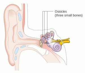

Middle ear parts

Middle ear parts


They are precisely shaped to vibrate in response to the movements of the tympanic membrane – and to transmit and focus those vibrations so that they become even clearer.


These bones contact the eardrum, or tympanic membrane, on the outside of the middle ear. They then transmit its vibrations through their specially-shaped bone structures and ultimately into the oval window.


When you read about the oval window below, you’ll see why the actions of these bones is so important to the process of hearing!


The Oval Window


The oval window is a small membrane which lies at the border between the middle and inner ears. Just as the tympanic membrane receives vibrations from the ear canal, the oval window receives vibrations from the malleus, incus, and stapes.


However, there’s a very important difference between the oval window and the tympanic membrane. The oval window is much smaller than the tympanic membrane – and the purpose of the malleus, incus, and stapes is to focus sound vibrations so that this much smaller surface area receives the full force of the vibrations from the tympanic membrane.


It’s a similar principle to focusing light from a large lens to fall on a tiny area: the resulting light is much more intense, and you may be able to see much more detail as a result. The vibrations that the malleus, incus, and stapes transmit to the oval window can be twenty times stronger than the vibrations they received from the eardrum!


The oval window’s vibrations are transmitted directly into the cochlea, where sound vibrations are turned into nerve impulses for the brain to interpret.
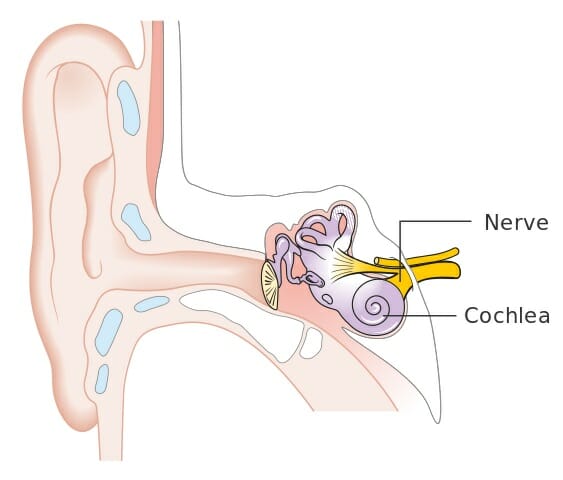

The Cochlea


The cochlea is filled with fluid, and “hair cells” that are extremely sensitive to vibration. The cochlea, and the auditory nerve which carries signals from the cochlea to the brain, are pictured here:
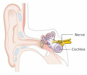

Inner ear parts

Inner ear parts


When hair cells are bent due to vibration of the fluid in the cochlea, the bending of the cells causes proteins called mechanically-gated ion channels to open. These ion channels allow positively charged particles such as potassium and calcium to enter the cell. This movement of charged particles across the cell membrane is quite similar to the firing of neural signals, or “action potentials,” by neurons cells.


Indeed, the movement of ions across the hair cell membranes cause electrochemical signals, which are ultimately sent to the auditory nerve. The auditory nerve then carries these signals to the brain, which analyzes information about which hair cells are being vibrated and turns this information into the experience of sound.


Just like cone cells in the human eye respond to different wavelengths of light, allowing us to see different colors, hair cells in the human ear can respond to different frequencies of sound. This allows us to distinguish the pitch of a sound.


The Semicircular Canals


The semicircular canals are similar to the cochlea in that they are bony canals which are filled with fluid and lined with hair cells. However, the hair cells in the semicircular canals are used for a different purpose from those in the cochlea. Instead of being turned into the sensation of sound, the signals from these hair cells are turned into information about movement and balance.


The hair cells of the vestibular system, or balance system. do not receive vibrations from the ear canal. Instead, they are bent by the movements of otoliths – tiny calcium carbonate crystals found within the semicircular canals.


Just like stones settle to the bottom of a river or lake, otoliths settle to the bottom of the semicircular canal. Of course, unlike a river or lake, our heads move quite a lot, which causes a jostling of our “stones.” The direction of settling of the otoliths, then, tells us which way is up, and which way our head is moving.


To maximize their ability to tell us about balance and movement, the semicircular canals are oriented in three different directions. Just like different hair cells are sensitive to different pitches of sound, these three different canals have maximum sensitivity to different kinds of movements and position changes


Most people are not aware of gaining this information from their semicircular canals. Our senses of movement and balance are simply always “there.”


Our brains use the signals from these hair cells to automatically adjust our movements. These movements include the movements of our eyes, which allow us to maintain a stable image of the world, even when our heads move; and the movements of our arms and legs, which are fine-tuned to keep us standing upright on two legs.


However, when the signals from the semicircular canals are disrupted, people notice very fast. Inner ear infections which temporarily disrupt these nerve signals render our eyes and bodies unable to automatically adjust for the movements of ourselves and our environments.


As a result, people with inner ear infections can experience dizziness; the illusion that the room is “spinning” when they move their head; and a “shaky camera” effect where their vision wobbles with every small movement of their heads. These people can also experience symptoms of “motion sickness” such as nausea and vomiting.


Fortunately, our semicircular canals work most of the time! Most inner ear infections last only a few days or weeks – just long enough to remind us of how amazing our bodies really are.


Quiz


1. What might explain the shape of the “pinna,” or the visible, outer part of the human ear?
A. The cup-like shape is left over from ancestors who could move their ears to focus sound.
B. The pinna acts like a funnel or satellite dish, gathering sound and directing it into the ear canal.
C. The cup-like shape is a freak mutation that occurs during embryogenesis.
D. None of the above.

Answer to Question #1

2. What is the function of the bones of the middle ear?
A. They have no function; they are vestigial.
B. They serve to support and protect the middle ear.
C. They amplify vibrations from the ear canal and transmit them to the inner ear.
D. None of the above.

Answer to Question #2

3. Which of the following could NOT occur as a result of damage to the inner ear?
A. The person may become unable to hear.
B. The person may become unable to keep their balance.
C. The person may get dizzy and feel as though the room is spinning when they turn their head.
D. None of the above.

Answer to Question #3

References


  • Standring, S. (2016). Gray’s anatomy: the anatomical basis of clinical practice. Philadelphia: Elsevier Limited.

  • Neuroanatomy 2nd Ed Neuroscience 4th Ed. (2009). Sinauer Associates Inc.

  • Understand the Types of Hearing Loss & Treatment Options. (n.d.). Retrieved August 21, 2017, from http://www.cochlear.com/wps/wcm/connect/us/home/about-us-and-hearing-loss/hearing-loss-explained


Ear

Thursday, September 7, 2017

Methanol

Methanol Definition


Methanol, sometimes called “wood alcohol,” is a clear liquid with the chemical formula CH3OH. It is clear liquid with polar properties, making it a good solvent. It is also highly flammable, and highly toxic to humans if ingested.


Historically, methanol was created when cellulose, the main sugar in wood and some other plants, was fermented by bacteria. This fermentation process led to a substance that was deadly to drink, but useful as a solvent for scientific and industrial purposes.


After scientists discovered methanol and its uses, humans began to produce methanol for industrial purposes using a much faster process of combining carbon monoxide, carbon dioxide, and hydrogen gases along with a copper-based catalyst that prompts these raw materials to combine to form methanol.


Methanol is used industrially as an ingredient in antifreeze, various chemical solvents, certain fuels, the creation of many plastics, and in blends of alcohol intended for medical or industrial use instead of consumption. “Denatured” alcohol used in medicine and industry often includes both ethanol (the same grain alcohol found in beer and wine) and methanol, which makes it toxic to consume.


Because it can be produced from the fermentation of plant matter, methanol has been the causes of many fatal cases of poisoning from drinking illegally produced alcohol.


During prohibition in the United States, amateur attempts at distilling liquor sometimes led to blindness, neuropathy, and death as a result of drinking methanol. In other countries today, major poisoning incidents still happen when unscrupulous sellers try to profit by selling home-brewed alcohol at lower prices than those offered by major retailers.


Like formaldehyde, methanol is a simple enough organic substance that it can be created by inorganic chemical reactions. For that reason, methanol has been discovered by telescopes in some regions of deep space where no life exists.


What is Methanol Used For?


Methanol has many industrial and scientific uses.


One of the most common uses of methanol is as an ingredient for formaldehyde. This chemical which can be derived from methanol is used extensively in the production of plastics, including those used in construction materials, car parts, paints, explosives, and wrinkle-resistant artificial fabrics. Formaldehyde is also used by morticians and scientists to preserve corpses and laboratory specimens.


Methanol can be used to make other useful solvents including acetic acid, dimethyl ether, and propylene, which is used in anti-freeze. Methanol itself can also be an ingredient in anti-freeze.


Fuel for both gasoline-powered and biodiesel vehicles can include methanol. Its highly flammable nature and usefulness as a solvent allows it to assist other fuels in blending and igniting.


Pure methanol has even been used by itself as a fuel for race cars. It produces high speeds – but also led to a devastating fire which killed two American race car drivers.


Methanol fires are especially dangerous because they are extremely easy to spark, and the flames are almost invisible. This allows the fires to spread out of control and catch other materials very quickly.


Today, both the United States and Europe have safety regulations on how much methanol car fuel is allowed to contain.


Methanol Structure


Methanol consists of an “OH” alcohol group attached to a single carbon atom. The carbon atom’s remaining bonding spots are occupied by three hydrogen atoms. This structure is illustrated below:


Methanol flat structure

Methanol flat structure


You may recognize that “methanol” shares a root word with the gas “methane.” The “meth” in both substances refers to this single carbon which is saturated with hydrogen atoms. In “methanol,” this carbon is attached to an alcohol group; in “methane,” the carbon with four hydrogens stands by itself.


Methanol is closely related to ethanol, or “grain alcohol.” Ethanol is the alcohol found in beer, wine, and liquor.


Where “meth” refers to a single carbon saturated by hydrogens, the prefix “eth” refers to a chain of two carbons saturated by hydrogens. Ethanol, then, has a chain of two carbons where methanol has one.


This extra carbon makes a very big difference to how our bodies metabolize the alcohols. While ethanol is safe to drink in moderation, methanol is broken down by our livers into formaldehyde – a highly toxic product that can cause blindness, nerve damage, and death.


Because methanol and ethanol are produced through similar chemical and microbial processes, great care must be taken when fermenting and distilling not to contaminate drinking alcohol with methanol.


Methanol Formula


The formula for methanol is CH3OH.


It is most commonly created by reacting precursor gases such as CO and CO2 with H2 hydrogen gas.


In the presence of a copper-based catalyst under the right conditions, the hydrogen atoms will bond to the carbon and oxygen atoms, displaying the double bonds between the C and O and resulting in a molecule that’s fully saturated with hydrogen.


This reaction can be seen here:


Reaction of production of formic with methanol oxidation


In the case of reacting CO2 with hydrogen, water is also created as a byproduct of the extra oxygen being saturated by hydrogen atoms.


Methanol Safety


One major risk of working with methanol is fire. Liquid methanol burns easily and can be set aflame by any stray spark or excessive heat. Methanol is also very dangerous to ingest.


Like formaldehyde, methanol is produced in tiny quantities by the activity of our own cells. However, also like formaldehyde, methanol is highly toxic to our cells and must be constantly removed and excreted through the liver and kidneys.


When ingested, methanol is metabolized by the liver into formaldehyde, and then into formic salts. These are highly toxic to the nervous system and can permanently destroy the optic nerve, causing blindness. The neurotoxic effects can also cause coma and death.


When methanol poisoning is treated properly, permanent damage can often be prevented. However, when left untreated, death from the neurotoxic effects of methanol can occur within hours of ingestion.


Quiz


1. What does the “meth” in “methanol” stand for?
A. “Meth” indicates that the chemical has a single carbon atom, just like the “meth” in “methane.”
B. “Meth” indicates that the substance is addictive, like the “meth” in “methamphetamine.”
C. “Meth” indicates that the substance is an alcohol.
D. None of the above.

Answer to Question #1

2. Which of the following is NOT a hazard of methanol?
A. It is highly toxic if consumed.
B. It is highly flammable.
C. It produces a thick, foggy vapor which can obscure your vision.
D. None of the above.

Answer to Question #2

3. What would you expect to happen if you reacted carbon dioxide with hydrogen gas and a copper catalyst?
A. Methanol would be created, with no other byproducts.
B. Methanol would be created, with water as a byproduct.
C. Methanol would be created, with formaldehyde as a byproduct.
D. None of the above.

Answer to Question #3

References


  • Methyl Alcohol (Methanol). (2012, March 15). Retrieved August 03, 2017, from https://www.cdc.gov/niosh/topics/methyl-alcohol/

  • References. Retrieved August 03, 2017, from http://www.marinemethanol.com/about-methanol/methanol-production

  • Lazonby, J. (n.d.). Methanol. Retrieved August 03, 2017, from http://www.essentialchemicalindustry.org/chemicals/methanol.html


Methanol

Tuesday, August 15, 2017

Abdomen

Abdomen Definition


The abdomen refers to the region between the pelvis (pelvic brim) and the thorax (thoracic diaphragm) in vertebrates, including humans. The space constituting the abdomen is termed the abdominal cavity. The borders of the abdominal cavity are comprised of the posterior peritoneal surface, the anterior abdominal wall, the inferior pelvic inlet, and the superior thoracic diaphragm. The abdomen functions to house the digestive system and provides muscles essential for posture, balance, and breathing.


Abdomen Anatomy


The abdomen is comprised primarily of the digestive tract and other accessory organs which assist in digestion, the urinary system, spleen, and the abdominal muscles (shown below). The majority of these organs are encased in a protective membrane termed the peritoneum. While the digestive organs and assessor organs are located within the peritoneum, the kidneys, ureters and urinary bladder are located outsider of the peritoneum, and thus, are considered by some scientists to be pelvic organs.


Abdominal Organs Anatomy


Digestive Tract


The organs of the digestive tract consist of the small and large intestines, the stomach, cecum, and the appendix. The stomach is located between the esophagus and the small intestine in the upper left region of the abdomen. The stomach is responsible for the secretion of digestive enzymes and gastric acid required to digest food products. The small intestine is situated between the stomach and large intestine and consists of the three segments (duodenum, jejunum, and ileum), each exhibiting distinct functional properties. The duodenum is situated around the top of the pancreas and receives the digested stomach contents known as gastric chyme. The duodenum functions to neutralize the acid contained in the gastric chyme, as well as break down proteins and fat via enzymes and bile. The jejunum is the middle segment of the small intestine and is responsible for the absorption of sugar, amino acids, and fatty acids into the bloodstream. The final segment of the small intestine is the ileum, which connects to the large intestine. The ileum is responsible for the absorption of vitamin B12, as well as any remaining nutrients. The large intestine consists of the cecum, colon, rectum, and anus and stretches the entire width of the abdominal cavity. The primary function of the large intestine is to absorb water and store the remaining food material as feces until it can be excreted from the body via defecation.


Accessory Digestive Organs


The organs which assist in digestion consist of the pancreas, liver, and gallbladder. These organs secrete various hormones (i.e., insulin), enzymes, and bile via specialized ducts to aid in digestion. In particular, the pancreas functions as an endocrine organ which secretes a variety of digestive enzymes as well as hormones which aid in the digestion of food passing through the digestive tract. The pancreas is located behind the stomach. The liver is located in the upper right quadrant of the abdomen and functions to produce bile, which is responsible for breaking down fats. The liver also functions to produce hormones, regulate the storage of glycogen, and detoxification of the blood. The gallbladder is responsible for the storage of bile produced by the liver until it is released into the small intestine. The gallbladder is situated in the abdomen just under the right lobe of the liver.


Spleen


The spleen functions as a secondary lymphoid organ and is responsible for the removal of red blood cells via active filtration. The spleen also acts as a reservoir of red blood cells and metabolizes hemoglobin obtained from old red blood cells. The spleen is located in the upper left quadrant of the abdomen.


Urinary System


The urinary system consists of the kidneys, ureters, and urinary bladder, which are responsible for the filtration and excretion of waste in the form of urine from the body. Since these organs are located outside of the peritoneum, they can also be considered pelvic organs by some researchers. In particular, the kidneys function to filter the blood of waste products, regulate blood pressure, and control the blood pH. The ureters are connected to the kidneys and are used to drain urine into the urinary bladder. The urinary bladder serves as to store the accumulated urine until it can be excreted via urination.


Abdomen Function


The primary functions of the abdomen consist of digestion, breathing, posture and balance, as well as movement. The major organs located in the abdomen are associated with digestion, for which the functions are described above. The abdomen is also required for breathing via the accessory muscles of respiration. Such muscles are also involved in postural support, movement, balance, coughing, urination, vomiting, singing, childbirth, and defecation.


Respiration


Although the diaphragm controls respiration under steady-state conditions, the accessory muscles of respiration assist in respiration when greater effort is required. These muscles include the scalene and sternocleidomastoid muscles which serve to raise the ribcage. When these muscles are engaged, it is typically a sign of respiratory distress, such as that observed during an asthma attack.


Movement and Posture


Abdominal muscles are also required for the maintenance of posture and balance, as well as movement. The transverse abdominis muscle and internal obliques affect posture by providing spinal support during rotation and lateral flexion, and stabilize the spine when standing. Both of these muscles are situated deep within the abdomen. The external obliques also function to support the lateral flexion and stabilize the spine when standing. Finally, the rectus abdominis functions to bend the spine forward.


Abdominal Muscles


The abdominal muscles consist of three distinct layers residing within the abdominal wall and extend to the pubis, iliac crest, lower ribs, and vertebral column. The muscle fibers merge at the midline, surround the rectus abdominus, and join on the other side at a point known as the linea alba. The abdominal muscle fibers criss-cross each other for added strength, with the transverse abdominal muscle extending horizontally forward, and the internal and external obliques running upward and downward, respectively towards the front (shown below).


Muscles of the Abdomen


Rectus Abdominis


The muscles comprising the rectus abdominis are long and flat, with three tendinous intersections crossing over the muscle. As described above, the three muscles forming the lateral abdominal wall enclose the rectus abdominis in a sheath. The rectus abdominal muscles begin at the pubis bone, line the sides of the linea alba and attach to the lower ribs. The inguinal canal passes through the lower layers of the rectus abdominis muscles in the groin accommodate the attachment of the uterus in females and the dissention of the testes from the abdominal wall in males.


Transverse Abdominal Muscle


The transverse abdominal muscle is a flat, triangular muscle composed of horizontal fibers that is situated between the internal oblique and transverse fascia. The transverse abdominal muscle attaches at the inner lip of the ilium, the lumbar fascia, and the inner surface of cartilage on the six lower ribs. The transverse abdominal muscle passes behind the rectus abdominis to meet the linea alba.


Pyramidalis Muscle


The pyramidalis muscle is a small, triangular-shaped muscle situated in front of the rectus abdominis in the lower portion of the abdomen. The pyramidalis muscle stretches from the pubic bone to the linea alba, joining before the umbilicus. The pyramidalis muscle functions to contract the linea alba (shown below).


Anatomy of the Human Body


Quiz


1. Which of the following statements is NOT true regarding the pancreas?
A. The pancreas is situated behind the stomach.
B. The pancreas secretes insulin.
C. The pancreas is an endocrine gland.
D. The pancreas is a secondary lymphoid organ.

Answer to Question #1

2. A primary function of the spleen is:
A. Regulation of blood pressure.
B. The production of digestive enzymes.
C. Secondary lymphoid organ.
D. All of the above are primary functions of the spleen.

Answer to Question #2

3. The major muscles providing spinal support required for posture in humans are:
A. Rectus abdominus
B. Transverse abdominus
C. Linea alba
D. Pyramidalis

Answer to Question #3

4. Which of the following abdominal organs is NOT required for digestion?
A. Liver
B. Gallbladder
C. Spleen
D. Pancreas

Answer to Question #4

References


  • Bilal M, Voin V, Topale N, Iwanaga J, Loukas M, and Tubbs RS. (2017). The Clinical anatomy of the physical examination of the abdomen: A comprehensive review. Clin Anat. 30(3):352-356.

  • Ghamkhar L and Kahlaee AH. (2015). Trunk muscles activation pattern during walking in subjects with and without chronic low back pain: a systematic review. PM R. 7(5):519-26.

  • Stensby JD, Baker JC, and Fox MG. (2016). Athletic injuries of the lateral abdominal wall: review of anatomy and MR imaging appearance. Skeletal Radiol. 45(2):155-62.


Abdomen