Showing posts with label 6k. Show all posts

Tuesday, January 23, 2018

Anatomy

Anatomy Definition


Anatomy is the branch of biology which studies how various parts of an organism are connected, and how they are related to other body parts both spatially and functionally. Anatomy has many sub-disciplines, and is used in many different fields. In general, there are two main types of anatomy: gross or macroscopic anatomy, and microscopic anatomy. However, most biology specialties require knowledge of both types of anatomy.


Types of Anatomy


Macroscopic Anatomy


Commonly called gross anatomy, macroscopic anatomy involves studying the structures and forms which can be seen on organism with the naked eye. The type of organism does not matter. A botanist may study the macroscopic anatomy of a plant, such as the shape and size of its leaves. A doctor might study the proportions of his patients, measuring their weight and height. Both of these scientists are using skills of gross anatomy.


Many branches of biology use gross anatomy to evaluate their subjects. While this is often combined with microscopic anatomy and physiology, sometimes the macroscopic anatomy is the only observable system. This definitely true of archeology and evolutionary biology. Both of these branches of biology use evidence from the fossil record to establish relationships between extinct animals. Soft tissue does not often fossilize, thus these scientists must have a comprehensive knowledge of skeletal anatomy. Different species and fossils can be compared using comparative anatomy, which recognizes similarities between specimens.


For instance, a scientist using comparative anatomy could hypothesize the evolutionary relationships between a bat, a blackbird, and an ostrich. At first glance, the blackbird and the bat may be more related based upon size. But the scientist would quickly notice that the bat is covered in hair, while the blackbird has feathers. Upon examination of the wings and their bones, the scientist would find that the bat wing resembles an outstretched hand, while the blackbird bones have fused into a large bone that extends the length of the wing, with the feathers and skin supporting the rest of the wing. Even though the ostrich cannot use its wings to fly, the structure of the bones are the same. They might be different sizes, but it is clear that the blackbird and ostrich are more closely related to each other than either is related to the bat. This simple exercise in gross anatomy provides the basis of the classification of many organisms.


Microscopic Anatomy


While gross anatomy provided the basis for many modern sciences, modern technology has revolutionized the study of microscopic anatomy. Starting with the invention of light microscopy and carrying through modern day inventions such as the electron microscope, the inner workings of cells and organisms are becoming increasingly understood. Entire new worlds of organisms, such as bacteria and single-celled eukaryotes, have been opened up for study. Cellular biology is an entire field dedicated to the study of cells, their organelles, and how they function. Microscopic anatomy is central to this study.


Microscopic anatomy covers everything from tissues, which are groups of similar cells, down to the inner workings of the molecules which direct the cell’s activities. A histologist studying muscle tissue, for example, would examine how the cells are held together in the tissue. Looking further into the cells using an electron microscope, he would see the complex arrangement of proteins in the cell which allow it to contract. He may also notice the nucleus, which contains the DNA coding for all of the proteins and products the cell produces.


Microscopic anatomy is often paired with biochemistry, molecular biology, and other disciplines to fully understand the organism or tissues being studied. Science knew for decades that cells contained many organelles. However, it was not until recent advances in DNA processing and protein analysis that the function of the many different organelles was understood. Using microscopic anatomy, scientist can also study the cells during the development of an organism. This is called embryology, and has developed into a wide field covering everything from human development to evolutionary relationships of organisms based on their developmental processes.


History of Anatomy


Anatomy is a science older than science itself. The first anatomists where the first humans, categorizing and recognizing the other organisms in their environment using skills of gross anatomy. Vision is fundamental to humans, and is the basis of our understanding of the world. As we advanced in thought and organization, early thinkers began to try to classify organisms. Without any other information, anatomy was often the only evidence available to bind organisms into groups. Aristotle was among the first to attempt serious organization of living things and used many attributes of their anatomy to group them together. His two main groups were plants and animals, two groups we can still easily distinguish today based on their gross anatomies.


Early medicine advanced quickly once the moratorium on dissection was lifted. Often frowned upon in early society, early anatomists like Leonardo Da Vinci often received scrutiny from the public or the church for their scientific inquiry. However, an understanding of the human body arose from these early pioneers, upon which is built the medical knowledge of today. Many of the first works of human and animal anatomy were published during the Renaissance. Many authors showed an advanced, if slightly lacking or skewed view of anatomy as we know it today. But, without any way to understand the workings of the body further, gross anatomy was stranded by itself.


Fast forward several hundred years and the “Father of Taxonomy” Carl Linnaeus was still mainly focused on gross anatomy as a starting point for classification. Darwin’s idea of evolution and common ancestors became accepted at the end of the 1800’s. Still, there were not many methods to evaluate the relationships between animals further. With the advent of better imaging technology, the 1900’s brought the emergence of microscopic anatomy, and really started to change biology. Once it was understood that DNA was the principle mode through which organisms inherited traits, revolutions in many disciplines occurred. Medicine saw a rapid increase in understanding, thanks to the discovery that bacteria and other microbes can cause disease. The inner workings of the cell were being pieced together, and the functions of the many different organelles understood. Many aspects of evolutionary biology were rediscovered or overturned as microscopic anatomy and DNA revealed different relationships than were once assumed. This revolution continues today, as new developments in microscopic anatomy and physiology continually reshape our understanding of organisms.


Careers


Many careers in the biological sciences require some knowledge of both gross and microscopic anatomy. Some professionals, such as a doctor, require specific anatomical knowledge of one species: humans. Human anatomy is the study of both the macroscopic and microscopic portions of the human body. Human anatomy is essential for professionals in the medical field as they must be able to discern between the many types of tissues in the body, and understand their relationship to each other. Ergonomics is the study of the physical stresses on the human body, and relies on a detailed understanding of its various components.


Other scientists focus on the anatomy of other species, or groups of species. A mammologist understands mammal anatomy, where a herpetologist understands the anatomy of reptiles and amphibians. An evolutionary biologist must understand the complex anatomies of many groups, and uses the information to understand their hereditary relationships. Archeologists study mainly gross anatomy of fossilized organisms, whereas cellular biologists and bacteriologists must rely on microscopic anatomy as their organisms are unicellular.


Degrees in anatomy can be obtained at the bachelors, graduate, and doctoral levels, with a wide variety of concentrations. Many schools offer concentrations and courses in human anatomy as a prerequisite for medical school. Other schools and programs focus on general anatomy, necessary for veterinary science, zoology, advanced biology degrees and other specialties that may rely heavily on anatomy. As a professor of anatomy, one would study and teach about the various aspects of anatomy. Many colleges have researchers who incorporate different aspects of anatomy into their research.


If you are good at anatomy or are interested in career paths with anatomy involved, try to find which branch of anatomy you enjoy the most. If gross anatomy suits you, then you may want to pursue a job as a surgeon or evolutionary biologist. If microscopic anatomy is more up your alley, you could become a microbiologist or study internal medicine. Anatomy is extremely important in many fields, especially when it is coupled with other disciplines of science, such as chemistry and physics. It can yield great insights into the world in branches of biology as far ranging as human medicine and evolution.


Reference


  • De luliis, G., & Pulera, D. (2007). The Dissection of Vertebrates. Amsterdam: Academic Press.

  • McMahon, M. J., Kofranek, A. M., & Rubatzky, V. E. (2011). Plant Science: Growth, Development, and Utilization of Cultivated Plants (5th ed.). Boston: Prentince Hall.


Anatomy

Friday, September 8, 2017

Stomach

Stomach Definition


The stomach is a muscular organ that is found in our upper abdomen. If we were to locate it on our bodies, it can be found on our left side just below the ribs. In simple terms, the stomach is a kind of digestive sac. It is a continuation of the esophagus and receives our churned food from it. Therefore, the stomach serves as a kind of connection between the esophagus and the small intestine, and is a definite pit stop along our alimentary canal. Muscular sphincters, which are similar to valves, allow some separation between these organs.


The stomach’s functions benefit from several morphological attributes. The stomach is able to secrete enzymes and acid from its cells, which enables it to perform its digestive functions. With its muscular lining, the stomach is able to engage in peristalsis (in other words, to form the ripples that propel the digested food forward) and in the general “churning” of food. Likewise, the abundant muscular tissue of the stomach has ridges in its linings called rugae. These increase the surface area of the stomach and facilitate its functions, which we will describe in more detail below.


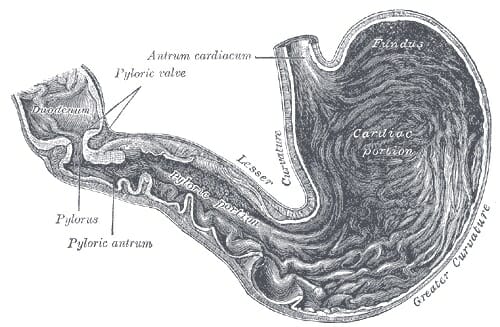
Stomach diagram

The image illustrates the esophagus, stomach, and intestinal regions of the human body


Functions of the Stomach


As mentioned before, the stomach is first and foremost a principal site of digestion. In fact, it is the first site of actual protein digestion. While sugars can begin to be lightly digested by salivary enzymes in the mouth, protein degradation will not occur until the food bolus reaches the stomach. This breakdown is carried out by the stomach’s pepsin enzyme. The stomach’s roles can essentially be distilled down to three functions.


Much like an elastic bag, the stomach will provide a place for varied amounts of swallowed food to rest and digest in. Hence, the stomach is a storage site. The stomach will also introduce our swallowed food to essential acids. The cells in the stomach’s lining will excrete a strong acidic mixture of hydrochloric acid, sodium chloride, and potassium chloride. This gastric acid, or colloquially known as gastric “juice,” will work to break down the bonds within the food particles at the molecular level. Pepsin enzyme will have the unique role of breaking the strong peptide bonds that hold the proteins in our food together, further preparing the food for the nutrient absorption that takes place in the small (mainly) and large intestines. This brings us to the third task the stomach has, which is to send off the churned watery mixture to the small intestine for further digestion and absorption. It takes about three hours for this to occur once the food is a liquid mix.


The stomach’s main roles:


  1. Food storage

  2. Acidic breakdown of swallowed food

  3. Sends mixture on to the next phase in the small intestine

Structure of the Stomach


Stomach

The archaic illustration depicts the different regions of the stomach


Although we have briefly discussed the location and physical traits of the stomach, it is important to detail the structure of the stomach, as well. The stomach begins at the lower esophageal sphincter that discerns the cut-off point of the esophagus. The stomach itself is very muscular. When the muscularis externa layers are dissected, one can visualize three distinct layers coined the longitudinal, circular, and oblique layers. The first region of the stomach is called the cardia. It is the layer closest to the esophagus and it contains cardiac glands that secrete mucus. Mucus protects the delicate epithelial lining of many tissues in the human body. This region is followed by the fundus, which is the superior arch of the stomach. Importantly, the fundus has the special function of containing gastric glands that release a cocktail of gastric juices. This region is followed by the body of the stomach, which is coated with rugae and is the largest region. Rugae, in turn, help facilitate digestion by increasing the site’s surface area. Finally, this section is followed by the pylorus region, which is closest to the exit into the duodenum of the small intestine and is pinched off by the pyloric sphincter.


Four regions of the stomach:


  • Cardiac

  • Fundus

  • Body

  • Pylorus


Common Stomach Issues


Almost every person has experienced a stomach related issue at one point in their lives. Perhaps the most common ones are indigestion and heartburn. These issues can be resolved quite easily with over-the-counter tablets (i.e. tums), but there is no denying that they are unpleasant experiences. However, there are more chronic illnesses that afflict many people. One of these is the gastroesophageal reflux disease, commonly known as GERD or Acid Reflux. GERD afflicts up to 3 million Americans each year, and is a physiological result of when the lower esophageal sphincter will not close properly. The principal function of this sphincter is to prevent food and stomach acids from regurgitating up the esophageal canal. While a healthy stomach has tons of mucus and barriers strong enough to prevent stomach acids from wreaking havoc on the epithelium, the esophagus is not quite so lucky. This, of course, has the long-term implications of damaging those delicate epithelial cells. When a patient does not have the sufficient barriers to prevent damage within the stomach, a medical issue that arises are peptic ulcers. Ulceration refers to the sores that pierce through an organ. When the stomach is not sufficiently protected from contact with these highly acidic acids, we do run into the issue of perforating the tissue and potentially having the stomach juices leak – which by all means requires urgent medical attention. These sores are very painful and recurrent in patients with peptic ulcer disease.


There are other red flag symptoms that present in the urgent or emergency care setting that indicate a stomach issue. These include a burning sensation in the chest (heartburn), piercing or diffuse abdominal pain, blood in the stool, and vomiting or diarrhea.


Quiz


1. How many hours does it take for the stomach to release the food to the small intestine?
A. 1
B. 2
C. 3
D. 4

Answer to Question #1

2. Which of the following is the largest region of the stomach?
A. Cardia
B. Fundus
C. Body
D. Pylorus

Answer to Question #2

3. Which region of the stomach releases gastric juice?
A. Cardia
B. Fundus
C. Body
D. Pylorus

Answer to Question #3

References


  • Hoffman, Matthew MD (2017). “Picture of the Stomach: Human Anatomy.” Web MD. Retrieved on 2017-08-26 from http://www.webmd.com/digestive-disorders/picture-of-the-stomach#1

  • MedicineNet (2017). “Medical Definiton of Stomach.” MedicineNet. Retrieved on 2017-08-27 from http://www.medicinenet.com/script/main/art.asp?articlekey=5560

  • Medline Plus (2017). “Stomach Disorders.” Medline Plus. Retrieved on 2017-08-28 from https://medlineplus.gov/stomachdisorders.html


Stomach

Uterus

Uterus Definition


The uterus, otherwise known as the womb, is the female sex organ that carries a huge significance in many species’ survival – ours included. The uterus itself is a hollow organ that is shaped in the form of a pear, and interestingly enough measures about that size. It is neatly tucked into the pelvic area of most mammals and, of course, in humans. It is important to dissect the anatomy of the human uterus. In the female body, the upper end of the uterus, called the fundus, will join the fallopian tubes at either side while the lower end will open into the vagina. The wide portion at the top of the uterus is called the fundus, and will be the superior-most region that will host a fertilized embryo as it grows into a baby. A little below the fundus lies the muscular corpus region.


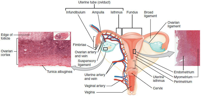
The corpus, in turn, is composed of three tissue layers. Post-pubescent women will have an innermost endometrium, which is the layer of muscle that is shed when the menstrual cycle commences in non-pregnant women. The endometrial tissue will thicken as the month’s cycle goes by in preparation for a fertilized egg to implant itself there. But in the absence of a fertilized egg, this layer will simply be shed away in what we know as menstruation. The middle muscle layer is called the myometrium, and is the layer that will expand during pregnancy and contract during childbirth. The outermost layer, the parametrium, will likewise expand and contract at these stages. Expanding will allow the uterus to house a growing baby, while the contractions will facilitate the newborn’s exit from the womb.


Uterus diagram

The image above depicts a diagram of the Uterus, with labeled tissue and arterial landmarks.


Function of the Uterus


Perhaps the principal, albeit lofty function of the uterus is to preserve life. It is the site of nourishment for the growing baby, making it one of the most important reproductive organs in the female body. This all begins when an egg, or ovum, is fertilized by a sperm and will make its downward trek in search of a better home. The tight fallopian tubes will not provide enough space to house the growing embryo! This is where the uterus meets all of these requirements, and more! The uterus’s thick, muscular nature will allow it to contract and expand to make room for the developing baby. The uterus is also rich in vasculature. There are many blood vessels supplying the muscle layers at any given time. This especially applies to the endometrium which is highly vascular and will come to nourish the embryo. In fact, many of the endometrial vessels that will come to supply the embryo will form just for this purpose. All of this explains why the fertilized ovum will choose to implant itself in the uterine lining – coined, the “site of implantation.” Thus, the uterus is the site that allows ours, and many species, to continue reproducing!


Location of the Uterus


The uterus measures about three inches long and two inches wide, and has a thick muscular lining within its walls. The lowest tip of the uterus will dip into the vagina in the area of the cervix, while the top most part will connect with the fallopian tubes through which the eggs travel. But a better way to pin its location is by describing its region as the area that lies between the belly button and the hip bones.


Abnormalities of the Uterus in Pregnancy


Nothing quite demonstrates the reproductive role of the uterus as the difficulties that arise from having an abnormal uterus. While the normal uterus will roughly measure three by two by one inches, some women will have uteruses that differ in shape and size. Many species’ evolution, including our own, has depended on having these precise dimensions to best support the growing embryo and to bring it to full term. But women with uterine abnormalities may realize they have this only once they have attempted and failed to conceive. These complications will surface either while trying to become pregnant, or after experiencing miscarriage. Examples of physical deformations of the uterus may include having a uterus with two inner cavities or vaginas (affecting roughly one in 350 women), having only one fallopian tube that will connect to the uterus, or having a heart shaped uterus instead of a pear-shaped one that is evolutionarily optimized to bear a child. Moreover, some afflicted women will have a septum that parts the uterus, or a slight indentation at the top of the uterus that will likewise compromise the ability for these women to have a baby. These abnormalities, however, are not all an absolute guarantee of infertility or miscarriage, but instead may lessen the probability of carrying a child to full-term.


Quiz


1. Which of the following is the outermost muscle layer of the corpus?
A. Endometrium
B. Parametrium
C. Myometrium
D. None of the above

Answer to Question #1

2. Which uterine layer nourishes the growing embryo?
A. Endometrium
B. Parametrium
C. Myometrium
D. None of the above

Answer to Question #2

3. Which of the following provides the best explanation for why the uterus is the main female reproductive organ?
A. It is the site of fertilization of the ovum
B. It is muscular and able to contract and expand
C. Its myometrial layer supplies the growing embryo
D. Both A and B

Answer to Question #3

References


  • MedicineNet (2017). “Uterus.” Medicine Net. Retrieved on 2017-08-26 from http://www.medicinenet.com/script/main/art.asp?articlekey=5918

  • BabyCentre Medical Advisory Board (2016). “Abnormalities of the uterus in pregnancy.” Retrieved on 2017-08-26 https://www.babycentre.co.uk/a551934/abnormalities-of-the-uterus-in-pregnancy

  • Danielsson, K. “Abnormal Uterus Shapes and iscarriage Risk.” Very Well. Retrieved on 2017-08-27 from https://www.verywell.com/abnormal-uterus-and-miscarriage-risk-2371694


Uterus

Tuesday, July 25, 2017

Foot

Foot Definition


The foot is a part of vertebrate anatomy which serves the purpose of supporting the animal’s weight and allowing for locomotion on land. In humans, the foot is one of the most complex structures in the body. It is made up of over 100 moving parts – bones, muscles, tendons, and ligaments designed to allow the foot to balance the body’s weight on just two legs and support such diverse actions as running, jumping, climbing, and walking.


Because they are so complicated, human feet can be especially prone to injury. Strains, sprains, tendonitis, torn ligaments, broken bones, fallen arches, bunions, corns, and plantar warts can all occur. Here we will talk more about the anatomy of the human foot and its many moving parts.


The complexity of the human foot may stem from the fact that it evolved from hand-like, grasping feet like those we see in apes today. Our ancient ancestors were tree-dwellers, and needed to be able to hang onto branches tightly with all four limbs. This caused them to evolve extraordinarily intricate hands and feet, which were capable of grasping, rotating, and gripping with dexterity that engineers are still trying to replicate in fields like robotics today.


Scientists are not sure why our ancestors eventually developed to walk upright, which caused the “fingers” of our feet to fuse and create a flat surface for walking on. It may have been because our ancestors began living on treeless grasslands, where standing tall to be able to see over the grass was more important than climbing. It could also have been because, as we began using tools, the ability to walk on two feet while using our hands to carry items became important.


Feet are present in other species too; especially mammals, birds, reptiles, and amphibians. Invertebrates such as mollusks and insects may have “feet” that they use to walk or move, but these are not complex bony structures like those found in vertebrates.


Here we will discuss the anatomy of the human foot, and some things that can go wrong to cause injuries or disorders.


These descriptions are meant for informational purposes only. You should always see a doctor if foot injury is suspected, as prompt and proper treatment can make for a faster, easier recovery! It is especially important to see a doctor if a suspected foot injury involves numbness, bleeding, or inability to move the foot, as these may be signs of serious complications.


Proper diagnosis and treatment takes a trained professional; improper diagnosis and treatment may lead to longer-lasting problems!


Foot Anatomy


The foot contains 26 bones, 33 joints, and over 100 tendons, muscles, and ligaments. This may sound like overkill for a flat structure that supports your weight, but you may not realize how much work your foot does!


The foot is responsible for balancing the body’s weight on two legs – a feat which modern roboticists are still trying to replicate. This requires strong, subtle muscles which can keep the foot standing firm even as we move our body’s weight around at different positions and angles.


The many bones work together to allow to allow this fine, delicate movement by subtly shifting inside the foot. They also allow us to perform intricate actions such as standing, climbing, and “grasping” at the ground with our feet on moving or uneven surfaces.


Here we will discuss the most important parts of the anatomy of the foot, and some injuries and disorders that can occur when these parts are damaged.


Of note, here we will make general statements about how different foot injuries and disorders may be treated by doctors. This is not a substitute for medical advice.


See a doctor about any suspected foot injury or disorder, as prompt diagnosis and treatment can make for a faster, easier recovery, while improper treatment may lead to long-term damage.


Foot Bones


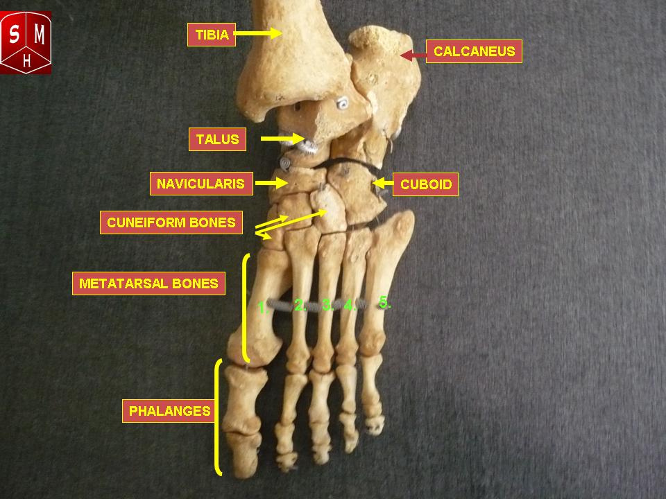
There are 26 bones in the foot. These include:


  • The phalanges, which are the bones in your toes

  • The metatarsals, which run through the flat part of your foot

  • The cuneiform bones, the navicularis, and the cuboid, all of which function to give your foot a solid yet somewhat flexible foundation

  • The calcaneus, which is the bone in your heel

  • The talus, which is the bone in your ankle

  • The talus connects to the tibia, which is the main bone in your lower leg
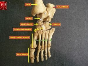

Foot bones


While you may not notice these bones in action every day, you’ll notice quickly if something is wrong with one of them. These bones allow your feet to execute the delicate shifts which enable you to keep your balance while walking, running, jumping, climbing, dancing, and playing sports!


Injury to a bone in the foot often results in a sharp or throbbing pain, especially when you move in a way that causes your weight or a nearby muscle to put pressure on the bone.


The most common broken bones in the foot are broken toes, which may occur after hitting a toe on a hard or sharp surface while walking, running, swimming, or playing sports.


Broken bones in the foot usually call for rest, ice, compression, and elevation to reduce any swelling. It is helpful to remember the acronym “RICE” for Rest, Ice, Compression, and Elevation. This combination of at-home treatments is a good first-line response for many leg and foot injuries.


Supportive wraps or protective casts may be used to reduce pain and keep bones properly aligned. Sometimes, crutches or other means of keeping weight off the foot entirely might be prescribed. In rare cases where a bone breaks into two or more pieces and these pieces become misaligned, surgery may be required to move the pieces back into alignment so they can heal.


Physical therapy may also be suggested to help regain healthy use of the muscles after the injury.


Another possible problem with bones in the foot is the problem of bunions, or bone spurs.


Bone spurs occur when extra bone growth occurs, usually near the end or joint of a bone. This can be caused by chronic irritation of the joint, such as rubbing against another bone or joint. The most common types of bone spurs in feet occur in the big toe, and these are called “bunions.”


Bunions and bone spurs can cause significant pain. Internally, they can rub against other bones, muscles, and nerves beneath the skin. Externally, they can change the shape of the foot, resulting in pain and discomfort from wearing normal shoes.


Mild bunions can be treated by wearing more comfortable shoes or shoe inserts, taking over-the-counter anti-inflammatory medications, applying rest, ice, compression, and elevation, and taping, or splinting the affected area. All of these measures might reduce swelling and prevent the bunion from causing pain.


If pain is not relieved by these activities, surgery may be required to remove some of the bunion tissue.


The risk for bunions is increased if you wear tight, narrow shoes, which may force bones to rub against each other. The risk is also increased if you have arthritis or a history of injuries to the foot.


Foot Ligaments


Ligaments are bands of very strong, flexible tissue that perform the important job of connecting bones together. Ligaments are very strong and difficult to injure, but ligament injuries can be serious when they do occur. This is because ligaments do not receive much blood flow like bones and muscles, so they are slow to repair themselves.


There are a lot of bones in the foot, so you might guess correctly that there are a lot of ligaments. In fact there are so many ligaments that we need three different diagrams to show them all to you!


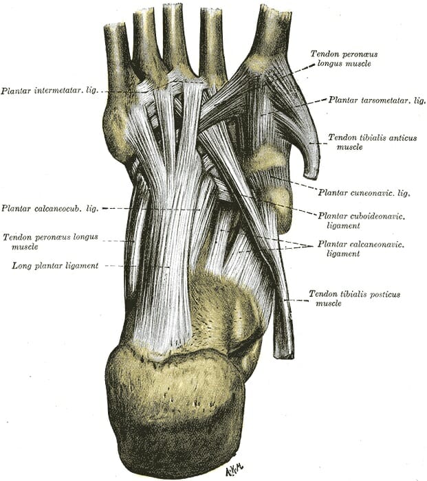
This diagram shows the sole of the foot. You can see the toes on the top and the heel on the bottom, while the arch and sole of the foot are made up of a thick web of ligaments holding the bones together:
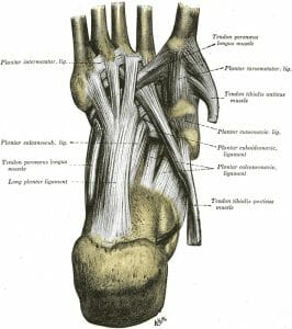

Ligaments of the sole of the foot


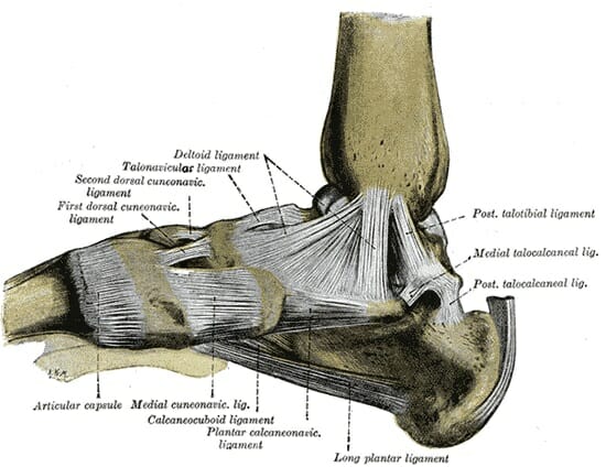
This diagram shows the “medial aspect” of the foot. This term comes from the terms “medial,” meaning “center,” or “in the middle,” and “aspect,” meaning “face.” In other words, this is the “face” that the foot shows to the center of the body. It is the side of the foot that faces inward.
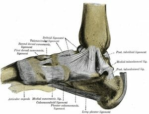

Ligaments of the medial aspect of the foot


This diagram shows the heel on the right, while the toes reach off the screen to the left.


Here you can see that the ankle is also a thick web of ligaments, where the tibia is connected to the bones of the ankle and the core of the foot. You can also see the bands of ligaments where the metatarsals and phalanges are connected to each other.


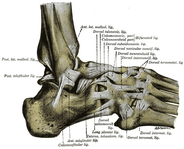
Lastly, this diagram shows the “lateral aspect” of the foot, with “lateral” meaning “to the side.” This is the view of the foot from the side of the body, then; the view of the part of the foot that faces outward.
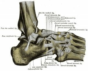

Ligaments of the foot from the lateral aspect


On the left side of the image, above the heel, you can see the delicate leg bone called the fibula. The fibula is smaller than the tibia and runs alongside it. Having two separate bones instead of one connecting the foot to the leg gives the foot and leg extra balance and maneuverability.


You can also see the thick web of ligaments on the top of the foot, where the bones of the foot’s core are connected on the top side.


Now you can begin to see why the middle of your foot feels solid, even though it’s made up of many bones. The many bones are bound together tightly by strong, flexible ligaments, which allow the center of your foot to shift subtly while remaining solid and stable.


Although ligaments are strong, they can be injured – especially in an area like the ankle, where the whole weight of your body hinges on a single joint.


Sprains occur when a body part is wrenched or twisted, resulting in damage to a ligament. Such damage can cause swelling and significant pain. Because ligaments do not receive much nourishing blood flow from the body, sprains can take a long time to heal, and long-term damage can result from continued stress on a sprained ligament.


Like broken bones, sprains are often treated with rest, ice, compression, and elevation; and a supportive wrap or cast to take stress off the sprained area. Sometimes, crutches or other means of keeping weight off the foot entirely might be prescribed.


Physical therapy can be especially helpful in the case of sprains, where it can ensure that the injured ligament is strengthened gradually and is properly supported by surrounding muscles.


A torn ligament occurs when the foot is wrenched or twisted so violently that the ligament actually snaps. This condition can be serious as ligaments which are completely torn may not heal themselves the way a bone or muscle would.


Torn ligaments can sometimes be treated in the same way as strains, but may require surgery if the tear is severe or if there is lasting damage to foot function. With surgery, doctors can join the two ends of a damaged ligament together, or replace a damaged ligament with a healthy one from another part of the body.


Foot Muscles


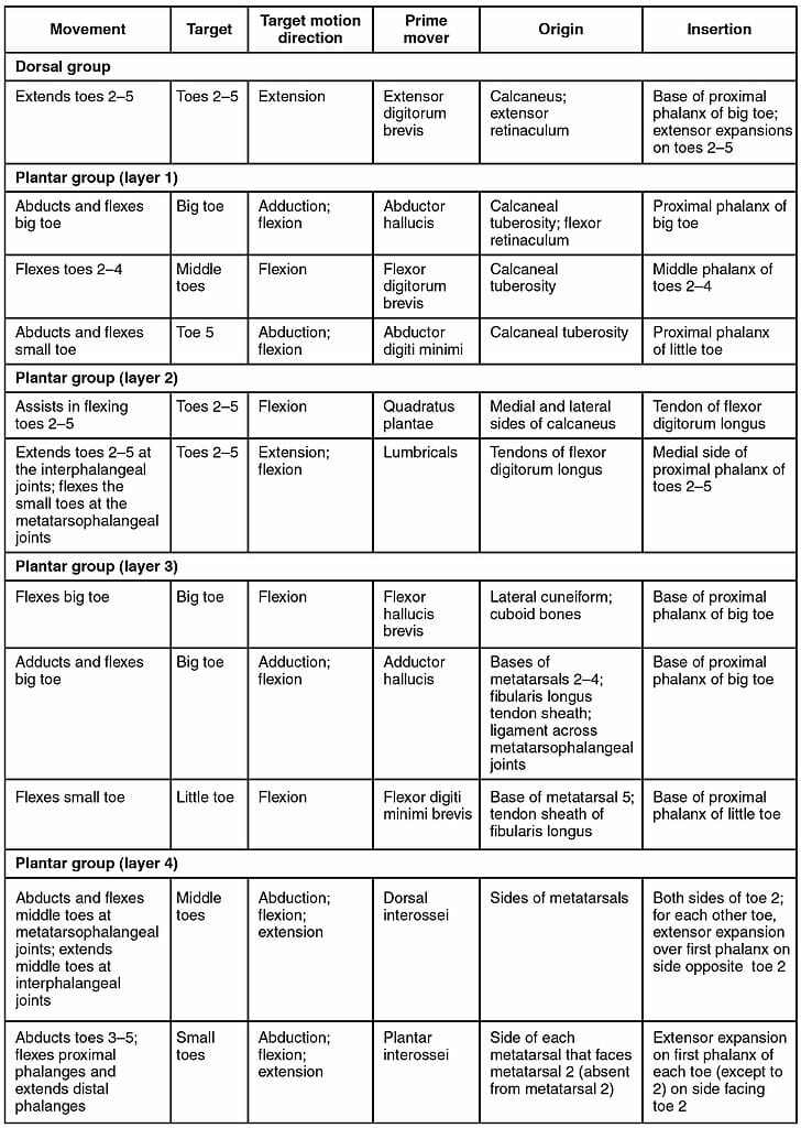
Just as there are many bones and ligaments of the sole of the foot, there are also many muscles. These can be divided up into four major groups:


  • The central muscles of the sole of the foot

  • The lateral muscles of the sole of the foot

  • The medial muscles of the sole of the foot

  • The muscles of the dorsum (top) of the foot


You can learn more about each individual group of muscles in the foot using this table:
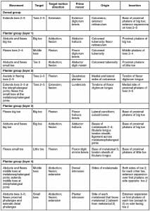

Intrinsic Muscles in the Foot


If muscles are overworked or overstressed, they can become torn or strained. Strains usually manifest as pain, especially with movement or pressure.


Mild strains often go away in days or weeks if the muscle is rested and not subjected to further stress. More serious muscle tears, however, may take months.


It is a good idea to see a doctor if a severe strain is suspected, as severe muscle strains can lead to serious complications.


The most severe form of muscle overuse – rhabdomyolysis – occurs when muscles are so stressed that their cells rupture and release toxic chemicals. This can actually be fatal if left untreated.


Rest, ice, compression, and elevation to reduce swelling are recommended to treat mild to moderate strains. Supportive wraps or casts and crutches or braces may be recommended if the strain is especially severe.


Physical therapy may also be suggested to help regain healthy use of the muscles after the injury.


Foot Tendons


Tendons are thick bands of tissue that connect muscles to bones. By connecting our rigid bones to our powerful muscles, tendons allow us to move. Movement occurs when our muscles pull on our bones, relocating them.


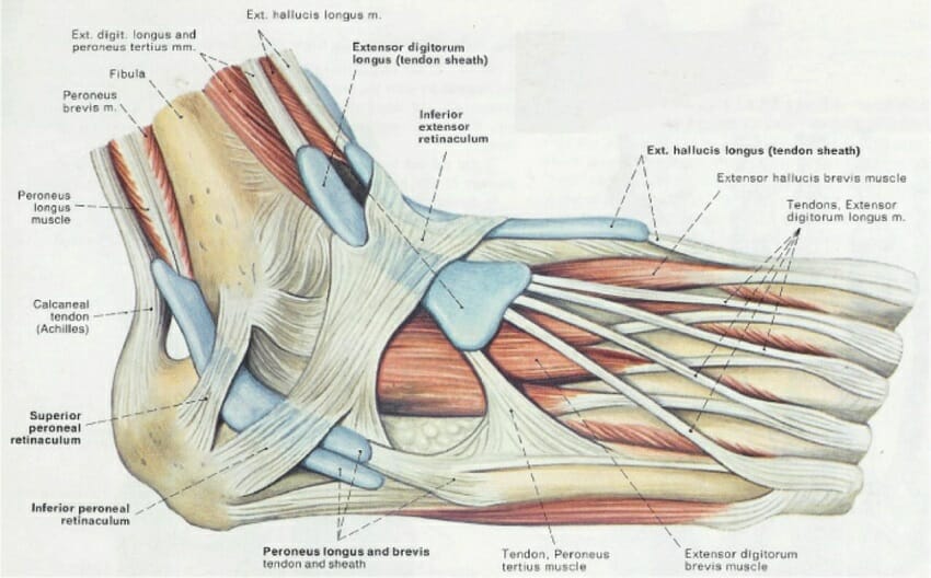
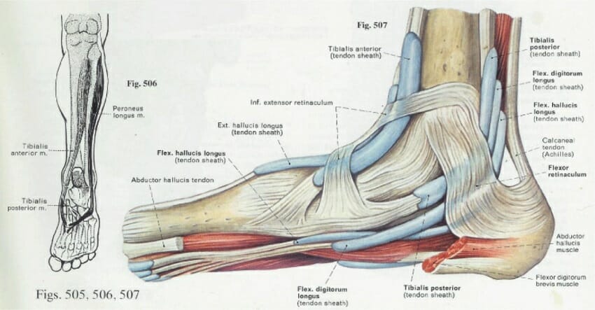
The following diagram shows the tendons of the lateral aspect of the foot – that is, the aspect that faces outward, away from your body:
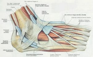

tendons


Here you can see the tendons that extend down the top of your foot toward your toes, allowing you to curl your toes upward if need be.


You can also see what is arguably the most important tendon in the foot – the calcaneal, or Achilles tendon, which allows the muscles of your calf to control the movement of your foot.


The Achilles tendon gets its name from the mythical Greek hero Achilles, who was invulnerable – except for his ankle. An injury to his ankle – possibly to the Achilles tendon – left him unable to stand and fight.
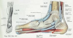

Medial aspect of the foot


This image of the medial aspect of the foot shows tendons that run along the bottom of the foot. It is these tendons that allow you to curl your toes and grip surfaces with your feet, by permitting muscles on the bottom of the foot to pull tight.


Injuries may happen to any tendon in the foot, and these may cause pain or impair balance. Achilles tendon injuries are one of the most common tendon injuries that can occur, as the body relies on the Achilles tendon to support its weight.


Lesser injuries to tendons can be treated with rest, ice, compression, elevation, and over-the-counter anti-inflammatory medications. Doctors may recommend prolonged periods of rest, and prescribe a supportive wrap or cast for substantial tendon injuries.


Severe injuries to the Achilles tendon that may occur while playing sports can require surgery to repair.


In addition to acute injuries like strains and tears, tendons can become irritated due to chronic stress.


Tendonitis occurs when a tendon – a touch cord of tissue which attaches a muscle to a bone – becomes irritated over time. This can occur from overuse or misuse if a person is moving in a way that causes stress to the tendon.


Tendonitis often appears slowly, manifesting as a sharp pain when a person performs a certain movement. People with tendonitis in the foot may find that it is painful to put weight on the foot, despite the absence of a clear injury like trauma or strain.


Tendonitis can be treated with RICE and over-the-counter anti-inflammatory drugs. Physical therapy can also be extremely beneficial, as this can gently exercise and stretch the tendon, and correct any movement habits that may have caused the irritation.


Foot Arches


Normally, tendons in the foot pull the bones of the foot in toward each other, resulting in distinctive arches between the heel and toes, and between the inner and outer toes. This arch is important for ensuring that weight is properly distributed among the strongest muscles of the leg and foot, and to ensure we can shift our weight as needed to keep our balance or move quickly.
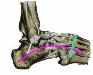

Foot Arches


Fallen arches, or “flat feet,” can occur when the tendons of the foot do not pull the foot’s bones together with a normal amount of strength. This results in the foot becoming “flat,” which can lead to pain, balance problems, and tiredness in the leg or foot.


Flat feet can occur as a result of injury, or some people’s tendons simply never pull together properly. Rarely, other health problems such as arthritis or problems with the nerves going to the feet can cause flat feet.


Flat feet may ache and tire easily. Back pain and leg pain may also result as muscles in the back and legs may work to overcompensate for the normal balancing functions of the arch.


Treatment for flat feet may depend on the cause. If you believe you have flat feet, see a doctor to find out what is the best treatment for you!


Skin and Toenails


The internal parts of the foot are not the only important parts! The skin on the bottom of our feet protects our muscles, bones, tendons and ligaments from injury. It also prevents infection.


Toenails protect the top of our toes, which, as we all know, can sometimes be vulnerable to being stubbed, stepped on, or having things dropped on them.


However, there are things that can go wrong with each of these and lead to problems.


Plantar warts – Plantar warts are growths that appear on the bottom of the foot, and may become painful. They are caused by a strain of human papillomavirus that infects skin of the feet and causes unusual growth of skin and blood vessels.


The strain of human papillomavirus that causes plantar warts and other warts is very common in the environment. It is not known why some people develop warts and others don’t. Avoiding sharing shoes and socks with people who have plantar warts may help protect against them, but many people develop plantar warts with no known instances of person-to-person transmission.


If plantar warts remain small, they might not cause pain, and no treatment may be needed. If they become painful, however, they may need to be removed. Several options exist for doing this, including over-the-counter applications, and procedures to freeze the wart tissue which can be performed by a doctor.


Corns and calluses are hard areas of skin which build up as a result of frequent friction against the skin. The body creates corns and calluses to “toughen” the skin against repeated stress.


People who work with their hands such as carpenters, gardeners, and musicians often develop calluses on their hands in areas where they frequently rub against their instruments. People who walk often or whose feet rub against the insides of their shoes may develop corns and calluses on their feet.


People with conditions that cause fragile skin or impaired blood flow to the feet, such as diabetes, should talk with their doctor as soon as corns or calluses develop. This may be a sign of an underlying problem, and treatments that are appropriate for healthy people may cause harm to people with these conditions.


For people who do not have such health conditions, over-the-counter corn-removal and exfoliation treatments can help relieve discomfort caused by corns and calluses. Changing one’s shoes or walking habits may also prevent them from forming in the future.


Once, human toenails served a similar function to those of fingernails or animals’ claws. However, the foot has undergone some important changes in evolutionary history. Toenails have not always kept up.


Ingrown toenails occur when a toenail inappropriately curves, causing it to stab into the flesh of the toe. This is a painful condition, and may become serious if injury and infection occur.


Ingrown toenails can sometimes be managed at home through frequent clipping. But in serious cases, medical attention may be necessary to avoid dangerous infections.


See your doctor immediately if an ingrown toenail causes severe pain, or if a toe with an ingrown toenail becomes red and swollen.


Quiz


1. Why is it important to see a doctor if a foot injury is suspected?
A. Because different types of injuries such as broken bones, sprains, and strains may have similar symptoms but require different treatments
B. Because prompt treatment can make for a slower, easier healing process than if an injury is ignored or treated improperly at first
C. Because doctors can prescribe helpful devices like supportive wraps, casts, and crutches that decrease pain and help with healing
D. All of the above

Answer to Question #1

2. Which of the following is NOT a bone found in the foot?
A. The calcaneus
B. The cuboid bone
C. The metacarpals
D. The phalanges

Answer to Question #2

3. Which of the following is not a foot-related health problem?
A. A broken toe
B. An Achilles tendon injury
C. A sprained ankle
D. Carpal tunnel syndrome

Answer to Question #3

References


  • Hoffman, M. (n.d.). Picture of the Feet. Retrieved July 05, 2017, from http://www.webmd.com/pain-management/picture-of-the-feet#1

  • Gray, H. (2012). Anatomy of the human body. London, England: Bounty.

  • Foot Injuries | Foot Disorders | MedlinePlus. (n.d.). Retrieved July 05, 2017, from https://medlineplus.gov/footinjuriesanddisorders.html

  • Topographic anatomy of the lower extremity, part II: knee, leg, ankle, and foot.[Video file]. (n.d.).

  • Muscles of the foot. (n.d.). Retrieved July 05, 2017, from https://www.kenhub.com/en/videos/muscles-foot

  • Calluses and Corns – Topic Overview. (n.d.). Retrieved July 05, 2017, from http://www.webmd.com/skin-problems-and-treatments/tc/calluses-and-corns-topic-overview#1


Foot