Uterus Definition
The uterus, otherwise known as the womb, is the female sex organ that carries a huge significance in many species’ survival – ours included. The uterus itself is a hollow organ that is shaped in the form of a pear, and interestingly enough measures about that size. It is neatly tucked into the pelvic area of most mammals and, of course, in humans. It is important to dissect the anatomy of the human uterus. In the female body, the upper end of the uterus, called the fundus, will join the fallopian tubes at either side while the lower end will open into the vagina. The wide portion at the top of the uterus is called the fundus, and will be the superior-most region that will host a fertilized embryo as it grows into a baby. A little below the fundus lies the muscular corpus region.
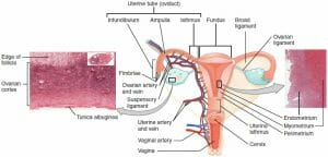
The corpus, in turn, is composed of three tissue layers. Post-pubescent women will have an innermost endometrium, which is the layer of muscle that is shed when the menstrual cycle commences in non-pregnant women. The endometrial tissue will thicken as the month’s cycle goes by in preparation for a fertilized egg to implant itself there. But in the absence of a fertilized egg, this layer will simply be shed away in what we know as menstruation. The middle muscle layer is called the myometrium, and is the layer that will expand during pregnancy and contract during childbirth. The outermost layer, the parametrium, will likewise expand and contract at these stages. Expanding will allow the uterus to house a growing baby, while the contractions will facilitate the newborn’s exit from the womb.

The image above depicts a diagram of the Uterus, with labeled tissue and arterial landmarks.
Function of the Uterus
Perhaps the principal, albeit lofty function of the uterus is to preserve life. It is the site of nourishment for the growing baby, making it one of the most important reproductive organs in the female body. This all begins when an egg, or ovum, is fertilized by a sperm and will make its downward trek in search of a better home. The tight fallopian tubes will not provide enough space to house the growing embryo! This is where the uterus meets all of these requirements, and more! The uterus’s thick, muscular nature will allow it to contract and expand to make room for the developing baby. The uterus is also rich in vasculature. There are many blood vessels supplying the muscle layers at any given time. This especially applies to the endometrium which is highly vascular and will come to nourish the embryo. In fact, many of the endometrial vessels that will come to supply the embryo will form just for this purpose. All of this explains why the fertilized ovum will choose to implant itself in the uterine lining – coined, the “site of implantation.” Thus, the uterus is the site that allows ours, and many species, to continue reproducing!
Location of the Uterus
The uterus measures about three inches long and two inches wide, and has a thick muscular lining within its walls. The lowest tip of the uterus will dip into the vagina in the area of the cervix, while the top most part will connect with the fallopian tubes through which the eggs travel. But a better way to pin its location is by describing its region as the area that lies between the belly button and the hip bones.
Abnormalities of the Uterus in Pregnancy
Nothing quite demonstrates the reproductive role of the uterus as the difficulties that arise from having an abnormal uterus. While the normal uterus will roughly measure three by two by one inches, some women will have uteruses that differ in shape and size. Many species’ evolution, including our own, has depended on having these precise dimensions to best support the growing embryo and to bring it to full term. But women with uterine abnormalities may realize they have this only once they have attempted and failed to conceive. These complications will surface either while trying to become pregnant, or after experiencing miscarriage. Examples of physical deformations of the uterus may include having a uterus with two inner cavities or vaginas (affecting roughly one in 350 women), having only one fallopian tube that will connect to the uterus, or having a heart shaped uterus instead of a pear-shaped one that is evolutionarily optimized to bear a child. Moreover, some afflicted women will have a septum that parts the uterus, or a slight indentation at the top of the uterus that will likewise compromise the ability for these women to have a baby. These abnormalities, however, are not all an absolute guarantee of infertility or miscarriage, but instead may lessen the probability of carrying a child to full-term.
Quiz
1. Which of the following is the outermost muscle layer of the corpus?
A. Endometrium
B. Parametrium
C. Myometrium
D. None of the above
2. Which uterine layer nourishes the growing embryo?
A. Endometrium
B. Parametrium
C. Myometrium
D. None of the above
3. Which of the following provides the best explanation for why the uterus is the main female reproductive organ?
A. It is the site of fertilization of the ovum
B. It is muscular and able to contract and expand
C. Its myometrial layer supplies the growing embryo
D. Both A and B
References
- MedicineNet (2017). “Uterus.” Medicine Net. Retrieved on 2017-08-26 from http://www.medicinenet.com/script/main/art.asp?articlekey=5918
- BabyCentre Medical Advisory Board (2016). “Abnormalities of the uterus in pregnancy.” Retrieved on 2017-08-26 https://www.babycentre.co.uk/a551934/abnormalities-of-the-uterus-in-pregnancy
- Danielsson, K. “Abnormal Uterus Shapes and iscarriage Risk.” Very Well. Retrieved on 2017-08-27 from https://www.verywell.com/abnormal-uterus-and-miscarriage-risk-2371694
Uterus
No comments:
Post a Comment