Sunday, October 7, 2018

Competition

Competition Definition in Biology


Competition is a relationship between organisms in which one is harmed when both are trying to use the same resource related to growth, reproduction, or survivability. Competition stems from the fact that resources are limited. There are simply not enough of some resources for all individuals to have equal access and supply. Competition can occur between organisms of the same species, or between members of different species.


Competition between species can either lead to the extinction of one of the species, or a decline in both of the species. However, this process can often be interrupted by environmental disturbances or evolution, which can change the rules of the game. Competition is often involved when species are limited in their range, often by direct competition from other organisms.


Examples of Competition


Intraspecific Competition


Intraspecific competition is a density-dependent form of competition. “Intra” refers to within a species, as opposed to “inter” which means between. Intraspecific competition can be summed up in the image below.


Intraspecific competition

Intraspecific competition


In this image, two wild dogs known as Dholes fight over a carcass. The carcass is a resource, something both organisms need to survive. Intraspecific competition is density dependent for one reason. The more dholes you have, the less food each one gets. To the individual dhole, food is everything. With very few predators of their own, the most successful dholes (the ones who survive and reproduce the most) often are simply the ones who eat the most.


Thus, while these dholes may have coordinated to take down this deer, they are now competing to see which one will get to eat first. The one that eats first will get more, and be more likely to survive and reproduce. The other one (or the last one if there are many) will not get as much. This will lower its survivability and the chances it will get to reproduce. Since evolution relies mainly on which organisms reproduce, this form of competition can quickly lead to changes in a population if only a few of the individuals are surviving and reproducing.


Interspecific Competition


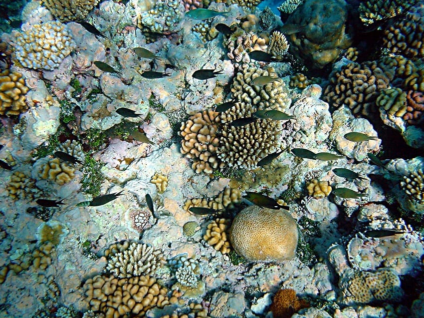
Interspecific competition is between individuals which are different species. This could be between any two species, as long as they are competing over a resource. An interesting example of interspecific competition is found in coastal marine environments, like the coral reef in the picture below.


Coral reefs with fishes


In this picture, there are dozens of species. There are several species of fish. Behind them, as a backdrop many people would ignore, is a canvas of dozens of species of coral. Coral, while it may look like some sort of rock or plant, is actually a colony of tiny animals. These tiny animals filter organic material from the water, and use stored bacteria to photosynthesize sunlight for additional energy. Thus, each coral species is competing with not only the other corals, but also with the fish for available nutrients and sunlight.


While corals might not seem like a competitive bunch, they are actually directly competitive with other corals. When an enemy coral is encroaching on their space, they can deploy chemical warfare to counter their rival. Often, coral fights end in one of the corals being killed by the other. While the corals are not predators of each other, the competition still ends in the death of one of the corals. The victorious coral was simply fighting for the resources it needs.


Direct and Indirect Competition


There is also another aspect of competition that can be applied to scenarios of limited resources, and that is the idea of direct vs indirect competition. Direct competition is like both of the scenarios above, and there are many more examples of it. Any time two or more animals fight or have a symbolized confrontation, this is probably some sort of competition for a resource.


However, indirect competition is when the two animals do not interact, but the presence of both animals in the same territory causes the competition. Think of the fish in the example above. If those fish feed on the same resources used by the corals, then the fish are in competition for the limited resources. Coral, being more or less anchored to the ocean floor, have little chance of directly attacking the fish. Instead, this would be referred to as an asymmetrical indirect competition. The fish eat as much of the food as they want, and the coral are limited to scraps. The coral have no way of competing. Luckily for most coral reef systems around the world, the ocean has plenty of food for most.


Outcomes of Competition


Competition is not a static process. Once set in motion, it can go a number of different ways. While the models may show that it will eventually drive one species to extinction, in reality a number of things can happen. First, an environmental disturbance, such as a fire or large wave, can upset the ecosystem and destroy the advantage the best competitor had. Typically, a pinewood forest is made mostly of pine trees because they are the best competitors in the environment. However, after a forest fire the most populous plants are small, opportunistic plants that grow quickly. The fire causes a change in the environment, which completely changes the dynamics of competition.


Further, most competition is also an evolutionary pressure on both parties. Animals from both sides that compete the best are able to survive and reproduce. Thus, over time the competition tends to resolve itself. More often than not, the competition can devolve as the species adapt to use different resources or change the way it uses a resource. This is known as character displacement. It is most well-documented in finches. When two different species of finch live on separate islands, their beaks are the same size because they prefer similar seeds. When they occupy the same island, one of their beaks gets smaller while the other gets larger. This separates the resources they consume and alleviates the competition.


Quiz


1. Which of the following represents competition?
A. Two swans (male and female) doing a mating dance
B. A lion defends its kill from a pack of hyenas
C. A lion stalks a buffalo, ready to pounce

Answer to Question #1
B is correct. In this case, the lion’s kill represents the resource. It needs the food to survive and reproduce. But so do the hyenas. Because they are fighting for it, it makes it direct competition. When the lion was stalking the buffalo that would be predation, which is different than competing for a resource. Two swans doing a ritualistic dance would be a form of intraspecies communication.

2. What is the difference between intra- and interspecific competition?
A. Intraspecific is between members of the same species
B. Interspecific is between members of the same species
C. They are the same

Answer to Question #2
A is correct. While the different forms of competition can be about similar resources, they are often carried out in different ways. Intraspecific competition often occurs when the resource is within the species, such as access to females. Interspecific competition is easiest remembered as different predators fighting over the same piece of meat.

3. A bald eagle is flying over a field, and sees a smaller hawk. The hawk has a fresh kill, but the bald eagle swoops in, threatens the hawk, and steals it. Which term best describes this scenario?
A. Predation
B. Intraspecific, indirect competition
C. Interspecific, direct competition

Answer to Question #3
C is correct. In this case, the bald eagle directly attacks and steals a resource from another species. If the species of hawk could no longer compete, because the eagles had already eaten all of the prey, that would be indirect competition. There are many other forms of indirect competition, but in this case the animals compete for the food directly.

References


  • Cain, M. L., Bowman, W. D., & Hacker, S. D. (2008). Ecology. Sunderland, MA: Sinauer Associates, Inc.

  • Feldhamer, G. A., Drickamer, L. C., Vessey, S. H., Merritt, J. F., & Krajewski, C. (2007). Mammology: Adaptation, Diversity, Ecology (3rd ed.). Baltimore: The Johns Hopkins University Press.

  • Kaiser, M. J., Attrill, M. J., Jennings, S., Thomas, D. N., Barnes, D. K., Brierley, A. S., & Hiddink, J. G. (2011). Marine Ecology: Processes, Systems, and Impacts. New York: Oxford University Press.


Competition

Lytic Cycle

Lytic Cycle Definition


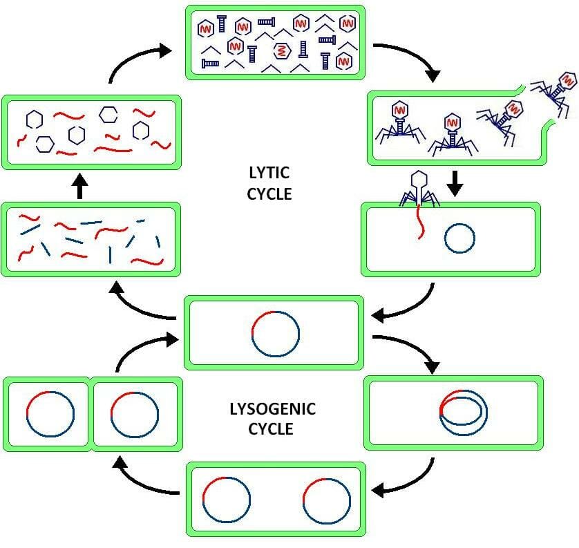
The lytic cycle is named for the process of lysis, which occurs when a virus has infected a cell, replicated new virus particles, and bursts through the cell membrane. This releases the new virions, or virus complexes, so they can infect more cells.


Lytic cycle


As seen in the graphic above, the lytic cycle is often accompanied by the lysogenic cycle in many bacteria viruses, known as bacteriophages. After the virus injects its DNA or RNA into the host bacteria, the genetic material can enter either the lytic cycle or the lysogenic cycle.


In the lysogenic cycle, the bacteriophage DNA lies practically dormant. However, whenever the bacteria divides, the DNA of the virus is inadvertently copied. In this way, the virus can continue replicating within its host. As long as the bacteria are successful, the virus may remain dormant. At a certain point, conditions may change, and the virus will enter the lytic cycle.


In this cycle, the viral DNA or RNA is expressed by the host organism’s cellular mechanisms. In other words, the viral genes use the proteins within the cell to replicate themselves and produce viral proteins. These proteins and copies of the DNA will become new virions. The cell, helpless to its viral hijacker, simply waits until the pressure of these new virions is too high. Then, the cell membrane breaks. This lysis of the cell releases the virions created in the lytic cycle. Their final destination is a new cell, in which the lytic cycle can take place again. If conditions are favorable and the cell is dividing, the virus may stay in the lysogenic cycle for a time. Ultimately, to infect a greater number of cells, more virus genomes will enter the lytic cycle and produce thousands or millions of copies of themselves in a shorter amount of time.


Steps of the Lytic Cycle


Bacteriophage lysogenic and lytic cycle

Bacteriophage lysogenic and lytic cycle


Adsorption and Penetration


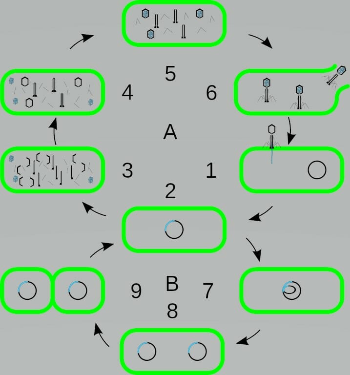
Adsorption is the process through which a bacteria gets its DNA or RNA into the host cell. This is labeled as 1 in the image above. The capsid, or protein coat around the viral genome, consists of very specific proteins. This sheild of proteins not only comes together to protect the viral genes, it serves as a sort of “key” to unlock a cell. The surface of the proteins are shaped to interact with proteins on the surface of the host cell.


When the “lock and key” align, the virion is bound to the cell membrane. When this happens, it also changes the shape of the capsid. This tears a hole or injects the viral DNA into the host cell. Here, it may travel into the nucleus or replicate in the cytoplasm. This depends on the virus itself, what type of genome it has, and the conditions of the cell.


Replication


During the lytic cycle, the replication of viral genes is carried out a number of times by a hijacked cellular system. Remember that the virus itself has imported few, if any, supporting proteins. Thus, the viral DNA must produce these in order to hijack the cell’s processes. The first proteins created are often created as the cell reads its own DNA and produces proteins. The viral genes simply sneak into the process. This creates what are called viral early proteins.


These early proteins have important functions (to the virus) of commandeering the cell’s machinery. They clear the cell’s normal metabolic agenda, and turn many of its activities toward the replication of viral genes and the production of viral proteins. The virus uses the raw products the cell has assembled (amino acids and nucleic acids) as building blocks for the parts it needs.


While this may seem like an overly complex process for such a small virus genome, consider first that there are really only a handful of proteins. Most viruses produce and code for only a handful of proteins. Unlike cells, a virus doesn’t need the complex proteins required to metabolize energy. As obligate parasites, a virus is dependent upon its host cell’s ability to provide raw materials. This makes it one of the most efficient forms of DNA replication that we know of.


Assembly and Release


As these parts are built, their natural evolutionary shapes help them come together in the proper way. Since most of the components are proteins, they have formed over evolutionary time to be able to come together with very little outside influence. The assembly of new virions is a hallmark of the lytic cycle. The other viral life cycle does not include producing and assembling new virions.


In this way, the lytic cycle resembles a small virus factory. All of the parts of the virus are produced independently, then assembled, and finally released into the environment. While the image above shows only 3 assembled virions at stage 6, in reality there would be millions. Compare the lytic cycle to the lysogenic cycle below it, in which an accurate 2 copies are shown after 1 bacterial division.


Quiz


1. Which of the following represents the lytic cycle?
A. Viral DNA is replicated as the host cell divides.
B. The viral genome takes over the host cell, and creates a virus factory.
C. The viral genome is mostly dormant.

Answer to Question #1
B is correct. Remember that the lytic cycle is like a factory. It takes over the cell and tries to make as many virions as fast as possible. Eventually, this overwhelms the cell and causes it to lyse, or break open. This is the reason it is called the lytic cycle. The lysogenic cycle is a much more dormant version of the viral life cycle. It is a passive state in which the viral genes get replicated as a byproduct of cell division.

2. Which life cycle, the lytic cycle or the lysogenic cycle, produces the most virions?
A. It depends
B. The lytic cycle
C. The lysogenic cycle

Answer to Question #2
A is correct. While the lytic cycle is a factory for new virions and is a clear answer, the question did not specify a time frame. If a virion infects a cell, instantly enters the lytic cycle and kills the cell, then it has only produced a million virions and now needs a new host. A bacteriophage genome which enters the lysogenic cycle may be inadvertently copied into millions of cells itself. Then, if even only a few of these enter the lytic cycle, that bacteriophage will far outnumber the previous example. In other words, it depends entirely on how long the lysogenic cycle is allowed to continue.

3. Based on what you now know about the lytic cycle, why is it so hard to eradicate the common cold?
A. It shouldn’t be!
B. The virus changes too much.
C. In targeting the mechanisms viruses use, you target the mechanisms every cell uses.

Answer to Question #3
C is correct. In most forms of medicine, a basic treatment to a disease is to take away the underlying cause. However, because viruses use the same machinery your healthy cells use, there is no way to target them specifically. If we did, we might accidently destroy all the cells in our body. I would rather get a cold.

References


  • Brusca, R. C., & Brusca, G. J. (2003). Invertebrates. Sunderland, MA: Sinauer Associates, Inc.

  • Lodish, H., Berk, A., Kaiser, C. A., Krieger, M., Scott, M. P., Bretscher, A., . . . Matsudaira, P. (2008). Molecular Cell Biology (6th ed.). New York: W.H. Freeman and Company.

  • McMahon, M. J., Kofranek, A. M., & Rubatzky, V. E. (2011). Plant Science: Growth, Development, and Utilization of Cultivated Plants (5th ed.). Boston: Prentince Hall.


Lytic Cycle

Lytic Cycle

Lytic Cycle Definition


The lytic cycle is named for the process of lysis, which occurs when a virus has infected a cell, replicated new virus particles, and bursts through the cell membrane. This releases the new virions, or virus complexes, so they can infect more cells.


Lytic cycle


As seen in the graphic above, the lytic cycle is often accompanied by the lysogenic cycle in many bacteria viruses, known as bacteriophages. After the virus injects its DNA or RNA into the host bacteria, the genetic material can enter either the lytic cycle or the lysogenic cycle.


In the lysogenic cycle, the bacteriophage DNA lies practically dormant. However, whenever the bacteria divides, the DNA of the virus is inadvertently copied. In this way, the virus can continue replicating within its host. As long as the bacteria are successful, the virus may remain dormant. At a certain point, conditions may change, and the virus will enter the lytic cycle.


In this cycle, the viral DNA or RNA is expressed by the host organism’s cellular mechanisms. In other words, the viral genes use the proteins within the cell to replicate themselves and produce viral proteins. These proteins and copies of the DNA will become new virions. The cell, helpless to its viral hijacker, simply waits until the pressure of these new virions is too high. Then, the cell membrane breaks. This lysis of the cell releases the virions created in the lytic cycle. Their final destination is a new cell, in which the lytic cycle can take place again. If conditions are favorable and the cell is dividing, the virus may stay in the lysogenic cycle for a time. Ultimately, to infect a greater number of cells, more virus genomes will enter the lytic cycle and produce thousands or millions of copies of themselves in a shorter amount of time.


Steps of the Lytic Cycle


Bacteriophage lysogenic and lytic cycle

Bacteriophage lysogenic and lytic cycle


Adsorption and Penetration


Adsorption is the process through which a bacteria gets its DNA or RNA into the host cell. This is labeled as 1 in the image above. The capsid, or protein coat around the viral genome, consists of very specific proteins. This sheild of proteins not only comes together to protect the viral genes, it serves as a sort of “key” to unlock a cell. The surface of the proteins are shaped to interact with proteins on the surface of the host cell.


When the “lock and key” align, the virion is bound to the cell membrane. When this happens, it also changes the shape of the capsid. This tears a hole or injects the viral DNA into the host cell. Here, it may travel into the nucleus or replicate in the cytoplasm. This depends on the virus itself, what type of genome it has, and the conditions of the cell.


Replication


During the lytic cycle, the replication of viral genes is carried out a number of times by a hijacked cellular system. Remember that the virus itself has imported few, if any, supporting proteins. Thus, the viral DNA must produce these in order to hijack the cell’s processes. The first proteins created are often created as the cell reads its own DNA and produces proteins. The viral genes simply sneak into the process. This creates what are called viral early proteins.


These early proteins have important functions (to the virus) of commandeering the cell’s machinery. They clear the cell’s normal metabolic agenda, and turn many of its activities toward the replication of viral genes and the production of viral proteins. The virus uses the raw products the cell has assembled (amino acids and nucleic acids) as building blocks for the parts it needs.


While this may seem like an overly complex process for such a small virus genome, consider first that there are really only a handful of proteins. Most viruses produce and code for only a handful of proteins. Unlike cells, a virus doesn’t need the complex proteins required to metabolize energy. As obligate parasites, a virus is dependent upon its host cell’s ability to provide raw materials. This makes it one of the most efficient forms of DNA replication that we know of.


Assembly and Release


As these parts are built, their natural evolutionary shapes help them come together in the proper way. Since most of the components are proteins, they have formed over evolutionary time to be able to come together with very little outside influence. The assembly of new virions is a hallmark of the lytic cycle. The other viral life cycle does not include producing and assembling new virions.


In this way, the lytic cycle resembles a small virus factory. All of the parts of the virus are produced independently, then assembled, and finally released into the environment. While the image above shows only 3 assembled virions at stage 6, in reality there would be millions. Compare the lytic cycle to the lysogenic cycle below it, in which an accurate 2 copies are shown after 1 bacterial division.


Quiz


1. Which of the following represents the lytic cycle?
A. Viral DNA is replicated as the host cell divides.
B. The viral genome takes over the host cell, and creates a virus factory.
C. The viral genome is mostly dormant.

Answer to Question #1
B is correct. Remember that the lytic cycle is like a factory. It takes over the cell and tries to make as many virions as fast as possible. Eventually, this overwhelms the cell and causes it to lyse, or break open. This is the reason it is called the lytic cycle. The lysogenic cycle is a much more dormant version of the viral life cycle. It is a passive state in which the viral genes get replicated as a byproduct of cell division.

2. Which life cycle, the lytic cycle or the lysogenic cycle, produces the most virions?
A. It depends
B. The lytic cycle
C. The lysogenic cycle

Answer to Question #2
A is correct. While the lytic cycle is a factory for new virions and is a clear answer, the question did not specify a time frame. If a virion infects a cell, instantly enters the lytic cycle and kills the cell, then it has only produced a million virions and now needs a new host. A bacteriophage genome which enters the lysogenic cycle may be inadvertently copied into millions of cells itself. Then, if even only a few of these enter the lytic cycle, that bacteriophage will far outnumber the previous example. In other words, it depends entirely on how long the lysogenic cycle is allowed to continue.

3. Based on what you now know about the lytic cycle, why is it so hard to eradicate the common cold?
A. It shouldn’t be!
B. The virus changes too much.
C. In targeting the mechanisms viruses use, you target the mechanisms every cell uses.

Answer to Question #3
C is correct. In most forms of medicine, a basic treatment to a disease is to take away the underlying cause. However, because viruses use the same machinery your healthy cells use, there is no way to target them specifically. If we did, we might accidently destroy all the cells in our body. I would rather get a cold.

References


  • Brusca, R. C., & Brusca, G. J. (2003). Invertebrates. Sunderland, MA: Sinauer Associates, Inc.

  • Lodish, H., Berk, A., Kaiser, C. A., Krieger, M., Scott, M. P., Bretscher, A., . . . Matsudaira, P. (2008). Molecular Cell Biology (6th ed.). New York: W.H. Freeman and Company.

  • McMahon, M. J., Kofranek, A. M., & Rubatzky, V. E. (2011). Plant Science: Growth, Development, and Utilization of Cultivated Plants (5th ed.). Boston: Prentince Hall.


Lytic Cycle

Friday, September 28, 2018

Elf


In the woods you happen across a clearing full of strangely beautiful people. At first you think they may be human, but upon closer examination you discover that their features are too fair and perfect to have come from mortal man. You walk into the clearing to get a better look and introduce yourself, but happen to step inside a ring of mushrooms and flattened grass. Their expressions quickly turn from curiosity to wrathful anger.

Suddenly you begin to feel ill. You reach out to apologize and explain your actions, but they are already disappearing into the depths of the forest. It is too late. As punishment for your offense, you have been elf-shot.


What is an Elf?


An elf is a mythical creature that appears to be human in nature, but has magical powers and does not age (or at least ages very slowly). It appears that elves have their origins in Germanic lore, but they are also commonly found in other European folklore.


Elf


The elves are seen as a ‘luminous’ group of people who are known to have fair complexions far more perfect than even the most beautiful human features. They are sometimes known as ‘the white people.’ It is thought that this is either a reference to their pure morality or, perhaps as a reference to their beauty and pale features.


Elves were seen as both a point of fear and curiosity in early societies. They were often known to be sociable and even friendly to humans, but they were still greatly feared because of their temper. If they perceived a human to have harmed or offended them in any way, they were quick to retaliate with a punishment. Common punishments included illness, night terrors, and cruel tricks and attacks directed towards the victim. It is noted, however, that the elves did sometimes help cure sicknesses in some instances.


Elves and Birth


The elves are a curious legend indeed. Although they are thought to never age (or to live for hundreds of years depending on which storyline you follow), the elves were thought to need human assistance to bring their children into the world. This help was often thought to be needed from mid-wives who could safely deliver the children and from wet-nurses. While midwives have a pattern of being married to preachers, the wet-nurses were normally women who had recently given birth. This caused great fear in many early European households.


While both of these groups of women had something to fear from traveling to the Elven world, the wet-nurses were especially terrified of their fate. They were normally taken from their newborns and families to the Elven world to take care of the newborn Elf babies. While this doesn’t seem to be nefarious, it would have been frightening for any woman who was put in this situation. It was rumored that eating any food offered in the Elven world or taking any hospitality from them would keep a person from ever returning to the human world. It is unclear if these rules held to the wet-nurses – who obviously had to spend a prolonged period of time in their world, but the fear of being barred from returning to their families would have been enough to give them great fright.


It was also common for midwives to be called upon to help bring elven babies into the world. It is unknown why a midwife was needed, but it was common knowledge at the time that a human midwife was needed to deliver a elf child. Usually, a midwife who was married to a preacher was called upon to perform the necessary duties. It was important that the midwife who was called did not eat or drink while in the Elf world for the above reasons. Some stories say that the midwives (who usually had time to prepare unlike the kidnapped wet-nurses) would sometimes pack food and water to take with them so that they could keep themselves from becoming hungry. There are several tales of midwives being summoned that were widely accepted in their day.


Peter Rahm’s Wife is Summoned


A clergyman by the name of Peter Rahm was known to have told a tale of his wife being summoned to help a mythical being give birth. She went with the creature and performed her duties. After the child was born, the grateful couple offered the midwife food and drink. She kindly refused. They offered her other forms of hospitality which she refused as well. She was sent on her way and returned home. The next day she found a pile of silver pieces – a gift from the new parents for delivering their child.


A Danish Story of An Elf and his Earthwife


A Danish tale tells of an Earthman (an elf) who sought the help of a midwife on Christmas Eve. He took the midwife underground and had her attend to his Earthwife during labor. When the child was delivered, the elven husband took the child away – seeking to steal the good fortune of a newly wed couple for the child. While he was gone, the elf wife gave the midwife a warning. She warned the woman against eating any food or taking any drink while she was under the surface of the earth. She told the midwife that she too had been a Christian woman before she was invited into the realm of the elves but had made the mistake of eating their food while she visited. Because she had accepted their hospitality, she was unable to return to her own world. When the husband returned, the midwife refused the offers of hospitality. Because of this, she was allowed to return to her home.


Elves and Their Relationships with Humans


While there seemed to be a great many ways that elves could threaten the safety of a human being, there were some elves who lived in peace with humans and even formed relationships with them.


Human and Elf

Human and Elf


While there are some accounts of elves who tried to seduce humans into having sexual relations with them, it appears that there were some humans and elves who had children consensually. These children were known to be especially beautiful and often went on to do great things.


More often than not, half-elf half-human children appeared to be human in their features (though they were often very beautiful) and were capable of great magic feats. They sometimes went on to become magicians, sorcerers, and healers.


There are several ballads and stories concerning elves who mate with humans. They usually involve some sort of riddle that the person must solve in order to become their lover. Some stories also require the character to rescue a human-turned-elf in order to win their lover’s hand in marriage.


The Elfin Knight


The Elfin Knight is a story that can be told in two manners. The first is that the knight threatens to steal a woman away to be his lover unless she can complete an impossible task. The second is that a woman must complete an impossible task in order to win the Knight’s hand in marriage. Over time, the second has become more popular.


The tale starts with the Knight blowing into a magic horn that causes desire to emerge in the heart of the maiden. She makes a wish that she could marry the Knight. Suddenly the Elfin Knight appears and tells her that he will marry her if she can perform several tasks – all impossible.


In return, the maiden responds with several impossible tasks of her own that the Knight must complete and wins the hand of Knight in marriage.


The Tale of Tam Lin


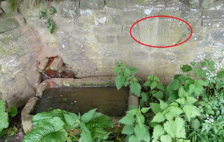
The tale of Tam Lin begins with a warning that Tam Lin is an elf who takes a possession or the virginity of any maiden that travels through the forest of Carterhaugh. One day, a young woman travels through the forest of Carterhaugh and plucks a double rose from the ground. Tam Lin appears and asks her why she has entered his forest and taken his possessions. She replies that Caterhaugh belongs to her – it was a gift from her father. The young maiden goes on her way, only to discover later that she had become pregnant.

Tam Lin

Tam Lin’s Well, Carterhaugh


She returns to the forest and plucks another set of roses from the ground. Tam Lin appears again to challenge her actions. She asks him if he had ever been a human, or if he has always been an elf. He tells the maiden that he had once been a mortal but was captured by the Queen of Fairies and turned into an elf. He also reveals that he is afraid that he will be sacrificed in a tithe to Hell this year if she does not save him.


Together they devise a plan to rescue Tam Lin from the Fairy Queen. They enact the plan and the maiden wins the love of Tam Lin. The Queen of Fairies acknowledges her defeat and releases Tam Lin.


Lady Isabel and the Elf Knight


Unfortunately, not all relationships between elves and humans end well. The tale of Lady Isabel and the Elf Knight starts the same as ‘The Elfin Knight’ with the Knight blowing into a horn that causes desire to arise in the heart of Lady Isabel. She wishes that she could marry the Knight and he appears and tells her that she will be his wife if she will come with him to the greenwood.


Upon arriving at the greenwood, Lady Isabel is shocked when the Knight reveals that he has killed the daughters of seven kings to steal their treasures and possessions and intends to make her the eighth. Fortunately, Lady Isabel is a quick thinker and tells the Knight to put his head on her knee to rest together before she is to die. She then lulls him to sleep with a charm, binds him with his own belt, and kills him.


What Do Elves Look Like?


The majority of elves that are described in folklore are female, though there were certainly male elves as well. The description of an elf’s appearance varies depending on the time period and the location that the story takes place in. It appears that the majority of female elves are known to be fair creatures. They often have blonde hair and blue or grey eyes (these are also the features that they value in humans) and are known to have characteristics that are similar to humans but much more perfect in nature. There are, of course, some variations in their appearance. These characteristics however, are the most commonly used in fairy tales.


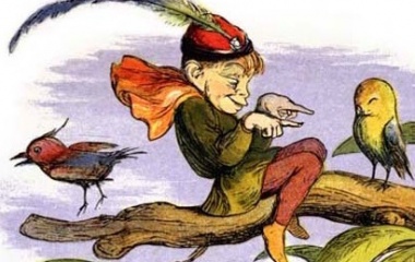
Male elves were often described as looking like old men, though this is not the case for all the elves that appeared in literature. There are also extremely handsome elves that appear and seduce women like the elves from Tam Lin and The Elfin Knight.


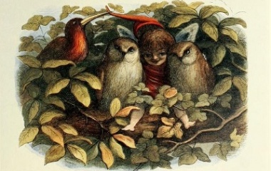
Most literature will describe elves as being human in shape and size. They are known to have especially fair features and are sometimes described as being even taller than the average human. Writers in Shakespeare’s time however, took a different approach in describing elves. They were transformed into tiny beings who often had wings and were surprisingly similar to fairies. This version of elf was known to enjoy playing tricks on humans.


Modern literature tends to take a blended perspective on elves. While you can still find the occasional reference to elves being small beings, there are also plenty of storylines that present elves as being roughly human-sized.


Where Do Elves Live?


The majority of storylines will claim that elves either inhabit homes that are deep inside the woods, carved into hollowed trees, or underneath the earth (often in a hill). This was fairly standard for the majority of supernatural creatures in early Europe.


Most people have lost their faith in the existence of the elves, but there still remains a large population who maintain their beliefs – or are at least open to the possibility – of the existence of elves in Iceland. The people of Iceland have taken special precautions to ensure that the homes of their beloved huldufolk are protected in modern day.


The Huldufolk Stop Construction Near Alftanes


In the Alfantes peninsula, road work has been proposed in an area that was thought to be frequented by the elves of Iceland. There were many protests against the construction on the grounds that it would ruin the habitat of the elves as well as the natural landscape. The protest caused the entire project to be halted until the Supreme Court of Iceland rules on the case.


This is not an isolated occurrence. Other roads that were proposed to be built in areas where elves were thought to live were stopped by strange equipment malfunctions or tools suddenly being stolen from the worksite. Some think that these incidents were caused by elves themselves.


Interestingly enough, a law was passed in 2012 forbidding construction in any area that was believed to be inhabited by elves or was culturally or historically significant otherwise to Iceland.


Dangers Posed by Elves


Elves and Sickness


Elves were often thought to be the cause of many sicknesses that could not be diagnosed or properly treated by a doctor. It was thought that elves were capable of living parallel to the human world (though invisible to the human eye) and would make a person sick if they felt offended by the individual. This was said to be accomplished in several ways. Sometimes a person became sick if an elf casted a spell over them. Other times, however, an elf could shoot a man with an invisible arrow that carried sickness. Therefore, it was not uncommon for people to be diagnosed as being ‘elf-shot’ when they were ill.


Elves were also thought to bring about other misfortunes that were connected with health – specifically sleeping. The German word Alpdruck means ‘nightmare’, but the literal translation means ‘elf oppression.’ From this, it can be assumed that elves were thought to be the cause of nightmares and night terrors – likely as a punishment for an offense that had been made towards them or as a cruel joke.


Curiously, it seems that elves were often blamed when an individual became afflicted with epilepsy. This is possibly because of the complicated nature of the illness and the lack of medical resources available to treat such an ailment.


Elves and Alchemy


Elves were known to be magical beings, so it is no surprise that they were often credited with several types of magic. There are many different types of magic credited to elves, but one of the most popular by today’s standards was alchemy.


Elf markwoman

Elf markwoman


Alchemy was a scientific and philosophical practice that was aimed at purifying different elements. One of the most popular types of alchemy recognized by humans was the practice of taking an earth element (usually some type of metal) and attempting to transform it into a precious metal like silver or gold.


The elfin tie to alchemy is likely why there are so many stories that reference an elf giving a human something that appeared to be worthless (like charcoal) that had magically transformed into gold by the time they had returned home.


Elves and Seduction


Another common threat that elves were thought to hold over humans was seduction. For some reason, elves had many desires to lure humans into sexual relations with them and were often warned against in tales.


Some elves were recorded to be very forceful with this desire. At some point, the notion arose that male elves would force themselves on women while they were sleeping. This issue was reported in the Scottish witch trials, and the elves in the tales were interpreted as being an alias for the Devil himself.


Elves and Changelings


Curiously enough, it was thought that elves sometimes valued human babies over their own kind. There was much speculation for why this may be, but there were two main theories. The first was that the elves were fond of the human babies that they stole because of their fair hair and blue or grey eyes. The second was that the children were stolen to use in a tithe to Hell so that the elves would not have to sacrifice one of their own. Regardless of what their motivation was, human parents came to dread the thought of losing their child to the elves.


It was thought that when an elf came to steal away a child, it left one of it’s own in place of the stolen infant. This elf child was known as a ‘changeling.’ These infants appeared to be human, but were often afflicted with unexplained illnesses. To discover a changeling in place of a human baby was a serious situation. Often, due to the many issues that came with changelings and their noticeable need to eat more than a human baby, changelings were killed before they had a chance to reach early childhood. It was thought that to let the changeling live was to put the resources of the entire family in jeopardy, making infanticide the best option for the unlucky couple.


Origin of the Elf Myth


Arisen from Cain’s Murder of Able


Some sources seem to think that the elves may have arisen from Cain’s murder of Able. This is the case in Beowulf, which clearly states that elves came to be a race because of this unfortunate event.


Semi-Banished Angels


Other sources seem to think that elves may have been the angels who chose to stay neutral in the fight for Heaven with God facing off against Lucifer. They were banished from Heaven because they did not help God, but because they did not betray him they were not sentenced to Hell. Instead, they were banished to Earth, where they would be come to known as elves.


Lost Children Of Eve


An old Icelandic tale suggests that elves could be the lost children of Eve. The tale states that one day when God was walking through the Garden of Eden, Eve was embarrassed that her children were dirty. She told them to go and hide from God so that she would not have to be embarrassed by God seeing them in their condition.


When God walked up to Eve and asked her where her children were, she lied and said she didn’t know. Angry that she would dare to lie to him, God said, “That which man hides from God, God will hide from man.” From that day forward, Eve never saw her children again.


The tale goes on to suggest that the children became the huldufolk (the hidden people [elves]) of Iceland.


Reborn into Elves


The Germanic tales which are thought to inspire the first stories of the elves suggest that these beings could be created by the rebirth of the dead. The old Norse texts seem to imply that worship of the elves and worship of dead ancestors are one and the same. This suggests that a person can be reborn into a supernatural creature like the elf when they pass on to the afterlife.


Evidence of this belief can be found in tales like ‘The Saga of Olaf the Holy’ in which the king’s ancestor has a burial mound that is marked as ‘Olaf, the Elf of Geirstad.’ This reflects the belief that the king’s ancestor became an elf in the afterlife.


The Explanation of Strange Occurrences


Last but not least, it is certainly possible that elves were simply created as a way to explain the unexplainable at the time. This is evidenced by some of the most common events that were blamed on elves – like elf-locks (when a strand of hair was found to be knotted).


This would have also helped people to reconcile with unfortunate events like the birth of a deformed baby. It certainly would have been easier to dispose of a child that put the new family at risk if it was thought to be an elf changeling instead of their own offspring.


Elf

Tuesday, September 18, 2018

Transferrin

Transferrin Definition


Transferrin is a crucial glycoprotein that shuttles iron in the blood. It would be an understatement to say that iron is vital for most life-sustaining processes. Transferrin has become an important biomarker for good health in the clinical setting, as it can reveal if a patient has functional iron depletion. This bio-marker, of course, will give a physician insight into a patient’s pathology, as well as which treatment plan will be most suitable moving forward.


Transferrin Structure


Transferrin

Transferrin


The image above is a 3-D depiction of human transferrin protein.


Structurally speaking, Transferrin is a polypeptide chain consisting of two carbohydrate chains and almost seven hundred amino acids. Transferrin has two homologous globular lobes, the N- and C- terminals comprised of alpha helices and beta sheets, with an iron binding site in between. The site itself is a six iron coordinate site occupied by a carbonate anion and four residues.


Each lobe is further divided into two clefts, or domains. Importantly, this structure lends transferrin the ability to undergo large conformational changes upon needing iron to be taken up or released. This is made possible by the rotating domains that rotate around a screw axis. Through x-ray crystallography, scientists have uncovered the mechanism for iron-release. This lies in how two of the basic residues from two of the domains will create a special hydrogen bond under neutral pH; however, this bond will break and thus release iron in the acidic pH of the endosome at its delivery site. Each transferrin molecule is able to carry two iron molecules in the bloodstream, and we will discuss in more detail the importance of sheltering iron until it is needed.


Transferrin Function


Iron is found everywhere on earth, and so it is no surprise that it also vital to sustaining life. Humans use iron for many cellular processes but perhaps the most important is iron’s ability to bind oxygen. As we know, oxygen is fundamental to cellular respiration and it is therefore necessary to transport oxygen from our lungs to each individual aerobic cell – without letting radical oxygen roam freely and ravage our cell’s membranes! Safe shuttling through our circulatory system is the answer. While humans contain about 3.7 grams of iron in our bodies, much of which comes from our diets, 2.5 grams will be “locked” inside hemoglobin with iron. Hemoglobin can then assume its role in transporting oxygen through the blood. However, just as importantly, we have evolved a way of recycling and storing this iron for future use. This is where transferrin comes in.


Plasma transferrin is a crucial player in iron metabolism. Transferrin essentially limits the levels of free iron in the blood. Free iron is dangerous in that it carries the risk of triggering free radical reactions, which sets off lipid oxidation and the destruction of thousands of molecules. Free radicals are defined as having at least one unpaired electron and they will thus be driven to steal electrons from every cell tissue including the heart, pancreas, brain, etc. Iron-triggered free radical damage can thus contribute to heart and liver disease, neurological issues, and more. Thankfully, transferrin binds essentially all circulating plasma iron. This chelation makes iron soluble and non-toxic as it is being delivered to tissues, accordingly serving the functions of rendering iron soluble, preventing iron-triggered free radical damage, and transporting iron. Transferrin, in fact, is the most valuable source of iron for red blood cells, with the highest turnover. The transferrin that circulates the blood is made and secreted by the liver. As previously mentioned, Transferrin can bind two iron ions. This is accomplished thanks its built-in iron (Fe3+) binding sites which have an extremely high affinity for iron. Lending to this affinity is an anion cofactor (preferably carbonate anion), that in its absence will make iron and transferrin binding negligible. The remaining four coordination sites are those from the transferrin molecule including an aspartic carboxylate oxygen, two tyrosine phenolate oxygens, and a histidine nitrogen. At any given time, about one third of the transferrin’s binding sites are filled. Upon radioactively labeling transferrin, it was found that about eighty percent of its iron was delivered to the bone marrow and then integrated into newly formed red blood cells. Other sites of delivery included the liver and spleen, which are major storage sites. It is said that of the 3 grams of iron found in adult human males, only about 0.1 percent of it ends up circulating in the plasma.


Clinical Significance of Transferrin


Tests measuring the levels of transferrin saturation are ordered when a healthcare provider suspects a patient has anemia. Symptoms may include pale coloration, fatigue, irritability, and shortness of breath. Anemia is defined as having low numbers of red blood cells, however one type is categorized by iron-deficiency. When iron levels run low in our bodies’ stores, our livers will upregulate transferrin synthesis in the healthy individual. Iron is necessary for hemoglobin synthesis, and thus having low levels of accessible iron will impede this process. Of course, there are multiple causes for anemia, which brings us to the Transferrin Saturation or Total Iron-Binding Capacity (TIBC) blood test. This test will determine if the underlying problem lies at the level of transferrin. This test checks how many of the possible transferrin binding sites end up “saturated,” or filled. In healthy individuals, transferrin levels range between 170 to 370 mg/dl and the percent saturated should lie between twenty and fifty percent. However, in severe iron-deficient cases this percentage may fall to under ten percent. Transferrin-iron saturation percentage will be low in patients with iron deficiency, and treatment options may include iron supplements or even blood transfusions.


Quiz


1. Which of the following best describes a main role of transferrin?
A. Systemic transportation of oxygen
B. Initiating radical pathways
C. Reducing levels of free iron
D. Preventing all anemia types

Answer to Question #1
C is correct. Like its name indicates, Transferrin transports and transfers iron. In doing so, this chelation will reduce levels of free iron which has the essential function of preventing secondary radical oxidative stress. While iron does help red blood cells carry oxygen, hemoglobin is the molecule that transports oxygen.

2. Which of the following was discussed as being necessary for transferrin-iron binding?
A. Oxygen
B. Carbonate
C. Calcium
D. Copper

Answer to Question #2
B is correct. While transferrin and iron can bind without assistance, the presence of a carbonate anion will lend transferrin its impactful fullest high affinity binding and is thus necessary.

References


  • Mizutani, Kimihiko et al. “X-ray structures of transferrins and related proteins, Biochimica et Biophysica Acta (BBA) – General Subjects, Volume 1820, Issue 3, 2012, Pages 203-211, ISSN 0304-4165. https://doi.org/10.1016/j.bbagen.2011.08.003.

  • Goodsell, David (2002). “Ferritin and Transferrin: molecule of the month.” PDB-101. Accessed 1 May 2018 from <http://pdb101.rcsb.org/motm/35>

  • Harvard BWH (2001). “Iron Transport and Cellular Uptake.” Accessed 1 May 2018 from <http://sickle.bwh.harvard.edu/iron_transport.html>

  • Iron Disorders Institute (2009). “How Iron Triggers Free Radical Activity.” Last accessed 2 May 2018 from <http://www.irondisorders.org/iron-tiggers-free-radical-activity>

  • University of Rochester Medical Center (2018). “Transferrin.” URMC Health Encyclopedia. Last accessed 2 May 2018 from <https://www.urmc.rochester.edu/encyclopedia/content.aspx?contenttypeid=167&contentid=transferrin>


Transferrin

Lipolysis

Lipolysis Definition


Lipolysis is the process by which fats are broken down in our bodies through enzymes and water, or hydrolysis. Lipolysis occurs in our adipose tissue stores, which are the fatty tissues that cushion and line our bodies and organs. In fact, fats can be thought of simply as stored energy. Fats are ready and available for when our glucose stores run low between meals, and it makes sense for lipolysis to occur as it will facilitate the movement of these stored fats through our bloodstream. Breaking down this “potential energy” into free moving fatty acids can then allow them to be repurposed or expended as fuel!


Lipolysis actually has links to various processes within our bodies. Free fatty acids are vital cell-to-cell communicators, are a staple ingredient of gluconeogenesis and cellular respiration, and can upregulate the transcription of proteins like the uncoupling proton channels that line our mitochondrial membrane – which will inhibit ATP synthesis without disrupting the respiratory chain. In sum, lipolysis is a key life-sustaining biological process; although, as of late, it’s taken on new meaning at cosmetic clinics around the world for its promise to zap unwanted fat! While for their namesake, both processes technically “lyse” or break fats, the way in which they accomplish this is obviously different – the latter utilizing cool lasers or heat to reduce fat cells.


Lipolysis Mechanism


Triglycerides are undoubtedly the main energy molecule in eukaryotic cells. Triglyceride is a glycerol derivative that is stored as lipid droplets within our fatty tissues, and herein lipolysis takes place. Let’s begin by describing lipolysis in big picture scope. These lipid droplets are first targeted by lipolytic enzymes that are highly regulated and will access these droplets in the event of phosphorylation.


These lipases will ensue to sequentially hydrolyze our triglycerides into their glycerol and fatty acid components until we are left with sole glycerols, and this takes place with three enzyme reactions. The breakdown of fats is termed beta-oxidation, or “fatty acid” oxidation because the triglycerides are being oxidized into their most basic functional parts. We are thus left with free fatty acids and glycerol that can enter other metabolic pathways or find new purpose. Let’s dive into specifics.


Lipolysis Mechanism

Figure 1


The image depicts the Lipolysis mechanism, breakdown of triglycerides into fatty acids and glycerol.


The first and rate-limiting step of lipolysis involves the enzyme, adipose triglyceride lipase (or ATGL), which is sensitive to hormones. The ATGL will hydrolyze our triacylglycerol into a diacylglycerol, losing a free fatty acid that will be free to mobilize in our bloodstream. The resultant diacylglycerol will then be acted upon by hormone-sensitive lipase (HSL), which will remove another fatty acid to give a monoacylglycerol molecule. Finally, monoacylglycerol lipase (MGL) will break the monacylglycerol further down to a single glycerol molecule.


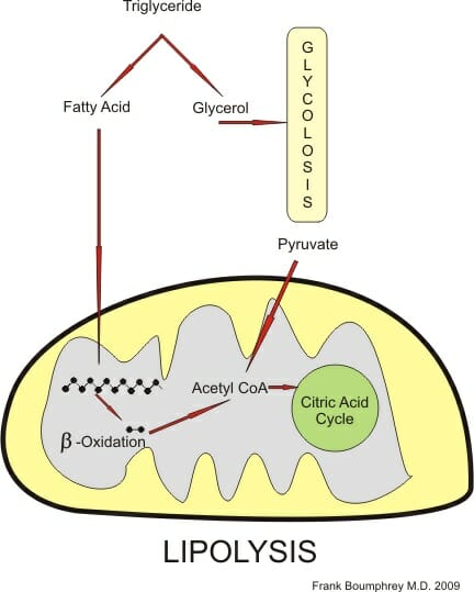
The figure below illustrates the main “destinies,” if you will, of the resulting fatty acids and glycerol. Fatty acids can undergo beta-oxidation and repurpose to create Acetyl-CoA. Of course, Acetyl-CoA is best known as a vital starting molecule that initiates the Krebs’s cycle in cellular respiration. This repurposing is vital when glucose stores are low in times of starvation, or even between meals, as cellular respiration can continue to run and sustain life. Similarly, the free glycerol can enter glycolysis. Normally glucose is converted to G6P at the first step of glycolysis. In the event that glucose levels are low, glycerol will be converted to dihydroxyacetone phosphate and will enter glycolysis at the second control point to keep glycolysis running. Thus, fats make the best energy store as they will ensure that cellular respiration continues to run and ATP is produced.


Lypolosis

Figure 2


The figure illustrates Lipolysis and the pathways the fatty acids and glycerol components take.


Lipolysis Regulation


Like every vital biological process, lipolysis is regulated to meet our needs. At any given time, it would be extremely harmful to have tons of free fatty acids flowing through our bloodstream. Anyone with high cholesterol or arterial plaques will attest to that. Thus, lipolysis – and its inverse process, lipogenesis – need to be counter-regulated and highly sensitive to the levels of specific hormones and proteins. For example, stimulatory hormones like, epinephrine, norepinephrine, cortisol, glucagon, and growth hormone induce lipolysis. Key hormones glucagon and epinephrine will use the same pathways to induce lipolysis with minor differences.


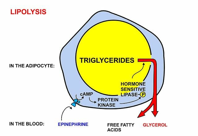
Both glucagon and epinephrine will serve as ligands that will bind to G-protein coupled receptors on the surface of fat cells. The G proteins will then activate adenylate cyclase and upregulate their conversion of ATP to cAMP. We might recognize cAMP as the famously ubiquitous secondary messenger of so many other biological pathways. Likewise, here the cAMP will activate protein kinase A (PKA), which will expend an ATP molecule in phosphorylating and upregulating the hydrolysis activity of our HSL enzyme – otherwise known as our second enzyme in the lipolysis pathway. As a result, we are left with free fatty acids and glycerol that can then enter metabolic pathways to counter the low sugars in our blood, for instance. Understandably, HSL was thought to be the rate-determining enzyme of lipolysis for some time before TAG lipase (or ATG, our first enzyme) was uncovered to be the key initiative lipolytic step. Let’s quickly take a look at why it makes sense for glucagon and epinephrine to trigger lipolysis.


Glucagon-induced Lipolysis


Glucagon is a peptide hormone that is synthesized by pancreatic cells in the event that glucose and thus insulin levels drop. Glucagon will then trigger our liver to break down its glycogen stores and release much needed glucose into our blood. Conversely, when our glucose and insulin levels are high, insulin in healthy individuals will allow glucose to exit the bloodstream and be taken up by insulin-dependent tissues. Of course, in diabetics, the tissues will no longer respond well to insulin and this sugar will not reach the tissues and instead cause havoc in the bloodstream.


Shifting back our focus to lipolysis, glucagon stores are small and will be expended quickly. Fat stores, on the other hand, are vast and ready to use. Here, glucagon serves its key role. Glucagon will bind to Glucagon G-protein coupled receptors on fat cell membranes, and trigger the HSL-activating pathway described earlier. The glycerol that is released can then travel to the liver or kidney where it will be eventually converted to GA3P and enter glycolysis and our gluconeogenesis pathway to synthesis badly needed glucose (refer to figure 2).


Epinephrine-induced Lipolysis


Metabolism

Figure 3


The diagram specifically illustrates epinephrine-induced Lipolysis through a G-protein mediated pathway.


Epinephrine will also bind G-protein receptors on fat cell membranes, however they will specifically bind beta-adrenergic receptors. This binding will likewise lead to the cAMP/PKA-led phosphorylation of hormone sensitive lipase, that will ultimately drive the release of free fatty acids and glycerol. Epinephrine is known for its connection to our instinctual “fight or flight” response. This hyperarousal occurs when we perceive an attack or threat to our survival. Thus, it makes sense that epinephrine would trigger lipolysis and its resulting up-drive of metabolic processes. If we are ever starving, our body will certainly react to this threat and use our fatty energy stores to respond and sustain life at all costs.


Lipolysis in Popular Culture


As briefly mentioned above, a fun fact is that lipolysis has become a popular term in the cosmetic world. Not to be confused with the adipose lipolysis pathways detailed in this article, laser lipolysis and even injection lipolysis are clinically proven methods of reducing the number of fat cells without liposuction surgery. Noninvasive fat reduction has become a new cosmetic staple, and promises to target fat cells through the use of heat, cooling (via lasers or radiofrequencies), or less commonly deoxycholic acid injections without disrupting surrounding tissues.


Quiz


1. Which of the following enzymes is the rate determining enzyme in lipolysis?
A. HSL
B. ATGL
C. MGL
D. None of the above

Answer to Question #1
B is correct. As mentioned above, researchers uncovered that the first lipolysis step, mediated by ATGL, is coincidentally the rate determining step of lipolysis. It was previously thought to be HSL as it undergoes phosphorylation.

2. Which of the following will induce lipolysis?
A. High insulin/Low epinephrine
B. High insulin/High epinephrine
C. Low insulin/High epinephrine
D. Low insulin/Low epinephrine

Answer to Question #2
C is correct. Low insulin and high epinephrine will trigger lipolysis. This makes sense if our body is constantly responding to feedback. When glucose and insulin levels are low, we will need fats to sustain gluconeogenesis and cellular respiration. High epinephrine will occur in the face of a life threat that will require the orchestration of fat energy expenditure.

References


  • Binienda, Z et al. “Role of Free Fatty Acids in Physiological Conditions and Mitochondrial Dysfunction.” SCIRP: Food and Nutrition Sciences, Vol. 4 No. 9A, 2013. Retrieved <http://www.scirp.org/journal/PaperInformation.aspx?PaperID=36092>

  • American Society of Plastic Surgeons (2018). “Nonsurgical Fat Reduction: Minimally Invasive Procedures.” Plasticsurgery.org. Accessed 2018, May 29 from <https://www.plasticsurgery.org/cosmetic-procedures/nonsurgical-fat-reduction/laser-lipolysis>

  • Ward, Colin (2015). “Lipolysis and Lipogenesis.” Diapedia: 51040851148 rev. no. 17. Accessed 29 May 2018 from <https://www.diapedia.org/metabolism-insulin-and-other-hormones/51040851148/lipolysis-and-lipogenesis>

  • Engelking, Larry R. (2014). “Chapter 70 – Lipolysis.” Textbook of Veterinary Physiological Chemistry (3rd Edition), Pages 444-449. Accessed 30 May 2018 from <https://www.sciencedirect.com/topics/neuroscience/lipolysis>

  • Fruhbeck, G et al. “Regulation of Adipocyte Lipolysis.” Nutr Res Rev. 2014 Jun; 27(1): 63-93. Doi: 10.1017/S095442241400002X


Lipolysis

Spindle Fibers

Spindle Fibers Definition


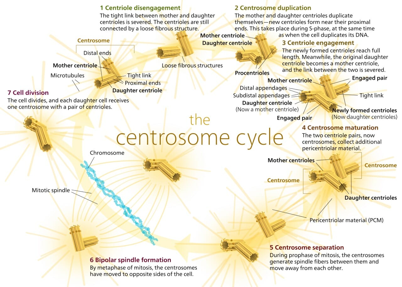
Spindle fibers are microscopic protein structures which help divide genetic material during cell division. The spindle fibers form out of the centrosome, also known as the microtubule-organizing center, or MTOC. Spindle fibers are formed from microtubules with many accessory proteins which help guide the process of genetic division. The spindle fibers form during cellular division near the poles of the dividing cell. As they extend across the cell, the search for the centromere of each chromosome.


Centrosome Cycle

Centrosome Cycle


Once attached, the spindle fiber is pulled back. With each fiber comes the chromosomes, which separates them along the poles. This process can be seen in the image above. The spindle fibers can be seen extending in all directions from the centrosomes. These spindle fibers are formed from several microtubules. The spindle fibers act like small machines during cell division. They carefully assemble and divide the chromosomes, and have been doing so for billions of year. But how does this complex process take place?


Structure of Spindle Fibers


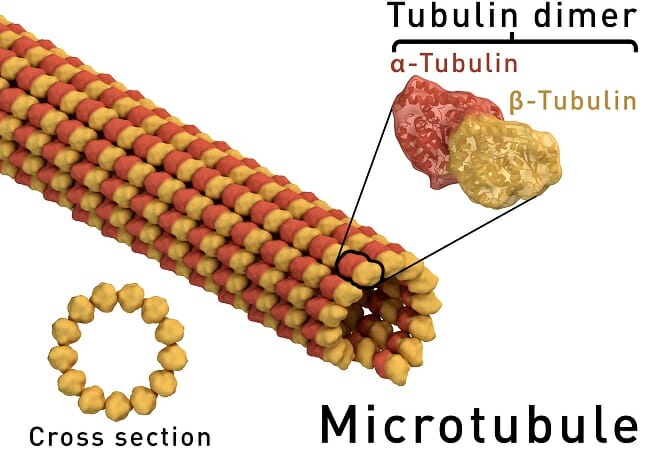
The centrosome, or MTOC, always has some microtubules preassembled. On the surface of the MTOC are small proteins, responsible for lengthening or shortening the microtubules. These proteins respond to signals from the cell, and when it is time for cell division, the begin lengthening the spindle fibers. To do this, they must add subunits of alpha-tubulin and beta-tubulin. Together, these two small proteins form the structure of a microtubule. Many individual microtubules together are called spindle fibers. A single microtubule can be seen in the graphic below.


Microtubule structure

Microtubule structure


Functions of Spindle Fibers


Shrinkage and Growth


The main feature of microtubules, and therefore of spindle fibers, is that the proteins which control them can extend or contract the microtubule by adding or removing tubulin dimers. At first the MTOCs must add many of these dimers to the microtubule, to extend it across the cell. As the microtubule travels, it eventually reaches a chromosome. Special proteins within the centromere of the chromosome can attach to the microtubule. Here, there are also proteins which can shorten and extend the spindle fibers.


This is one of the main ways that the chromosomes get aligned on the metaphase plate, a hypothetical middle of the cell. It is also the main way they are separated during anaphase. While the addition and subtraction of dimers is one of the main ways that spindle fibers help carry chromosomes about the cell, there are two other primary methods.


Sliding


When spindle fibers from opposite poles of the cell meet, they are bound together by a special protein. Instead of grabbing onto a chromosome, they more or less attach to each other via the protein. This protein is a specialized motor protein, which reacts to signals from the cell. At the appropriate time during cell division, the motor protein will begin crawling along each microtubule it is attached to. This “sliding action” causes pressure to be exerted against the poles, and helps drive the poles apart. This action of the spindle fibers is what forces the cell apart and allows for it to be divided in half during telophase.


Microtubule anchors


The final action carried out by some spindle fibers is that of anchoring to the cell surface. On the inside surface of the cell membrane, specialized proteins are placed to anchor the microtubules. While these anchors cannot assemble dimers into the microtubule, they can bind onto it. Then, when the MTOC starts removing microtubule dimers, the whole spindle fiber shortens. In this way it pulls the cell membrane toward the MTOC, and starts to define the area of the newly forming cell.


Quiz


1. Which of the following is NOT caused by the actions of spindle fibers?
A. The movement of chromosomes
B. The change in shape of the cell
C. The structure of the cell when not dividing

Answer to Question #1
C is correct. Spindle fibers form during cell division and are disassembled afterwards. While there are many different kinds of microtubules, they only act as spindle fibers during cell division. After cell division, the function of structure is carried out by more interspersed microtubules and other small structures. By using a completely different set of proteins, cell division and the organization of spindle fibers which is required can be completely regulated.

2. Microtubules form in a peculiar fashion. While the entire structure is just repeated units of the small tubulin dimer, the structure has polarity to it. That is, each side of the microtubule is different. On one side the beta-tubulin is more exposed, while on the other side the alpha-tubulin is more exposed. How must the proteins in the MTOC and the proteins on chromosomes be different in order to work?
A. They must be the same
B. They must be able to add dimers from opposite sides
C. They are completely different processes, therefore they are completely different proteins

Answer to Question #2
B is correct. The different sides of the microtubule (often referred to as + and –), have slightly different shapes which are just the opposite of each other. On one side, the protein must add or remove dimers with the alpha-tubulin facing in, while the others must do it with the beta-tubulin facing in. These two dimers are almost identical, so the change is small. But, it is still present and affects the way the cell’s machinery works.

3. Often, when products of an organelle are exported, they are contained within vesicles. These small compartments of cell membrane are then attached to a microtubule via a small motor protein. The protein works its way down the microtubule, like in the sliding example above. It carries the vesicle to another organelle or the cell surface. Here it can be expelled or absorbed. Are these microtubules considered spindle fibers?
A. No
B. Yes
C. Maybe

Answer to Question #3
A is correct. These are definitely not spindle fibers. Remember that spindle fibers are formed only during cell division and that their main purpose is dividing the genetic components of the cell. These are microtubules, but there are many uses for microtubules within the cell.

References


  • Lodish, H., Berk, A., Kaiser, C. A., Krieger, M., Scott, M. P., Bretscher, A., . . . Matsudaira, P. (2008). Molecular Cell Biology (6th ed.). New York: W.H. Freeman and Company.

  • McMahon, M. J., Kofranek, A. M., & Rubatzky, V. E. (2011). Plant Science: Growth, Development, and Utilization of Cultivated Plants (5th ed.). Boston: Prentince Hall.

  • Nelson, D. L., & Cox, M. M. (2008). Principles of Biochemistry. New York: W.H. Freeman and Company.


Spindle Fibers