Tuesday, September 18, 2018

Commensalism, Mutualism and Parasitism

Symbiosis describes several types of living arrangements between different species of organisms in an ecosystem. These relationships can be beneficial, neutral, or harmful to one or both organisms which are called symbionts. In the complex web of nature, species often have several symbiotic relationship at a time.


Symbiosis can take two forms known as obligatory and facultative. In obligatory symbiosis, one or both organisms are entirely dependent on the relationship and will die without it. Conversely, organisms in facultative relationships can live independently from each other.


Symbiotic relationships are also described by the physical relationship between the symbionts. Conjunctive symbiosis occurs when the symbionts have bodily contact with each other. In contrast, symbionts that do not have physical contact have a disjunctive symbiotic relationship. The term ectosymbiosis is when one organism lives on another, like a flea living in a dog’s fur. Endosymbiosis is a relationship where one symbiont lives in the tissues of another such as bacteria living in the human gut.


Commensalism, mutualism, and parasitism are the three main categories of symbiosis found in nature.


Commensalism


In a commensal relationship, one species benefits and there is a neutral effect on the other—it neither benefits nor is harmed. An example of this relationship is birds building nests in trees. The nests don’t interfere with photosynthesis and are light weight, so they don’t put a strain on the trees. The birds, on the other hand, benefit by having their young protected from predators on the ground and hidden by the leaves and branches of the tree. The tree may also provide an accessible food source for the birds such as berries, grubs, and insects. Other examples of commensalism are spiders spinning webs on plants and hermit crabs that use discarded snail shells to protect themselves.


Commensal relationships are sometimes hard to identify because it can be difficult proving that one symbiont does not benefit in some way from the relationship.


Mutualism


In this type of symbiosis, both organisms benefit from the relationship. A classic example of this is the relationship between termites and the protists that live in their gut. The protists digest the cellulose contained in the wood, releasing nutrients for the benefit of the termite. In turn, the protists receive a steady supply of food and live in a protected environment. The protists themselves also have a symbiotic relationship with the bacteria that live in their gut, without which they could not digest cellulose. This relationship between termites and protists is obligatory—the termites would die of starvation without the protists to digest their food.


Other examples of mutualism are the algae that live in the tissues of coral in reefs, clownfish that live in the tentacles of sea anemones, and the relationship between the Oxpecker bird and zebras and rhinoceroses on the African plains.


Parasitism


Parasitism is a relationship where one symbiont benefits (the parasite) and the other (the host) is harmed in some way and may eventually die. Parasites can damage their hosts or sicken them and make them weak. There is usually a built-in selection process that slows down the rate of damage to the host, giving the parasite time to complete its reproductive cycle and for its offspring to find a new host.


A tapeworm in the digestive tract of a human or other animal is an example of a parasitic relationship. The worm feeds on the food the person eats and grows within the intestines, sometimes reaching 50 feet in length. Other examples are the malaria parasite spread by mosquitoes, fleas and ticks, and aphids that suck the sap from plants.


References


  • Nelson, D. (2018, February 6). Mutualism, Commensalism, Parasitism: Types of Symbiosis with Examples. Retrieved May 23, 2018, from https://sciencetrends.com/comparing-examples-mutualism-commensalism-parasitism-symbiosis/

  • Symbiosis. (2018, May 9). In Wikipedia. Retrieved from https://en.wikipedia.org/w/index.php?title=Symbiosis&oldid=840414702


Commensalism, Mutualism and Parasitism

Sperm Motility

Sperm motility is the ability of sperm to move through water during external fertilization or within the female reproductive tract for internal fertilization to reach the egg. Motility also refers to the quality of the sperm motion, meaning that sperm which does not move properly can’t reach the egg and successfully fertilize it. Motility also encompasses the ability of the sperm to penetrate the egg once it reaches it.


Sperm Structure and Movement


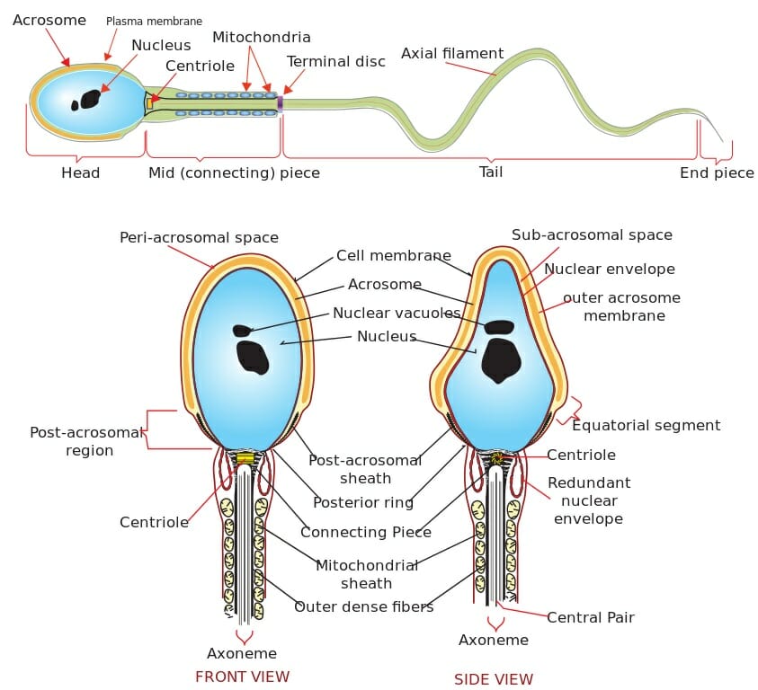
A sperm has four main sections: the head, midpiece, tail, and end piece (Figure 1). The head contains the nucleus and is surrounded by the acrosome (cap) and the plasma membrane. The acrosome contains the enzymes the sperm will need to penetrate the surface of the egg. The centriole (which the sperm will donate to the egg upon fertilization) joins the head to the midpiece which has a filamentous core composed of 11 tubules called the axoneme. The midpiece is surrounded by mitochondria that supply energy for the sperm in the form of adenosine triphosphate (ATP).


The tail or flagellum of the sperm is the longest section and the terminal disc separates it from the midpiece. The tail, powered by ATP made by the mitochondria in the midpiece, propels the sperm using a back and forth lashing motion. The motion is created by the rhythmical sliding of the tubules in the axoneme. The end piece contains the axoneme surrounded by the plasma membrane. It is located at the terminus of the tail and tapers down in diameter.


Changes in ion concentration and pH activate sperm movement and the requirements vary by species. For example, in some mammals, an increase in pH and calcium ions activates sperm. The ultimate effect is membrane hyperpolarization which activates the sperm.


Human spermatozoa diagram

Figure 1


The image above shows the detailed structure of a human sperm cell.


Evaluating Sperm Motility


The percentage of motile sperm is the most widely used measurement of semen quality. Normal or acceptable sperm motility varies among species. For example, human sperm motility greater than 50% is normal but only 30% is required in bulls, and as high as 70% is required in dogs. Sperm motility is described as non-motile, progressively motile, and non-progressively motile. Progressively motile sperm swim in a straight line while non-progressively motile means the sperm swim in an abnormal path such as around in circles. Test results usually report the percentage of progressively motile sperm.


There are three main methods for quantifying sperm motility. Some are more accurate than others and require more skill on the part of the operator and/or use more expensive equipment. In a manual motility estimate, a diluted sample of semen is placed on a pre-warmed slide and viewed under a microscope. The operator counts the number of non-motile, progressively motile, and non-progressively motile sperm in at least ten different fields on the slide. From this, an estimate of the percentage of motile sperm is calculated.


Track motility estimates use the same sample preparation as the manual motility estimate. The sperm are photographed using an exposure time of about 0.2 seconds which records their movement on the slide. Progressively motile sperm will leave tracks in a straight line and the non-progressively motile sperm will leave circles or tracks showing some other abnormal path of movement. Of course, non-motile sperm leaves no tracks.


The latest technology involves computer-aided motility analysis. The method is similar to the track motility test, but software detects and tracks the movement of each sperm in the sample and tabulates the data automatically. This method gathers additional data such as the velocity of the sperm and other details about their movement.


References


  • Sperm. (2018, May 15). In Wikipedia. Retrieved from https://en.wikipedia.org/w/index.php?title=Sperm&oldid=841448719

  • Sperm motility. (2017, August 5). In Wikipedia. Retrieved from https://en.wikipedia.org/w/index.php?title=Sperm_motility&oldid=793978230

  • Sperm Motility. (n.d.). Retrieved May 22, 2018, from http://www.vivo.colostate.edu/hbooks/pathphys/reprod/semeneval/motility.html


Sperm Motility

Monocot Root, Leaf, Flower and Plants

The term monocot is short for monocotyledon. The cotyledon is an embryonic leaf in a seed that is the first to emerge when it germinates. Monocot seeds have one cotyledon while dicotyledons, or dicots, have two. Monocots and dicots are two types of angiosperm plants which reproduce using seeds and fruits.


There are about 60,000 species of monocot plants. The largest family are the orchids which have over 20,000 species followed by grasses with 10,000 species. Scientists believe monocots evolved as early as 140 million years ago. Based on pollen grains in the fossil record, the earliest monocots lived in the early Cretaceous period about 120-110 million years ago.


Monocots are found in a variety of habitats. They grow primarily on land but also in rivers, lakes, and ponds, mostly rooted to the bottom but sometimes free-floating. Some also live in intertidal zones near the seashore and a few are marine plants rooted in shallow areas in the ocean.


Roots


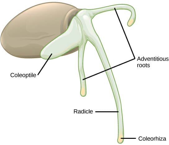
The roots of monocots cannot grow in diameter due to the lack of vascular cambium. Instead, they grow more roots at the shoot (radicle) and send out creeping shoots called runners or rhizomes (Figure 1). The coleorhiza is a tough sheath of tissue at the end of each root that protects it as it works its way through the soil. A structure called the coleoptile has the same function earlier in the growth of the root. This fibrous root system that originates from areas of the plant other than existing roots is called an adventitious root system.


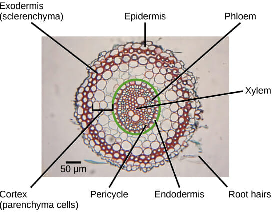
The tissue at the center of monocot roots consists of xylem and phloem (vascular bundle) and it is surrounded by the cortex which is made of parenchyma cells (Figure 2). The outermost layer of the root is called the epidermis followed by the exodermis or sclerenchyma. The endodermis is an inner layer of cells surrounding the vascular bundle. The endodermis and phloem are separated by a layer of cells called the pericycle where root branching occurs.


Figure 1

Figure 1


The image above shows the root structure of a germinating monocot seed.


Figure 2

Figure 2


The image above is the cross section of a monocot root.


Leaves


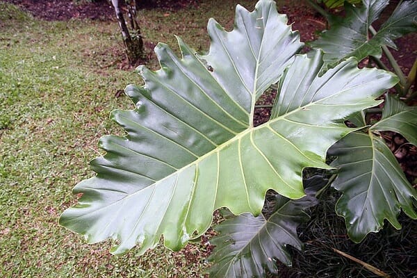
Monocot leaves are usually long and narrow or oblong with parallel veins running through them (Figure 3). However, the diversity of nature reveals many exceptions to this rule. There is usually one leaf per node on the stem because the base of the leaf takes up more than half of the circumference of the stem.


Monocot leaves have an equal number of stomata (pores) on the upper and lower leaf surfaces. They also have large vascular bundles and bulliform (bubble-shaped) cells on the upper surface. Both of these features help monocots retain water during dry or stressful environmental conditions. Also, the cuticle layer is thicker on the upper leaf surface.


Philodendron Wilsonii

Figure 3


The image above shows the parallel veins in Philodendron wilsonii which is characteristic of monocots.


Flowers


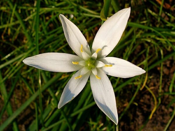
Monocots are identified by their flowers and flower parts that are in groups of three, also called trimerous (Figure 4). About two-thirds of all monocots are pollinated by animals including bats, monkeys, deer, rodents, and birds such as hummingbirds. Therefore, the flowers are often colorful and ‘showy’ to visually attract pollinators and they use pleasing aromas for chemical attraction.


Ornithogalum umbellatum

Figure 4


The image above shows the grass lily Ornithogalum umbellatum with its flower parts in multiples of three, characteristic of monocots.


Examples of Monocot Plants


Monocots are important plants around the world both economically and culturally. They account for many human and animal food staples like wheat, corn (Figure 5), barley, rice, and grasses. Other examples of monocot plants are bananas, sugarcane, palms, pineapples, orchids, and lilies. Monocots make up the most species grown in agriculture in terms of the amount of biomass produced.


References


  • Monocotyledon. (2018, May 16). In Wikipedia. Retrieved from https://en.wikipedia.org/w/index.php?title=Monocotyledon&oldid=841518727

  • Monocotyledon plant. (n.d.). Retrieved May 22, 2018, from https://www.britannica.com/plant/monocotyledon


Monocot Root, Leaf, Flower and Plants

Monocot vs Dicot

Angiosperms are plants that live on land and reproduce using seeds in flowers and fruits.


Monocotyledons and dicotyledons, also known as monocots and dicots, respectively, are two types of angiosperm plants. The Italian physician and biologist Marcello Malpighi (1628 – 1694) was the first to use the term cotyledon (the Latin word meaning seed leaf) and John Ray (1627 – 1705), an English naturalist, was the first to notice that some plants have one cotyledon and others have two.


The cotyledon part of angiosperms is an embryonic leaf that is the first leaf (or leaves) to appear when a seed is germinating. Cotyledons perform photosynthesis but are not true leaves because they are present in the seed before it germinates. True leaves grow after the seed has germinated. Cotyledons may last only a few days after the seed germinates (ephemeral) or last up to a year (persistent).


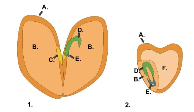
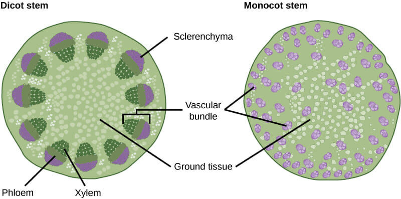
Monocots and dicots differ in several ways which help in their identification and understanding of their origins. Paleobotanists, scientists who study the origins of plants, hypothesize that dicotyledons evolved first, and monocots branched off about 140 to 150 million years ago either from the fusion of the cotyledons or as a separate line. See Figures 1 and 2 for illustrations of the different physical features discussed below.


Monocots


Monocot plants have one cotyledon. They also have long narrow leaves with parallel veins. Cutting a cross section from the stem of a monocot shows the vascular bundles scattered around in the plant tissue. The young plant stores food in the form of starches and other nutrients in a structure called the endosperm.


Another key characteristic for identifying monocots is by the number of flowers or flower parts which are arranged in groups of three. Also, the pollen grains of monocot plants have a single pore or furrow making them monosulcate (from the Greek word mono meaning ‘single’ or ‘one‘ and the Latin word sulcatus meaning ‘furrow’) and new roots originate from the stem of the plant. Some examples of monocots are lilies, orchids, corn, rice, wheat, barley, pineapple, sugar cane, bananas, palms, and grasses.


Dicots


As opposed to monocots, dicots (also called eudicots) have two cotyledons during germination which supply the young plant with food and nutrients. The leaves of dicot plants come in a variety of shapes and sizes and the veins form branching patterns. Microscopic examination of dicot seeds shows a structure called the hilum which is a scar on the seed coat where the ovary was attached. This feature is not seen in monocots. Also, different from monocots is the roots of dicot plants originate from the radicle.


Another way dicots are distinct from monocots is their flowers and flower parts are arranged in multiples of four or five. In addition, the cross section of a dicot stem shows the vascular bundles arranged in a circular pattern. Unlike monocots, the pollen grains of dicot plants have three pores and are called trisulcate. Dicot plants can also have bark and secondary growth increases the diameter (girth) of the plant. Examples of dicots include potatoes, tomatoes, apples, pears, peaches, cauliflower, peppers, broccoli, and cabbage.


MonocotsDicots
Direction of leaf veinsParallelBranched
Orientation of vascular bundlesScatteredArranged in circles
Number of flowersMultiples of 3Multiples of 4 or 5
Number of embryonic leaves12
Origin of new rootsFrom nodes in the stemFrom the radicle
Shape of true leavesMostly long and narrowWide variety of shapes
Secondary growthNoneYes. Plant girth increases each year
Forms true bark?NoYes
Number of furrows or pores in pollen grains1 (monosulcate)3 (trisulcate)
Food and nutrient storage locationEndospermCotyledons
Has a hilum?NoYes

Monocot dicot seed

Figure 1: The image above shows a generalized dicot seed (1) and a generalized monocot seed (2). The structures in each type of seed are: A = seed coat, B = cotyledon, C = hilum, D = plumule, E = radicle, and F = endosperm. Note that the dicot seed lacks endosperm, and the monocot does not have the hilum that is present in the dicot seed.


Dicot stem vs monocot stem

Figure 2: The image above shows a cross section of the stem of a dicot plant (left) and monocot (right). Note how the vascular bundles are scattered in the monocot stem and arranged in a circular pattern in the dicot stem.


References


  • Cotyledon. (2018, April 6). In Wikipedia. Retrieved from https://en.wikipedia.org/w/index.php?title=Cotyledon&oldid=834509052

  • OpenStax College. (2018). Concepts of Biology. Houston, TX. OpenStax CNX. Retrieved from http://cnx.org/contents/b3c1e1d2-839c-42b0-a314-e119a8aafbdd@9.39


Monocot vs Dicot

Lateral Meristem

Meristem is undifferentiated plant tissue found in areas of plant growth. The three types of meristematic tissue are intercalary, apical, and lateral. Apical meristem tissue is found in the tips of shoots and gives rise to leaves and flowers and is also found in the roots. The intercalary tissue in the middle of the plant is capable of rapid growth and regrowth. For example, the intercalary tissue at the base of a blade of grass allows it to regrow after being cut.


Plants use lateral meristem tissue to grow in diameter as part of secondary growth. There are two types of lateral meristematic tissue—the vascular cambium and the cork cambium.


Vascular Cambium


In plants, the vascular cambium is the main route by which the stems and roots grow. The tissue consists of xylem toward the outside and phloem inside. In woody plants, it forms a continuous ring of new wood around the stem. Herbaceous plants don’t have wood, so the vascular cambium forms bead-like bundles that create a ring around the stem. The two types of vascular cambium cells are fusiform initials which are tall and aligned with the axis of the stem and ray initials which are smaller than fusiform initials and rounder.


The vascular cambium has its own set of hormones that control growth, regulation, and maintenance activities in the tissue. The hormones belong to such families as auxins, gibberellins, and cytokinins, and chemicals like ethylene also have hormonal functions in the vascular cambium.


Xylem rays

The image above is the cross-section of a plant stem showing the vascular cambium, xylem cells, and xylem rays.


Cork Cambium


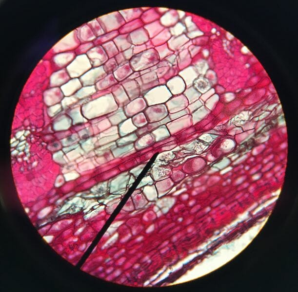
This tissue is present in mostly woody and some herbaceous plants and gives rise to the cork or bark layer on the outside of the stem and secondary growth in the epidermis of roots. This is accomplished by replacing the epidermal cells with the periderm which consists of three layers. The phelloderm is the innermost layer made of living parenchymal cells. On top of that layer is the cork cambium itself or the phellogen that gives rise to the periderm. The outermost layer is the cork or phellem (bark) which is made of dead, air-filled cork cells. The development and appearance of the cork cambium varies greatly among species. Some plants and trees have smooth bark while others are rough, scaly, and even naturally flake off from the tree.


Cork cambium

In the image above, the black pointer shows the location of the cork cambium in the cross-section of a woody plant stem.


References


  • Cork cambium. (2018, January 30). In Wikipedia. Retrieved from https://en.wikipedia.org/w/index.php?title=Cork_cambium&oldid=823080623

  • Vascular cambium. (2018, March 2). In Wikipedia. Retrieved from https://en.wikipedia.org/w/index.php?title=Vascular_cambium&oldid=828437156


Lateral Meristem

Marine Ecosystem Facts

Marine ecosystems include not just the oceans but also shorelines, tidepools, estuaries, barrier islands, mangrove forests, and salt marshes. Here are the top 5 facts about marine ecosystem.


The Marine Ecosystem is the Largest Ecosystem on Earth


The oceans alone cover about 70% of the Earth’s surface or 140,000,000 square miles. The average ocean depth is about 12,000 feet and the deepest point is the Mariana Trench in the Pacific Ocean with a depth of about 32,800 feet.


The Marine Ecosystem has the Greatest Biodiversity on Earth


Almost half of the known species on Earth live in marine ecosystems and scientists suspect there may be another 1 million yet to be discovered. Roughly 700,000 to 1 million species live in the oceans.


Phytoplankton in the Oceans Provide 50% to 85% of the Oxygen on Earth


Phytoplankton are tiny plants that live in the upper areas of the ocean and use photosynthesis to make their food. They are so abundant in the oceans that all together they account for about 50% of the photosynthetic activity and over 50% of the oxygen production on the planet.


Mangrove Forests are Diverse Ecosystems


Mangrove forests are found on tropical and subtropical marine coastlines and tidal areas. They contain small trees and shrubs tolerant of salt water. The root systems of the forests form tangled webs of habitat where many species of fish, invertebrates, seabirds, and waterfowl live, reproduce, and mature.


Oceans Regulate the Earth’s Climate


The oceans absorb most of the heat radiated from the sun especially around the equator. The ocean currents distribute the heat around the planet, but most of the heat is lost due to evaporation. The constantly evaporating ocean waters create rain, thunderstorms, and hurricanes by increasing the temperature and humidity of the air. Because the trade winds carry these storms over vast distances, most of the precipitation that falls on land originates in the oceans.


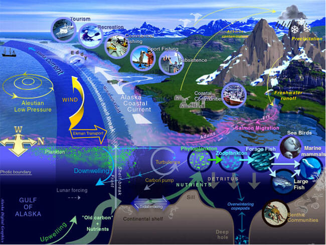
General characteristics of a large marine ecosystem

The image above shows the typical characteristics of the marine ecosystem in the Gulf of Alaska.


References


  • Facts and figures on marine biodiversity | United Nations Educational, Scientific and Cultural Organization. (n.d.). Retrieved May 16, 2018, from http://www.unesco.org/new/en/natural-sciences/ioc-oceans/focus-areas/rio-20-ocean/blueprint-for-the-future-we-want/marine-biodiversity/facts-and-figures-on-marine-biodiversity/

  • Ocean. (2018, May 15). In Wikipedia. Retrieved from https://en.wikipedia.org/w/index.php?title=Ocean&oldid=841293100


Marine Ecosystem Facts

How Climate Change Affects the Biodiversity of Marine Ecosystems

The biodiversity found in marine ecosystems is greater than in any other on Earth. Climate change causes wide-ranging effects including changes to water pH, nutrients, oxygen content, and stratification. These changes affect the biodiversity of communities, particularly in the polar regions of the planet.


Effects on Ice-Dominated Polar Ecosystems


Climate change is affecting the Earth’s northern and southern poles at a faster rate than anywhere else. The health of polar marine ecosystems is intimately tied to seawater temperature and the amount of sea ice present. These two factors influence the growth and reproduction of organisms, food sources, and the biogeochemical cycles of the region.


An example of the effects of climate change on the biodiversity in the polar regions is the reduced population of Adélie penguins. The loss of sea ice in the area along with reduced amounts of krill and an increase in late spring snowfalls has resulted in an 80% reduction in the Adélie penguin population in the region of Palmer Station in Antarctica. At the same time, species such as the Gentoo penguin and fur seals are migrating to this area to take advantage of the ecological niche that has opened up due to the decline in the penguin population.


Effects on Coral Reef Ecosystems


About 25% of all marine species are associated with coral reefs. These reefs are very sensitive to changes in the pH and temperature of ocean waters. For example, an increase in water temperature as little as 1°C causes coral bleaching, the loss of color due to the death of the zooxanthellae that live within the coral tissues. But, bleaching does not affect only the color of coral. Moderately bleached coral has lower growth and reproduction rates and severe bleaching kills them. Because of this high sensitivity, reef stress is an early warning sign of changes in water acidification and temperature. Besides climate change, coral reefs also suffer from pollution, overfishing, invasive species, and nutrient overenrichment.


Many organisms that live in coral reefs are negatively impacted when reefs are damaged by increased temperature and water acidification. Coral provides food, structure, mating/spawning areas, and cover for these creatures. With the loss of reefs, some species can migrate to rocky areas to live but others specialized to live in the reefs will die off. Scientists believe if conditions continue to deteriorate, there will be reduced diversity of fish and invertebrate species in these areas.


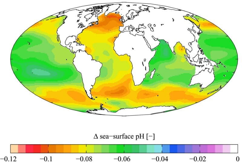
Estimated change in annual mean sea surface pH

The image above shows the change (delta) in the surface water pH of the world’s oceans. The acidification of the oceans is one of the key indicators of climate change.


References


  • Doney, S. C., Ruckelshaus, M., Duffy, J. E., Barry, J. P., Chan, F., English, C. A., … Talley, L. D. (2012). Climate Change Impacts on Marine Ecosystems. Annual Review of Marine Science, 4(1), 11–37. https://doi.org/10.1146/annurev-marine-041911-111611


How Climate Change Affects the Biodiversity of Marine Ecosystems