Showing posts with label 10k. Show all posts

Monday, April 2, 2018

Bilirubin

Bilirubin Definition


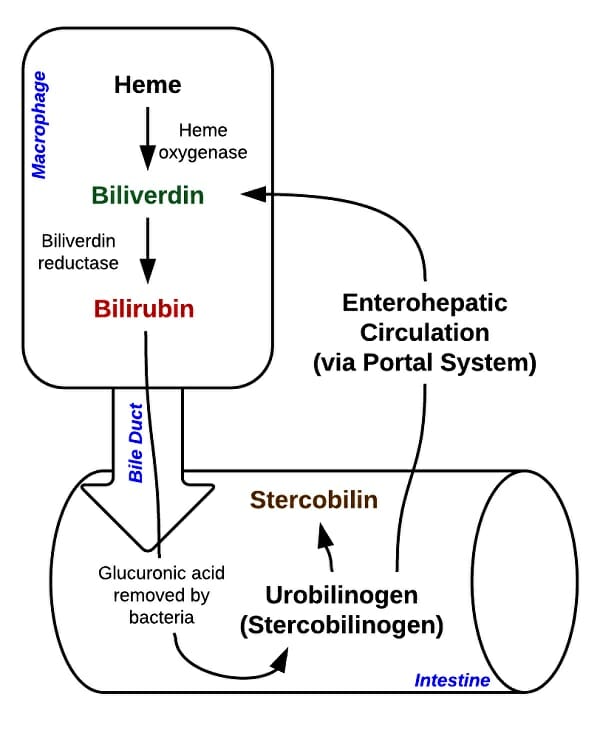
Bilirubin is a molecule formed from the breakdown of red blood cells, and other cells with porphyrins. Red blood cells carry special molecules, called hemes (a type of porphyrin), which hosts an iron atom. This arrangement allows the red blood cell to carry oxygen. When blood cells break down, they release the heme into the blood, which could do damage if not controlled.


Heme is actively broken down in the liver, as seen in the image below. It is first converted into biliverden. Another enzyme continues the process, and converts biliverden into bilirubin. Bilirubin is then deposited into the intestines, via the bile duct. From there it can be excreted in the urine or in the feces. In the intestines it is converted to stercobilin, a reddish-brown substance that gives feces its distinct coloration. In the urine, bilirubin becomes urobilin, a very yellow substance.


Heme Breakdown


Bilirubin, because it is related to porphyrin molecules, is a natural pigment. As it changes into different molecules and versions, it also changes color. Scientists can use these colors to detect disease. For instance, a bruise is caused by the pooling of blood under the skin. Pooling blood is a dark red or purple, and will slowly turn more brownish as the iron oxidizes. As the blood is removed and recycled, the heme groups must be broken down. Heme is first converted to biliverden, which has a greenish tint. If you’ve ever had a large bruise that appeared greenish, this is why. After this, it is converted to bilirubin, and has a distinctive yellow color. Old bruises will turn this distinctive yellow before disappearing.


The distinctive yellow coloration of bilirubin buildup is a common symptom of conditions like jaundice, in which bilirubin builds up all over the body. This can happen in babies, when the liver has not yet fully developed, or in adults with non-functioning livers. While the yellow coloration of the eyes and skin is a definite sign that something is drastically wrong, a bilirubin test can show the levels of bilirubin long before they become dangerous.


Bilirubin is also found in plants, as a breakdown product of chlorophyll, a molecule highly related to the heme molecules in animal cells. It is also very similar to the pigment phycobin, which is used by autotrophic bacteria to capture energy from sunlight.


Bilirubin Test


Conjugated versus Unconjugated Bilirubin


Bilirubin exists in multiple forms in your blood. It exists in a water-soluble form, which is attached to glucuronic acid in the liver by a special enzyme. This makes bilirubin easier to detect and is known as conjugated bilirubin. Unconjugated bilirubin, on the other hand, is fat-soluble, which makes it harder to detect in solution. As such, some of this bilirubin is undetectable. It bonds to itself and other fat-loving molecules.


Therefore, a test of a person’s total bilirubin includes the direct measurement of soluble bilirubin, plus the estimated indirect bilirubin. These two forms, and their abundance in the system, can tell a doctor a lot about which process may be getting interrupted in the liver. For instance, a healthy person will have a higher concentration of unconjugated bilirubin, but the exact levels depend on the person and circumstance.


Normal levels for total bilirubin can be anywhere from .1 to 1.2 milligrams per deciliter of blood, and the direct bilirubin will only be .1 to .4 mg/dL of that. Conditions of hyperbilirubinemia, or a really high bilirubin level, can happen for many reasons. The total blood bilirubin would have to be much higher than this before this condition would start to appear. Typically, around 10 mg/dL symptoms of bilirubin buildup, such as jaundice of the eyes and skin can be seen. This is around 10 times the regular levels.


It could be caused by rapid blood cell loss, a non-functioning liver, or a variety of other condition which stop the liver or other organs from properly processing bilirubin. This condition could also be caused by a blockage of the bile duct, hepatitis, or certain pharmaceuticals which are interfering with the passage or conversion of bilirubin. Your doctor will have many other tests to take into consideration, as your bilirubin levels can indicate many different conditions.


Urine Bilirubin Test


Healthy people do not normally excrete bilirubin in the urine. It is normally converted to another chemical, which is much more water soluble. Bilirubin in the urine is a symptom of a number of liver, kidney, and intestinal diseases in which a biological process is being obstructed. Finding bilirubin in the urine can help a doctor differentiate where in the excretory system the error is occurring. There are many factors which will help determine this, including the type of bilirubin found and its quantity.


Quiz


1. Why is bilirubin yellow and biliverden green?
A. The structure of their bonds
B. The energy they give off
C. It’s their favorite color!

Answer to Question #1

2. What do chlorophyll, heme, and bilirubin all have in common?
A. Nothing
B. They are all pigments
C. They are all porphyrin rings

Answer to Question #2

3. Newborn babies often become “jaundiced” or very yellow after birth. Why is this?
A. They genetically can’t process bilirubin
B. The liver is not yet fully developed
C. It is perfectly natural and not harmful

Answer to Question #3

References


  • Bruice, P. Y. (2011). Organic Chemistry (6th ed.). Boston: Prentice Hall.

  • Nelson, D. L., & Cox, M. M. (2008). Principles of Biochemistry. New York: W.H. Freeman and Company.


Bilirubin

Friday, September 8, 2017

Pituitary Gland

Definition of Pituitary Gland


The pituitary gland, also known as hypophysis, is a diminutive, pea sized gland located at the base of our brains. It is commonly referred to as the “master gland” of the human body, as it releases a ton of hormones that circulate our system and aid in maintaining our internal homeostasis. Moreover, the pituitary gland is also the “master” or dominant gland controlling the activity of other glands, as well. The pituitary gland is both responsible for producing and storing an assortment of important hormones that we will discuss in more detail.


Pituitary gland in brain

The image is an illustration of the pituitary gland as it is situated in the human brain. The depiction shows its relative size.


Pituitary Gland Location


The pituitary gland lies roughly in the center of the human skull. It rests below the hypothalamus of the brain and behind the bridge of our noses. Its setting actually makes sense, in light of the hypothalamus’s role in fine tuning the activity of the pituitary gland. This is made possible by the nerve fibers that span these two structures and allow for easy communication. Likewise, a thin vascular connection that is forged within the pituitary stalk, or infundibulum, facilitates the hypothalamus’s control. Further, the pituitary gland itself is supplied by branches off of the internal carotid artery. Its regulation is fine-tuned by a negative feedback relationship between the pituitary and hypothalamus.


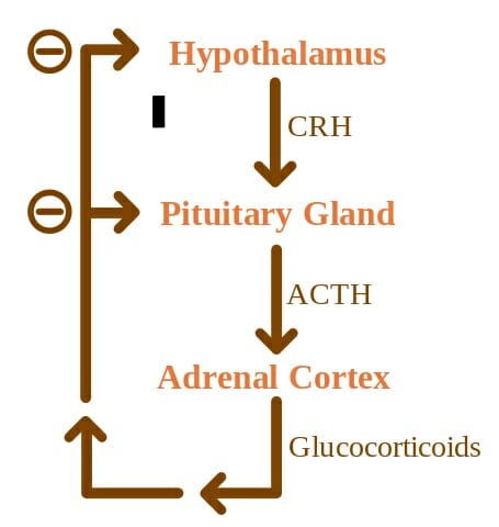
ACTH Negative Feedback

The concept map illustrates the complex regulatory relationship between the superseding hypothalamus and the pituitary gland. The relationship follows a negative feedback loop.


Structurally speaking, the pituitary gland is notably parsed into three sections: the anterior (front), intermediate, and posterior (back) lobes. Each can be described according to their unique functions. The anterior lobe has primary roles in the development of the human body. This involves secreting hormones that orchestrate our reproduction and sexual maturation. These hormones will control growth as well as activate the adrenal and thyroid glands and sexual organs. The intermediate lobe will secrete hormones that stimulate the cells in our body that produce pigment, called melanocytes. These melanocytes are the reason there is such a variation in our skin color. Lastly, the posterior lobe makes ADH, which is the hormone that allows our kidneys to reabsorb water into the bloodstream to prevent dehydration. Oxytocin is also made in the posterior lobe and will induce contractions during childbirth. While these hormones are supremely important to our species’ survival, they are few among the many hormones made by the pituitary gland.


Pituitary Gland Function


The main function of the pituitary gland lies in its ability to make hormones that retain many of our bodily functions. The front and back lobes are the primary secretory glands. As discussed before, the posterior lobe secretes oxytocin and ADH. Oxytocin not only stimulates uterine contractions to facilitate birth but also causes breast tissue to make milk, in preparation of caring for a child. The anterior pituitary gland has a bigger roster of hormones. It produces prolactin, which like the posterior’s oxytocin will trigger milk production post-partum. Follicle-stimulating hormone (or FSH) is released to stimulate sperm production and egg maturation in women that are able to produce estrogen. Likewise, luteinizing Hormone (LH) will stimulate testosterone release in men and egg release in ovulating women. One of the most important products of the anterior lobe is thyroid-stimulating hormone (TSH). The thyroid helps coordinate metabolic activity, and likewise, the TSH will stimulate thyroid activity. Therefore, TSH indirectly allow the thyroid to assume all of its roles. Adrenocorticotropic (ACTH) is released as well and will stimulate the creation of stress hormone, cortisol. Cortisol is essential to our survival and will keep our blood pressure and sugar levels at a healthy normal – of course, in healthy amounts. Any over or under expression has negative consequences. Lastly, the anterior pituitary lobe also releases growth hormone (GH), which is responsible for the muscle and bone mass growth that occurs during development. When growth is unaccounted for, as with a dysregulation of GH production, it can lead to serious illness, if not cancer as we will discuss below.


Posterior Pituitary Lobe:


  • ADH

  • Oxytocin


Anterior Pituitary Lobe:


  • Prolactin

  • Follicle Stimulating Hormone (FSH)

  • Luteinizing Hormone (LH)

  • Thyroid-Stimulating Hormone (TSH)

  • Adrenocorticotropic (ACTH)

  • Growth Hormone (GH)


Pituitary Gland Disorders


Deficiencies in any of the hormones mentioned above can cause illness, which vary in gravity. Starting with the posterior lobe, a deficiency of ADH will increase our thirst and urination. A lack of prolactin will quite predictably lead to an inability to lactate, which to this day cannot be treated. TSH deficiency has symptoms similar to those from a compromised thyroid gland, which includes fatigue, memory loss, and bodily weakness. A lack of LH or FSH will result in a decrease in libido, irregular menses, erectile dysfunction, and mood changes. ACTH deficiency will cause nausea, body aches, poor appetite, and even low blood sugar and pressure. Lastly, deficiency growth hormone will lower muscle mass and bone density, which has long term ramifications on the quality of our lives.


An overproduction of hormone has its own consequences. Too much growth hormone can lead to gigantism and acromegaly, or too much growth of bones and soft tissues leading to heart issues and sleep apnea. Too much TSH will result in shakiness, irritability, and high blood pressure. Too much prolactin will cause inappropriate expression of breast milk that can occur in women or men, and a weakening of bones. Excess ACTH will cause weight gain among brittle bones and mood instability. Lastly, excess FSH and LH are linked to infertility and irregular menstruation.


The most common type of pituitary gland disorder, however, are tumors. The grand majority of pituitary tumors are benign, or just a noncancerous swelling in the gland that may not cause any symptoms and may never be symptomatic. Unlike many types of tumors, most people afflicted with pituitary tumors have no prior family history of issues with the pituitary gland and is not usually genetically inherited. One of these exceptions is multiple endocrine neoplasia (or MEN) which is a set of inherited disorders that lead the body’s endocrine glands, including the pituitary gland, to overexpress hormones. But pituitary tumors, specifically, still remain by and large benign.


Tumors on the Pituitary Gland


Various types of pituitary tumors exist. In general, people with a pituitary gland tumor will experience a series of telltale symptoms. Most will have vision problems, headaches, menstrual changes, infertility, mood changes, fatigue, and even Cushing’s syndrome – which has its own set of symptoms including, but not limited to, high blood pressure and weight gain secondary to too much ACTH release.


The most common type of pituitary tumor is called a “non-functioning” tumor. The name derives from its inability to make hormones. These patients will have issues with their vision and headaches. Furthermore, pituitary tumors can be divided into three groups according to their problematic actions.


Hypersecretion refers to the making of too much hormone, and this is an issue afflicted by a secretory pituitary tumor. Hyposecretion, in contrast, is too little hormone production and is normally caused by a large pituitary tumor that will physically block the pituitary gland from making hormone. It can also result from surgical resection of a tumor. Lastly, tumor mass effects are the issues that arise from a growing pituitary tumor that is pressing against the pituitary gland and may result in compromised vision and headaches, as well.


Other pituitary conditions worth noting include craniopharyngioma. This is a type of cyst or tumor that is congenital, meaning it is present at birth. It can swell and fill with fluid, and may cause headaches and vision issues as well as sleep issues. ESS, or empty sella syndrome is a disorder that arises from an affliction in the bony structure that encases the brain and surrounds the pituitary. A primary ESS will be a small defect that give rise to high pressure in the bony base that causes the gland to flatten. This is linked to high blood pressure and obesity in females. On the other hand, secondary ESS will result from surgery or an injury that has caused the pituitary gland to regress. The symptoms will be related to pituitary function loss, such as infertility and fatigue.


Quiz


1. Which of the following is released by the posterior pituitary?
A. Prolactin
B. ACTH
C. Oxytocin
D. Growth Hormone

Answer to Question #1

2. Match the correct symptom with a deficiency in ACTH, per the article:
A. Erectile dysfunction
B. Nausea
C. Thirst
D. Reduced muscle mass

Answer to Question #2

3. Match the correct symptom with an overexpression of LH, per the article:
A. Infertility
B. Weakened bones
C. Weight gain
D. Acromegaly

Answer to Question #3

References


  • Hormone Health Network. “Pituitary Disorders.” Hormone Health Network. Retrieved on 2017-07-28 from http://www.hormone.org/diseases-and-conditions/pituitary

  • Cancer Editorial Board (2016). “Pituitary Gland Tumor: Syptoms and Signs.” Cancer.Net. Retrieved on 2017-07-29 from http://www.cancer.net/cancer-types/pituitary-gland-tumor/symptoms-and-signs

  • Health Line Medical Team (2017). “Pituitary Gland.” Health Line. Retrieved on 2017-07-28 from http://www.healthline.com/human-body-maps/pituitary-gland

  • Pituitary Foundation (2017). “What is the pituitary gland?” Pituitary. Retrieved on 2017-07-29 from https://www.pituitary.org.uk/information/what-is-the-pituitary-gland/


Pituitary Gland

Thursday, July 27, 2017

Blood

Blood Definition


Blood is the body fluid in humans and other animals that delivers the essential materials for life to the body’s cells. It has sometimes been called a fluid “tissue,” because like solid tissues it contains several types of cells which perform complex functions for the human body.


The components of blood are produced mainly in the bone marrow, where special cells produce red cells, white cells, and platelets. So-called “blood cancers” such as leukemia are actually cancers of the bone marrow. As cancerous tissue replaces healthy bone marrow tissue, healthy red blood cells, white blood cells, and platelets cannot be made.


Despite looking like a simple red fluid, blood is as complex as any tissue in the body. Here we will discuss its functions, its components, and some clinically important characteristics of blood.


Function of Blood


Important functions of the blood include:


Bringing Vital Substances to Cells


Complex multicellular organisms need complex circulatory systems; that’s because we have many cells, and these cells have high metabolisms.


Without highly efficient means of delivering vital substances like oxygen, water, and nutrients, complex and active life forms like ourselves could not exist.


Some of the vital substances which blood delivers to our cells include:


  • Oxygen – Near-constant supply needed for cellular respiration.

  • Water – Correct balance needed for enzymatic activity to proceed smoothly.

  • Nutrients – Fuel for cellular respiration, and necessary materials for cellular maintenance.

  • Biological building blocks – Molecules out of which replacement parts and daughter cells can be made.

  • Chemical messages from other cells – Allows body cells to alter their activity appropriately in response to environmental changes.


Blood also performs other important functions for our bodies, including…


Removing Dangerous Wastes


Most living things produce waste products that, at a certain concentration, become toxic to their own cells. Multicellular organisms with high metabolisms like us have had to find a way to deal with all those waste products in order to allow many cells to live together in a single organism.


We have our liver and kidneys, which break down toxic substances into harmless substances and expel them from the body in the form of urine. Our blood carries toxins from all of our tissues to these organs, where they are processed and removed.


Our blood also releases unwanted gases in the lungs, where they are exchanged for fresh oxygen.


The blood keeps our cells safe by carrying all of these waste products out of our tissues and to the correct processing and elimination organs. Some waste products our blood helps us get rid of include:


  • Carbon dioxide gas – Byproduct of cellular respiration, stops cellular respiration and causes acidification of blood if not removed.

  • Excess water, salt, and other substances – Too much of a good thing can be a bad thing.

  • Debris from dead cells – Cells die regularly, especially red blood cells which are not made to live longer than two weeks. Dead cells release toxic substances as they break down.

  • Toxic waste products of metabolism – Some forms of routine cellular metabolism produce highly toxic substances that can be safely swept away by the blood to the liver and kidneys.

  • Toxins we ingest in our food and water – Our liver and kidneys aren’t fool-proof, but they can handle some toxins we might encounter in the environment.


In addition to transporting substances to and from cells in other organs, the blood also contains its own cells and performs its own unique functions. These include:


Contains and Transports Immune Factors


Our blood contains antibodies and white blood cells which fight viruses, bacteria, and other invaders. Without these vital cells, we would quickly succumb to infections and die.


Our white blood cells even fight cancers that originate within our own bodies. It’s thought that most people develop cancerous cells at some point in their lives – but in most healthy people, the immune system destroys them before they are noticed. That’s why people with immune disorders have a higher chance of developing certain cancers than those with healthy immune systems.


The lymphatic system is also important for the movement, storage, and creation of immune factors.


Contains and Transports Clotting Factors


One of the most serious risks of injury to our body is the risk of blood loss. Because all of our organs, including our brains, rely on constant blood flow to stay alive, loss of large amounts of blood can be devastating. This is the most common cause of death from trauma.


Fortunately, our blood has a response system in place for when we are injured. A combination of cell fragments called platelets, chemicals called clotting factors, and other components of the blood work together to form blood into solid clots and scabs to stop bleeding.


Our blood clotting system cannot save us from large injuries, such as those that result in ruptures to our arteries. But their power can be seen in cases of people who do not have properly working blood clotting system.


People with certain cancers, vitamin deficiencies, and other diseases have blood that does not clot normally. These people can bruise and bleed with no apparent cause, and sometimes die from minor injuries or apparently spontaneous bleeding.


This happens because their clotting systems are not functioning properly. We can be thankful that most of our clotting systems do!


Components of Blood


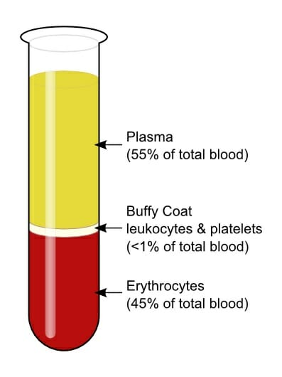
There are several major components of the seemingly uniform liquid that is our blood. When centrifuged, the components of different densities separate to look something like this:
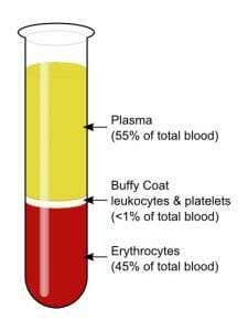

Blood centrifugation scheme


Here we will discuss the most vital components of blood, including serum, white blood cells or “leukocytes,” red blood cells, and platelets.


Plasma


Plasma is the liquid which carries the red blood cells, white blood cells, platelets, and other substances found in blood. More than half the volume of our blood is composed of this fluid.


Our blood plasma is mostly water, but it also contains salts proteins, and other substances, which can make it appear thick and syrupy even when the red and white cells have been filtered out.


One important protein, albumin, exists in part to keep the blood thick and syrupy. This ensures that the blood does not leak out of our vessels and into tissues, and slows bleeding when we are injured.


Other substances that can be found in the plasma include:


  • Antibodies, which are proteins that attack invading pathogens

  • Clotting factors, which prevent bleeding

  • Hormones, which are chemical messages sent between different tissues in the body

  • Electrolytes such as salt

  • Nutrients such as sugar, vitamins, and minerals

  • Lipids including cholesterol


So even this seemingly simple fluid is a veritable stew of the ingredients for life! But it could not do its job without…


Red Blood Cells


Red blood cells can be thought of as the cargo ships of the body. They are small, numerous cells which are specifically designed to carry oxygen from the lungs to cells, and carry carbon dioxide back to the lungs to be expelled when we exhale.


Red blood cells contain hemoglobin – a protein which is beautifully tailored to bind aggressively to oxygen in the lungs, and then release it and pick up carbon dioxide at a slow, steady rate as it passes through the body.


Hemoglobin is a pigment which changes color slightly, depending on whether it is bound to a molecule of oxygen or not. That’s why blood drawn from veins, which carry oxygen-depleted blood back toward the lungs, is a dark red that can appear almost brown. Blood drawn from arteries, which carry oxygen-rich blood from the lungs to the tissues, is a bright red.


White Blood Cells


White blood cells perform both immune and clean-up functions for the body. Like red blood cells, they are made by stem cells in the bone marrow.


There are many types of white blood cells, which play many different roles in immune response to infection and injury. Some types of white blood cells include:


  • Neutrophils – Target bacteria and fungi.

  • Eosinophils – Target larger parasites such as those which cause malaria. Also play a role in allergic inflammatory responses.

  • Basophils – Release chemicals that enhance inflammatory responses.

  • B Lymphocytes – Release antibodies and assist in activating T cell lymphocytes.

  • T Lymphocytes – Different subtypes help the immune system learn to “recognize” new infection so it can target it; help immune system to activate in response to infection, then return to normal after infection has passed; target virus-infected and tumor cells.

  • Natural Killer Lymphocytes – Target virus-infected and tumor cells for destruction.

  • Monocyte – Migrate into tissues and mature into macrophages, literally “big eaters,” which engulf harmful cells and cellular debris and destroy them; some mature into Kupffer cells, which live in the liver and break down and recycle dying red blood cells.


Platelets


Platelets are cell fragments – bits of membrane-bound cytoplasm – which stop bleeding by clumping together to form clots and scabs seal wounds. Like red and white blood cells, they are made in the bone marrow. Cancer of the bone marrow may prevent production of properly functioning platelets.


Platelets have two states: active platelets, which are prepared to create blood clots, and inactive platelets that do not clot. Under normal circumstances, the endothelial lining of healthy blood vessels produces chemical messages that tells platelets to remain in their inactive form, so that they don’t form clots inside of healthy blood vessels.


Under normal circumstances, platelets are activated when a nearby injury starts a chemical cascade that urges platelets and other nearby clotting factors to activate. These factors then release clot-promoting messages of their own, encouraging more clotting factors to join their growing clot.


Platelets can sometimes be incorrectly activated when endothelial lining is damaged and does not produce the usual inhibitory messages for platelets. This can happen in people with some metabolic disorders and some forms of cardiovascular disease.


Blood Types


In early medical history, it was thought that blood transfusions from one person to another might be impossible. This was because when this was attempted, most test subjects died.


In time, however, scientists discovered the existence of “blood types” – a few basic proteins that coat the surface of our blood cells, helping the immune system to differentiate between our own blood cells and foreign invaders.


Attempting to transfuse blood of an incompatible type into a person can cause a clotting reaction, which may be fatal. Fortunately, today doctors have rapid tests to determine a patient’s blood type, and store blood bags for transfusion sorted by type so that patients are assured of getting a compatible treatment.


The three common blood type protein markers recognized by science are called the A, B, and Rh proteins.


The A/B protein group can give rise to blood types A, B, AB, or O. There is no “O” blood type protein – instead “O” is the blood type used to describe people who have neither A nor B marker proteins.


Each of these blood types can also be positive or negative for the Rh protein, leading to blood types such as “AB+” or “AB-.”


The blood type “O negative” is known as the universal donor. Because it does not have A or B proteins and is negative for the Rh protein, people of any blood type can receive O negative blood without having an adverse immune response to foreign proteins.


Unfortunately, people with “O negative” blood type also have the narrowest selection of possible donors for themselves. O negative people cannot receive any blood that has A, B, or Rh proteins; they can only receive blood from other O negative people.


When platelets are deficient or absent, the results can be seen in an increased probability of catastrophic bleeding events, including spontaneous bleeding and bleeding from minor injuries.


Quiz


1. Which of the following is NOT a function of blood?
A. To transport oxygen to our cells
B. To transport water to and away from our cells, as needed
C. To break down toxins into harmless substances
D. To fight infections

Answer to Question #1

2. Which of the following is NOT likely to be found in a blood sample?
A. Red blood cells
B. Plasma
C. White blood cells
D. Bone marrow cells

Answer to Question #2

3. Which of the following is NOT likely to be found in blood plasma?
A. Salt
B. Digestive enzymes
C. Hormones
D. Albumin

Answer to Question #3

References


  • Laki, K. (1972). Our Ancient Heritage In Blood Clotting And Some Of Its Consequences. Annals of the New York Academy of Sciences, 202(1), 297-307. doi:10.1111/j.1749-6632.1972.tb16342.x

  • Haubrich, W. S. (2004). Kupffer of Kupffer cells. Gastroenterology, 127(1), 16. doi:10.1053/j.gastro.2004.05.041

  • Wilson, J. H., & Hunt, T. (2002). Molecular biology of the cell, 4th ed. New York: Garland.

  • American Society of Hematology. (2014, March 29). Retrieved July 11, 2017, from http://www.hematology.org/Patients/Basics/


Blood
Liver

Liver Definition


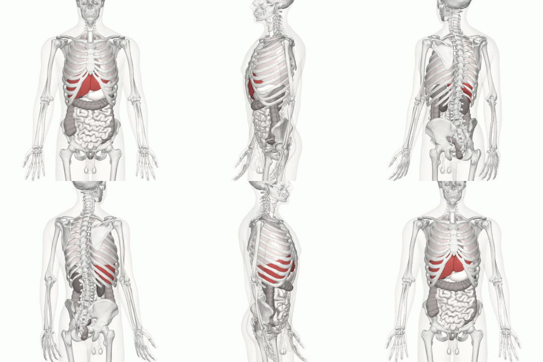
The liver is a vital organ found in humans and other vertebrates. It is a large organ, with its major lobe occupying the right side of the abdomen below the diaphragm, while the narrower left lobe extends all the way across the abdomen to the left. The liver is the dark pink organ in this image:


Liver animation

(click to play animated GIF)


The liver performs many vital functions without which humans cannot survive.


Presently, there is no way to replace a liver with an artificial one in the long-term, although partial liver donations in which a donor gives part of their liver to someone in need of a transplant have been successful. The liver has a remarkable regenerative capacity, and can grow back most of its tissue if it is removed or damaged.


Despite the success of partial transplants in some cases, liver failure is an important cause of death among people with chronic alcohol and drug use.


Liver failure can also occur accidentally as a result of an accidental overdose of acetaminophen – a medicinal compound found in many over-the-counter and prescription medications. Because acetaminophen is found in so many different medications, people often don’t realize that two or more medicines they’re taking together contain the same compound, which can overwhelm the liver and lead to potentially fatal liver damage.


This is why it’s always important to tell your doctor about all medications you are taking, and check the labels of different medications for common ingredients. We will talk more about acetaminophen toxicity, and how to avoid it, below.


Liver Function


The liver serves many vital functions in the body, including:


Detoxifying the Blood


The liver’s most well-known role is as a detoxifier of the blood. It contains cells with special enzymes that can break down toxic substances into non-toxic forms.


These enzymes explain why certain medications, foods, and supplements can interact with each other. Some liver enzymes break down multiple types of toxic substances; if the enzymes are “busy” with one substance, they might not be able to break down the other substance as they usually do.


This is why you should always tell your doctor about all medications and supplements you are taking, and why some medications require that you avoid certain substances like alcohol or grapefruit.


Without these enzymes to break down toxic substances, the body slowly poisons itself. It doesn’t even need to consume anything toxic from the environment – the chemicals produced by the body’s own cells are sufficient to cause fatal toxicity over time.


Fortunately, the liver is very good at what it does. We rarely have to feel the effects of these toxins, unless we ingest large quantities of substances that can damage the liver, such as alcohol, acetaminophen, or anti-freeze.


Our livers can also run into trouble if we contract viruses that damage liver cells, such as hepatitis.


Making Blood Clotting Factors


The liver uses Vitamin K to produce proteins that are important for blood clotting. Without these proteins, the multi-step process of blood clotting may not be able to get started.


This is why people with severe liver disease or Vitamin K deficiency often develop bleeding disorders. With the body unable to clot to repair even tiny, routine injuries, people with these conditions can appear to bruise and bleed for no reason.


This can be a very serious complication in the treatment of severe liver disease, since transplantation is a surgical procedure with a risk of severe bleeding.


Making Digestive Chemicals


The liver produces bile, which is a little-known but vital ingredient to the digestive process. Bile helps the body to break down and absorb fats, and also uses it to help get rid of certain waste products.


Problems with the liver are occasionally signaled by changes in fecal matter resulting from a lack of bile in the digestive tract. That’s one reason why it’s recommended to see a doctor if you experience changes to the color, consistency, or frequency of your bowel movements that last for several weeks.


Making Energy from Protein


Under normal circumstances, the body tries not to digest proteins for energy. That’s because there are so many better uses for protein and its building blocks, such as making enzymes and other essential cellular machinery.


However, under starvation conditions where there are not sufficient stores of carbohydrates or fats to meet the body’s needs, the liver can turn amino acids into fuel for our cells to make ATP.


The liver does us a double favor here. Not only does it make it possible for our cells to survive off of proteins – it also detoxifies the toxic byproduct of this process, which is ammonia. The liver turns ammonia into urea, which can be safely eliminated by the kidneys, before releasing it into the blood.


Storing Glycogen


The liver also serves another purpose that is useful when food is scarce. It stores carbohydrates in the form of a high-density, high-calorie substance called glycogen.


Our body’s normal order of priority for digestion is: carbs, fats, protein. Our body will metabolize any carbohydrates we eat first; if it runs out of carbs, then it will turn to metabolizing our long-term fat stores.


But in between those steps, it has the liver’s glycogen. This acts as a “quick release” fuel that is easier to release and replenish than fat. Only after depleting our liver’s glycogen reserves will our body normally start digesting fat.


Breaking Down Red Blood Cells


Red blood cells die more often than any other cell type in the body. This is because red blood cells do not have nuclei, so they cannot make their own proteins. When the proteins they had at maturity wear out, they break down and are replaced by new blood cells.


Without the liver, this process would lead to severe toxicity in the blood. Dying cells release toxic compounds, which must be processed by the liver in order to keep the body safe.


Thanks to the action of the liver’s cells and enzymes, the materials from dying red blood cells are broken down into harmless forms, or even recycled for later use in new red blood cells.


Producing Hormones


The liver produces several chemical messengers that help the body to coordinate its activities. These include:


  • Insulin-like growth factor 1 – A hormone that prompts tissue to grow, and is especially important in childhood.

  • Thrombopoietin – A hormone that tells the bone marrow how many platelets to produce to help with blood clotting.

  • Hepcidin – A hormone that tells the body whether iron should be absorbed, or eliminated as waste.

  • Angiotensinogen – A pro-hormone that increases blood pressure.

  • Carrier proteins – The liver also produces carrier proteins, which bond to other hormones including sex hormones, thyroid hormone, stress hormones, and vitamins and minerals to specified destinations.


Liver Location


The liver is found within the lower ribcage, under the diaphragm which separates the heart, lungs, and stomach from the abdominal cavity.


It is located in the upper abdomen, with the bulk of the liver contained on the right side of the body. Its smaller lobe extends all the way to the left side of the diaphragm.


The liver normally cannot be felt, as it is shielded by the lower ribs. However, blows to the solar plexus – the soft part of the upper abdomen where the ribcage parts – can injure the liver.


Liver pain may appear as pain in the upper abdomen. It is usually on the left side or in the center, but can sometimes appear on the right or even seem to come from lower in the abdomen.


Doctors will often examine the abdomen if liver abnormalities are suspected, as a swollen liver can often be felt despite the ribcage.


Acetaminophen Toxicity


Some medicinal compounds are considered so safe and effective that they are used in dozens of different medications. Acetaminophen is one of these. The main ingredient in Tylenol, it relieves pain and reduces fever with a very good safety profile – as long as you take it in the correct doses.


Unfortunately, because acetaminophen is so effective, it is found in many, many medications. One of the most common causes of liver damage in the U.S. is people taking two or more of these medications at once, not realizing that they all contain the same ingredient. This can result in an overdose, especially if multiple medications containing acetaminophen are taken over long periods of time.


Acetaminophen is processed by the liver into a toxic product, which can poison the liver. In the proper doses, the liver can eliminate this byproduct without any problem, but when people take more acetaminophen than they should over a period of days or weeks, the product can build up and kill liver tissue.


This is unfortunately common since acetaminophen is included in some medications which are advertised to treat joint pain, fever, heartburn, flu symptoms, cough, menstrual cramps, and more. People often don’t think that these medications are related or that they might contain the same ingredients.


When taking multiple medications on the same day, it is a good idea to check the labels for shared ingredients. Taking a proper dose of acetaminophen is safe and effective – but taking two or three times the proper dose can cause very big problems.


Medications which contain acetaminophen and should not be taken together include:


ActifedAlka-Seltzer PlusAnacin
BitartrateButalbitalCepacol
ContacCoricidinDayquil
DimetappDristanEndocet
ExcedrinFioricetFeverall
Formula 44Goody’s PowdersHycotab
HydrocetLiquiprinLortab
MidolMIDRINMucinex
NORCONyquilOxycodone
PanadolPercocetPhenaphen
RobitussinROXICETSaint Joseph Aspirin-Free
SedapapSingletSinutab
SudafedTapanolTramadol
TriaminicTylenol Brand ProductsTylox
UltracetVanquishVicodin
VicksZydone

Quiz


1. Which of the following is NOT a function of the liver?
A. Breaking down toxins into harmless substances
B. Breaking down old red blood cells
C. Metabolizing glucose for the body
D. Storing carbohydrates

Answer to Question #1

2. Which of the following is NOT a common cause of liver failure?
A. Chronic, heavy alcohol consumption
B. Viral hepatitis
C. Too much exercise
D. Acetaminophen toxicity

Answer to Question #2

3. Pain in which of the following areas might originate with the liver?
A. Right upper quadrant of the abdomen
B. Central upper quadrant of the abdomen
C. Right upper quadrant or lower abdomen
D. All of the above

Answer to Question #3

References


  • Boron, W. F., & Boulpaep, E. L. (2009). Medical physiology: a cellular and molecular approach. Philadelphia, PA: Saunders/Elsevier.

  • Common Medicines With Acetaminophen. (n.d.). Retrieved July 11, 2017, from http://www.knowyourdose.org/common-medicines/

  • Abdel-Misih, S. R., & Bloomston, M. (2010). Liver Anatomy. Surgical Clinics of North America,90(4), 643-653. doi:10.1016/j.suc.2010.04.017

  • Acetaminophen Toxicity. (2017, January 06). Retrieved July 11, 2017, from http://emedicine.medscape.com/article/820200-overview

  • Hoffman, M. (n.d.). Picture of the Liver. Retrieved July 11, 2017, from http://www.webmd.com/digestive-disorders/picture-of-the-liver#1

  • How does the liver work? (2016, August 22). Retrieved July 11, 2017, from https://www.ncbi.nlm.nih.gov/pubmedhealth/PMH0072577/


Liver

Spleen

Spleen Definition


The spleen is a small organ, typically located on the left side of the body, behind the ribcage and stomach. It is the largest organ in the body’s lymphatic system, which is responsible for promoting immune function, filtering the blood, and managing blood volume.


The lymphatic system is the system of ducts and lymph nodes which are found under the skin. They are a channel through which white blood cells can travel outside of the blood stream, and they are also a receptacle for bacteria, dead cells, debris, and extra fluid that may accumulate in the blood.


The spleen performs several helpful functions for the body, including making antibodies, removing bacteria, removing old red blood cells, breaking down waste products of red blood cells, recycling iron for use in new red blood cells, and holding a reserve of red blood cells and immune cells which the body can use in case of emergencies.


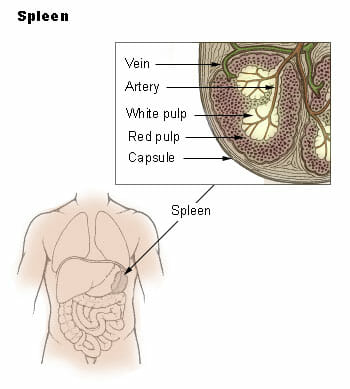
The spleen makes antibodies in a type of tissue called “white pulp.” Its tissue called “red pulp” contains a reservoir of immune cells that fight infection and promote tissue healing, which the spleen can release as needed.


The image below shows where the spleen is located in the body, along with the arrangement of its “red pulp” and “white pulp”:


Spleen


Though the spleen is useful to the body, it is not essential for survival. Some of its functions can be taken over by other organs if the spleen is removed; other functions of the spleen are helpful in case of emergency, but are not required for survival under most circumstances.


The spleen may need to be removed if it becomes injured or infected. In these cases the spleen may pose more of a risk than a benefit to the rest of the body, and doctors may elect to perform surgery to remove it.


The spleen can also be damaged by sickle cell disease, which can block blood flow to the spleen. This is one reason why people with sickle cell disease should get extra immunizations.


People without a spleen typically lead normal, healthy lives, though they are advised to take regular immunizations to protect against infection.


Interestingly, about 10% of people have an “accessory spleen” – a small extra spleen! This causes no problems or symptoms, and most people only find out they have an accessory spleen if they require imaging scans of their body for another reason.


Spleen Function


The spleen performs numerous helpful functions for the body, including:


  • Making antibodies

  • Storing emergency reserves of red blood cells that can be released in case of blood loss

  • Storing emergency reserves of white blood cells that can be released to fight infection and promote healing

  • Breaking down waste products from dead cells

  • Recycling useful components, such as iron, for use in future red blood cells


It’s easy to see how the spleen helps us to survive illness and injury, and helps us to salvage useful nutrients.


In the modern era, spleens are not necessary for survival. With thorough safeguards such as vaccinations and antibiotics and the ability to take iron supplements if necessary, people without spleens are simply advised to be a little more careful about getting vaccinated and avoiding disease.


The spleen is most commonly removed because of injury, such as injuries sustained during car crashes or playing sports. An injured or ruptured spleen cause massive blood loss which can quickly become life-threatening.


There is some research suggesting that the spleen might have a poorly-understood role in preventing cardiovascular disease, as one study found that people with their spleens removed were more likely to die of cardiovascular disease than a control group.


For this reason, some scientists advocate for better injury prevention, such as protective body armor for sports players, to reduce the possibility of spleen injuries that may result in the need for spleen removal.


Spleen Location


In most people, the spleen is located under the left side of the ribcage, next to the stomach. It normally cannot be felt or detected, as it is only about the size of the fist and is tucked away behind the stomach.


Its location makes the spleen fairly well-protected, but it can still be injured by blows to the chest and side, such as those that are sometimes sustained in sports or in car accidents.


A ruptured spleen can be very dangerous to the rest of the body. This is because the spleen processes a large blood supply, so rupture of the spleen may lead to rapid, massive blood loss. For that reason, spleens are sometimes removed if they are injured and there is concern that they may rupture.


Doctors can sometimes feel the spleen if it is abnormally swollen, as in the case of mononucleosis, blood cancers, and other conditions. If someone is suspected of having those conditions, doctors may feel the abdomen for signs of an enlarged spleen.


Spleen Structure


The spleen consists of two different types of tissues, red pulp and white pulp. These tissues are encased within a membrane of tissue. The spleen’s external surface can be divided into two portions: the diaphragmatic surface, and the visceral surface. Here we’ll discuss more about each of these important features.


Diaphragmatic Surface


The diaphragmatic surface of the spleen is the surface which faces the diaphragm. This is an upward-facing surface that curves outward, pressing against the diaphragm.


The diaphragm – a strong muscle located beneath the lungs and stomach, which enables us to breathe – lies between the spleen and the lung.


Visceral Surface


The visceral surface of the spleen faces down, toward the organs of the “viscera,” “gut.” It is divided by a ridge into two regions: the renal region, and the gastric region.


The “gastric,” or stomach region is the “anterior,” or front surface of the spleen. It faces forward and curves inward, like a soup spoon. This allows it to curve around the stomach, which nestles into the spleen. The very bottom of the gastric region touches the tail of the pancreas.


The “renal,” or kidney region, faces in toward the center of the body and downward. It comes into contact with the front of the left kidney and sometimes with the left adrenal gland.


Red Pulp


The red pulp of the spleen serves the circulatory system by filtering the blood, and acting as a recycling station for red blood cells and a storage point for other important blood components. It makes up the majority of tissue in most healthy spleens.


The red pulp consists of connective tissue called the “cords of Billroth.” These first filter the blood, removing pathogens and red blood cells that have reached the end of their useful lives. It then uses immune cells called macrophages to break down the useless or harmful components of these cells, while saving useful components such as iron for use in future red blood cells.


The red pulp also stores white corpuscles (another type of white blood cell), special cells called “splenic cells” which store, digest, and transport red blood cells, and platelets.


On occasion, platelet deficiency may occur of the spleen stores too many platelets and does not release enough of them into the blood.


White Pulp


The white pulp of the spleen is the tissue that performs the spleen’s immune functions. It consists of a layering of different tissues and nodules, each of which perform distinct functions for the immune system:


  • The periarteriolar lymphoid sheaths (PALS) serve as a reservoir for white blood cells called T lymphocytes. Some scientists have described this reserve of white blood cells as being similar to having a standing army, ready to mobilize to protect the body in the event of attack.

  • The lymph follicles contain a type of blood cell called B lymphocytes, which divide and reproduce inside the follicles. Antibody molecules that help the immune system to recognize and fight illness are also made in this tissue.Other tissues in the body also perform these functions, so they will not cease if the spleen is removed; but the spleen lends the immune system extra strength by producing these cells and antibodies.

  • The marginal zone contains a unique type of cell called “antigen presenting cells.” Antigens are molecules found on bacteria or viruses which antibodies and white blood cells recognize. The term literally comes from “anti-” for “antibody” and “gen” for “beginning.”

Antigen-presenting cells serve the immune system by eating invading pathogens, and then presenting molecules from the pathogens on the surface of their cells. This can “teach” and stimulate other immune cells to respond to new pathogens.


Quiz


1. Which of the following is NOT a function of the spleen?
A. To make antibodies.
B. To recycle iron for use in new blood cells.
C. To store emergency reserves of red and white blood cells.
D. To help digest food.

Answer to Question #1

2. Which of the following is a potential consequence of having one’s spleen removed?
A. One might need to take insulin shots to make up for lost insulin.
B. One might need to use a dialysis machine to clean toxins from the blood.
C. One might need extra immunizations to protect against infections.
D. None of the above.

Answer to Question #2

3. Which of the following is NOT true of the spleen?
A. It is located behind your stomach, on the left side of your ribcage.
B. It plays a vital role in filtering toxins from the blood.
C. It can pose a bleeding risk if injured, as it receives a massive blood supply.
D. Some people have two spleens.

Answer to Question #3

References


  • Mebius, R. E., & Kraal, G. (2005). Structure and function of the spleen. Nature Reviews Immunology, 5(8), 606-616. doi:10.1038/nri1669

  • Hoffman, M. (n.d.). Picture of the Spleen. Retrieved July 08, 2017, from http://www.webmd.com/digestive-disorders/picture-of-the-spleen#1

  • Szalay, J. (2015, January 29). Spleen: Function, Location & Problems. Retrieved July 08, 2017, from https://www.livescience.com/44725-spleen.html

  • Angier, N. (2009, August 03). Finally, the Spleen Gets Some Respect. Retrieved July 10, 2017, from http://www.nytimes.com/2009/08/04/science/04angier.html


Spleen

Wednesday, July 26, 2017

Placenta

Placenta Definition


The placenta is an organ which is responsible for nourishing and protecting a fetus during pregnancy. It is unique in that it is a temporary organ; it grows alongside the fetus during pregnancy, and then is expelled along with the fetus at birth. The placenta is also sometimes called “afterbirth,” as it is expelled through the vagina after the fetus has been delivered.


The placenta performs myriad functions to support fetal development, including facilitating blood flow, gas exchange, waste elimination, and serving as a protective barrier for the fetus against any infections the mother experiences during pregnancy.


The placenta is unique in that it is an organ which arises from the tissue of two genetically distinct organisms; part of the placenta develops from the tissue of the mother’s uterine wall, while another part develops from the fetus’ own tissue. After the blastocyst which will develop into the fetus makes contact with the uterine wall, blastocyst and maternal tissue grow together to form a single, cooperating organ that links the two together.


Mammals who give birth to live, fully-developed young, rather than laying eggs or carrying underdeveloped offspring in pouches, are often called “placental mammals.” The evolution of the placenta is one of the primary characteristics shared by all mammals except for marsupials and egg-laying mammals such as the platypus.


Some marsupials – close cousins to placental mammals like cats, dogs, and humans – have a primitive placenta in which blood vessels grow throughout the protective membranes surrounding the embryo. It is possible that placentas as we know them may have evolved from similar primitive structures in found the common ancestor of marsupials and placental mammals.


Pregnancies in which the placenta does not develop properly typically miscarry. This can occur either because of problems with the development of maternal uterine tissue, or because of problems with development of the fetal placental tissue. Fetuses which suffer from chromosomal problems may not be able to form a proper placenta and may miscarry, most often in the first trimester.


Many animals and some human cultures make a practice of eating the placenta after it is delivered. Scientists are divided on whether this is a good idea for humans. Some say it may contain valuable nutrients and even hormonal components that can be of assistance to the mother after pregnancy; others say that this practice may carry a risk of spreading infectious diseases, and that no benefits to eating the placenta have been proven in humans.


Function of Placenta


The placenta acts as a lifeline between the mother and fetus, ensuring that the fetus gets what it needs from the mother’s body to survive. At the same time, it acts as a protective barrier, shielding the fetus from some maternal infections. The functions of the placenta include:


  • Allows gas exchange so the fetus gets enough oxygen

  • Helps the fetus get sufficient nutrition

  • Helps regulate the fetus’ body temperature

  • Removes waste from the fetus for processing by the mother’s body

  • Filters out some microbes that could cause infection

  • Transfers antibodies from the mother to the fetus, conferring some immune protection

  • Produces hormones that keep the mother’s body primed to support pregnancy


The placenta, then, essentially serves the function of several organ systems for the fetus, since the fetus is unable to eat, breathe, or eliminate waste itself while it is inside the womb!


Unfortunately, the placenta is not fool-proof as an infection barrier, and some fetuses do become infected with diseases contracted by their mother. Toxins of sufficiently small molecular size can also pass through.


That’s one reason why pregnant women are advised to avoid all possible sources of disease and toxins – diseases and toxins which may not be dangerous at all to adult women may be devastating to a developing fetus.


How Does the Placenta Work


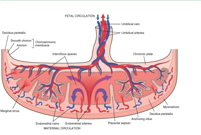
When it is delivered, the placenta looks like a flat, round organ that is suffused with thick blood vessels. The fetus’ umbilical cord attaches to one flat surface, while the reverse surface grows out of the mother’s uterus during pregnancy.


The placenta works mainly by allowing substances to be exchanged between maternal and fetal blood. This allows the fetus to obtain nutrients, oxygen, antibodies, and other vital substances without having to share the mother’s blood supply directly.


This is vital because fetuses do not always have the same blood type as their mother, and direct mixing of the bloodstreams could cause the mother’s immune system to attack the fetal blood supply. Even with the placenta separating the two, problems are occasionally caused by maternal antibodies attacking fetal blood supplies. Some women receive vaccines or other treatments to stop that form happening.


The diagram below shows how the fetal blood vessels infiltrate the placenta. It also shows how the mother’s arteries permeate the placenta. The placental tissue in between the two acts as a sort of filtration system, preventing most cells from passing through the barrier while allowing substances such as nutrients, antibodies, and gases to do so:


Placenta


Eating the Placenta


In recent years, the topic of placenta-eating has been in the news a great deal. Many celebrities have contracted with companies that promise to turn their placentas into pills or food, and touted it as a move that has great health benefits. But many doctors have cautioned that there’s no evidence that placenta-eating confers real health benefits, and that eating your placenta may actually make your baby’s health worse.


In nature, animals often eat their placentas. There’s good reason for this: in nature food is often scarce, and the placenta is rich in protein, iron, and other nutrients that can be difficult to procure in the wild. That means that placenta-eating is often worth the risk for animal moms who just gave birth, and now need to provide nutritious milk for their young.


However, for humans, the risk of disease may outweigh any benefits of placenta-eating. Because the placenta serves as a filter to prevent harmful bacteria and viruses from reaching the baby, it can contain bacteria from infections the mother had during pregnancy.


Even if these pathogens aren’t harmful to the mother – some viruses and bacteria hardly bother adults – they can still be passed on to the newborn baby through breast milk, if the mother consumes an infected placenta. Cases have been recorded of babies becoming sick with bacterial infections which were later traced to their mother’s placenta supplements.


For that reason, many doctors counsel that the placenta is just like any other human tissue – it should not be eaten by humans, because doing so could spread disease.


Because the placenta does not qualify as a medication, health food and birthing companies that promise a “healthy experience” if you pay them to prepare your placenta are often not subject to the same regulations for safety and effectiveness as medications are.


In conclusion, just because some famous moms have done it doesn’t mean it’s a scientifically supported idea!


Quiz


1. Why are mammals such as dogs, cats, and humans called “placental mammals?”
A. Because the placenta is the evolutionary adaptation we all share, while monotremes, marsupials, and non-mammals do not.
B. Because all placental mammals have placentas, while only some non-mammals have placentas.
C. Both of the above.

Answer to Question #1

2. Which of the following is true of the placental filtration system?
A. It is able to filter out all diseases and toxins, protecting the fetus.
B. It allows nutrients and oxygen to pass through, nourishing the fetus.
C. It is able to filter out some diseases and toxins, but not all of them.
D. Both B and C.

Answer to Question #2

3. Which of the following is NOT true of placental anatomy?
A. It contains many blood vessels from both the mother and fetus.
B. It is composed of both maternal and fetal tissue.
C. It allows the mother’s blood to flow into the fetus, nourishing it.
D. None of the above.

Answer to Question #3

References


  • Slater, D. (2017, April 03). The Myth Of The Placental Barrier. Retrieved July 08, 2017, from https://www.fitpregnancy.com/pregnancy/pregnancy-health/myth-placental-barrier

  • 10.2 Development of the placental villi. (n.d.). Retrieved July 08, 2017, from http://www.embryology.ch/anglais/fplacenta/villosite01.html

  • Should I Eat My Placenta? (n.d.). Retrieved July 08, 2017, from http://www.webmd.com/baby/should-i-eat-my-placenta


Placenta

Sunday, May 7, 2017

Fat

Fat Definition


Fat is a term used to describe a class of macro nutrients used in metabolism called triglycerides. These make up one of three classes of macronutrients including proteins and carbohydrates. Fats provide a means of storing energy for most eukaryotes, as well as act as a food source. Fats have the highest energy storage potential of the macronutrients, and are very chemically stable, making them ideal for storing energy for later use. Macronutrients does not refer to the size of the molecule, but to the amount needed to sustain life. Vitamins and minerals are considered micronutrients.


Chemistry of Fat


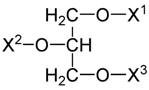
Triglycerides consist of a glycerol backbone bonded to three fatty acid chains via an ester bond. The ester bond allows for easy breaking via hydrophilic attack by water, rendering the fatty acid chains suitable for breakdown in regular metabolism.


Fatty acid chains are built by endogenous enzymatic machinery by linking malonyl-coenzyme-A units together. The chain is elongated on a single fatty-acid synthase until it reaches a length determined by the biochemistry of its producer. At the point of termination, the fatty acid is hydrolyzed off the synthase forming the carboxylic acid derivative. This produces a fatty acid of even carbon length. Most fatty acids are formed this way and therefore most fatty acids have an even number of carbons.


Triglycerides are formed by a phosphorylated glycerol molecule being nucleophilically attacked by a fatty acid-coenzyme A. This causes an esterification reaction that produces an ester bond between the carboxyl group of the fatty acid and one of the three hydroxyl groups of the glycerol. Synthases that catalyze these reactions are sterically specific to incorporate one type of fatty acid. They only produce one type of triglyceride.


Acylglycerine

Ester bonds in triglycerides


Types of Fat


Fat is organized into two subgroups: saturated fat, and unsaturated fat. Unsaturated fat is further classified as monounsaturated fat, polyunsaturated fat, and trans-fat. These different classifications determine the effects of these fats on an organism, and the roles that they have in metabolism.


Saturated Fat


Saturated fat, or animal fat, is composed of a glycerol backbone with three fully saturated fatty acids attached. Saturated refers to all the carbons in the backbone being sp3 hybridized, with two hydrogen atoms covalently bonded per carbon. This class of fats have higher viscosity and energy content than their unsaturated cousins. Due to poor solubility issues, this is the type of fat that is most commonly associated with heart disease.


Unsaturated Fat


Unsaturated fat, or vegetable fat, is composed of a glycerol backbone with three fatty acid chains where there is at least one sp2 hybridized carbon. This forms a double bond somewhere in the chain. Monounsaturated fats have one double bond in the chain, while polyunsaturated fats have two or more.


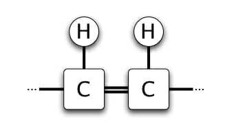
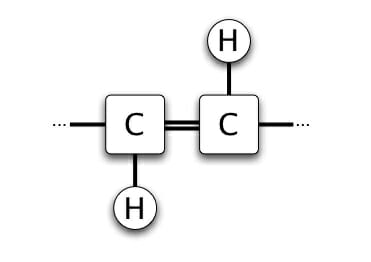
Naturally occurring unsaturated fats, since they are produced by enzymes, have specific stereochemistry. Natural fats always show the cis conformation, which has a higher solubility in water, and is easily broken down by the metabolic machinery. Artificially produced fats, since they are produced using organic synthesis techniques, contain a racemic mixture of trans and cis bonds. Trans fats are less soluble – like saturated fats. However, they are not readily metabolized by cellular machinery.


Fat Conformation Cis

Cis bond


Fat Conformation Trans

Trans bond


Types of Unsaturation:

The unsaturated fats are classified by the position of the unsaturation. This designation is denoted by the ω symbol, then the number of carbon with the unsaturation. For example, ω-3 fats have an unsaturation at the third carbon position. Ω-3,7 polyunsaturated fats have an unsaturation at the third and seventh carbon position. The position of the unsaturation determines the metabolic pathway that the fat will follow.


Examples of Fat


Saturated Fat


Examples of saturated fat include animal fat found on beef, pork, and chicken. Since these types of fats are also called “animal fats”, they are primarily found in animals. Saturated fat measurements are used in medical diagnostic tests as inverse indicators of a healthy lifestyle.


Unsaturated Fats


Monounsaturated fats are often referred to as “good” fats. They make up the oils and fats found in avocados and olive oil. Polyunsaturated fats are found in canola oil and other less viscous plant oils.


Trans Fats


Trans fats are chemically produced fats. They are generally considered to be unhealthy, and are found in mass produced oil found in processed and fried foods. Trans fats have a controversial history. They have even been banned for consumer use in some countries.


Quiz


1. Which is NOT a type of fat?
A. Polyunsaturated fat
B. Monounsaturated fat
C. Saturated fat
D. Supersaturated fat

Answer to Question #1

2. Which Greek letter is used to denote unsaturations in biological nomenclature?
A. π
B. η
C. ω
D. ψ

Answer to Question #2

3. Which is NOT one of the three types of macronutrients?
A. Fat
B. Vitamins
C. Protein
D. Carbohydrates

Answer to Question #3

References


  • Esposito, L. (2017, May 03). 8 Fast Facts on Trans Fats. Retrieved May 04, 2017, from http://health.usnews.com/wellness/food/articles/2017-05-03/8-fast-facts-on-trans-fats?src=usn_fb

  • Monounsaturated Fat: What Makes a Fat. (2017, February 24). Retrieved May 04, 2017, from https://paleoleap.com/monounsaturated-fat-what-makes-a-fat-good/

  • Commissioner, O. O. (n.d.). Consumer Updates – FDA Cuts Trans Fat in Processed Foods. Retrieved May 04, 2017, from https://www.fda.gov/forconsumers/consumerupdates/ucm372915.html


Fat