Hyphae Definition
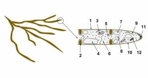
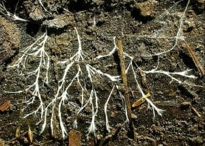
Hyphae are comprised of hypha, which are the long filamentous branches found in fungi and actinobacteria (shown below). Hyphae are important structures required for growth in these species, and together, are referred to as mycelium.
Hyphae Structure
Each hypha is comprised of at least one cell encapsulated by a protective cell wall typically made of chitin, and contain internal septa, which serve to divide the cells. Septa are important as they allow cellular organelles (e.g., ribosomes) to pass between cells via large pores. However, not all species of fungi contain septa. The average hyphae are approximately 4 to 6 microns in size.
Hyphae Growth
Hyphae growth occurs by extending the cell walls and internal components from the tips. During tip growth, a specialized organelle called the spitzenkörper, assists in the formation of new cell wall and membrane structures by harboring vesicles derived from the golgi apparatus and releasing them along the apex of the hypha. As the spitzenkörper moves, the tip of the hypha is extended via the release of the vesicle contents, which form the cell wall, and the vesicle membranes, which create a new cell membrane. As the hypha extends, new septa can be created to internally divide the cells. The characteristic branching of hyphae is the result of the formation of a new tip from a hypha, or the division of a growing tip (see diagram below).

(1- Hyphal wall 2- Septum 3- Mitochondrion 4- Vacuole 5- Ergosterol crystal 6- Ribosome 7- Nucleus 8- Endoplasmic reticulum 9- Lipid body 10- Plasma membrane 11- Spitzenkörper/growth tip and vesicles 12- Golgi apparatus)
Hyphae Function
Hyphae are associated with multiple different functions, depending on the specific requirements of each fungal species. The following are a list of the most commonly known hyphae functions:
Nutrient Absorption from a Host
Some hyphae of parasitic fungi are specialized for nutrient absorption within a specific host. These hyphae have specialized tips called haustoria, which penetrate the cell walls of plants or tissues of other organisms in order to obtain nutrients.
Nutrient Absorption from Soil
Some fungal species (e.g., mycorrihizae) have developed a symbiotic relationship with vascular plant species. The fungi forms specialized hyphae called arbuscules, which can be found in the roots or phylum of vascular plants, and function to absorb nutrients and water from the soil. In this manner, the hyphae aid the plants by increasing its access to nutrients in the soil while facilitating its own growth.
Trapping Structures
In some fungal species, hyphae have evolved into specialized nematode-trapping structures, using nets and ring structures to trap nematode species.
Nutrient Transportation
Several fungal species exhibit hyphae composed of chord-like structures, termed mycelial chords, which are used by fungi (e.g., lichens and mushrooms) to transport nutrients across great distances.
Hyphae Classification
In general, hyphae can be classified based on the following traits:
Hyphae Characteristics
Hyphae characteristics are an important method of classifying various fungal species. There are three main hyphae characteristics:
- Binding: Binding hyphae have a thick cell wall and are highly branched.
- Generative: Generative hyphae have a thin cell wall, a large number of septa, and are typically less differentiated. Generative hyphae may also be contained within other materials (e.g., gelatin or mucilage) and can also develop structures used in reproduction. All fungal species typically contain generative hyphae.
- Skeletal: Skeletal hyphae contain a long and thick cell wall with few septa. Skeletal hyphae can also be of a fusiform subtype, with a swollen midsection surrounded by tapered ends.
Hyphae Composition
Fungal species are also further classified based on the hyphal systems they contain. There are four general subtypes:
- Monomitic: While virtually all fungal species contain generative hyphae, those with only exhibit this type are referred to as monomitic (e.g., agaric mushrooms).
- Dimitic: A species that contains generative hyphae in addition to one other type of hyphae. The most common combination of dimitic fungi is generative and skeletal.
- Trimitic: Species which contain all three types of hyphae (generative, binding, and skeletal).
- Sarcodimitic and sarcotrimitic: Sarcodimitic hyphae are fusiform skeletal hyphae bound to generative hyphae. Sarcotrimitic species contain fusiform skeletal hyphae, as well as binding and generative hyphae.
Hyphae Refraction
Under a microscope, the appearance of oily or granular hyphae under a microscope is termed gloeoplerous. This term is also used to further classify the hyphae of various species.
Cell division
Hyphae can be classified based on the presence of internal septa (septate versus aseptate species). Hyphae can also be distinguished from species which produce pseudohyphae via cell division. Pseudohyphae is a form of incomplete cell division, in which the dividing cells do not separate. There are several yeast species which produce such pseudohyphae.
Quiz
1. Which of the following statements is TRUE regarding hyphae?
A. All fungi contain skeletal hyphae.
B. All hyphae contain septa.
C. Fungal species can exhibit both generative and binding hyphae.
D. Fusiform skeletal hyphae are a form of pseudohyphae.
2. Which of the following is NOT a primary function of hyphae:
A. Nutrient absorption from the soil
B. Nutrient transportation
C. Nutrient absorption from host tissues
D. All of the above
E. Only A and B are primary functions
References
- Fricker et al. (2017). The Mycelium as a Network. Microbiol Spectr. 5(3): doi: 10.1128/microbiolspec.FUNK-0033-2017.
- Lew, R. (2011). How does a hypha grow? The biophysics of pressurized growth in fungi. Nat Rev Microbiol. 9(7): 509-18.
- Steinberg et al. (2017). Cell Biology of Hyphal Growth. Microbiol Spectr. 5(3): doi: 10.1128/microbiolspec.FUNK-0034-2016.
Hyphae

No comments:
Post a Comment