Tuesday, February 28, 2017

RNA Polymerase

RNA Polymerase Definition


A ribonucleic acid polymerase, or RNA polymerase (RNAP), is a multi subunit enzyme that catalyzes the process of transcription where an RNA polymer is synthesized from a DNA template. The sequence of the RNA polymer is complementary to that of the template DNA and is synthesized in a 5’→ 3′ orientation. This RNA strand is called the primary transcript and needs to be processed before it can be functional inside the cell.


RNA polymerases interact with many proteins in order to accomplish their task. These proteins help in enhancing the binding specificity of the enzyme, aid in unwinding the double helical structure of DNA, modulate the activity of the enzyme based on the requirements of the cell and alter the speed of transcription. Some RNAP molecules can catalyze the formation of a polymer over four thousand bases in length every minute. However, they have a dynamic range of velocities and they can occasionally pause, or even stop at certain sequences in order to maintain fidelity during transcription.


Functions of RNA Polymerase


Traditionally, the central dogma of molecular biology has looked at RNA as a messenger molecule, that exports the information coded into DNA out of the nucleus in order to drive the synthesis of proteins in the cytoplasm: DNA → RNA → Protein. The other well known RNAs are transfer RNA (tRNA) and ribosomal RNA (rRNA) which are also intimately connected with the protein synthetic machinery. However, over the past two decades, it has become increasingly clear that RNA serves a range of functions, of which protein coding is only one part. Some regulate gene expression, others act as enzymes, some are even crucial in the formation of gametes. These are called non-coding or ncRNA.


Since RNAP is involved in the production of molecules that have such a wide range of roles, one of its main functions is to regulate the number and kind of RNA transcripts formed in response to the cell’s requirements. A number of different proteins, transcription factors and signaling molecules interact with the enzyme, especially the carboxy-terminal end of one subunit, to regulate its activity. It is believed that this regulation was crucial for the development of eukaryotic plants and animals, where genetically identical cells show differential gene expression and specialization in multicellular organisms.


In addition, the optimal functioning of these RNA molecules depends on the fidelity of transcription – the sequence in the DNA template strand must be represented accurately in the RNA. Even a single base change in some regions can lead to a completely non-functional product. Therefore, while the enzyme needs to work quickly and complete the polymerization reaction in a short span of time, it needs robust mechanisms to ensure extremely low error rates. The nucleotide substrate is screened at multiple steps for complementarity to the template DNA strand. When the correct nucleotide is present, it creates an environment conducive to catalysis and the elongation of the RNA strand. Additionally, a proofreading step allows incorrect bases to be excised.


Finally, RNA polymerases are also involved in post-transcriptional modification of RNAs to make them functional, facilitating their export from the nucleus towards their ultimate site of action.


Types of RNA Polymerase


There is remarkable similarity in the RNA polymerases found in prokaryotes, eukaryotes, archea and even some viruses. This points to the possibility that they evolved from a common ancestor. Prokaryotic RNAP is made of four subunits, including a sigma-factor that dissociates from the enzyme complex after transcription initiation. While prokaryotes use the same RNAP to catalyze the polymerization of coding as well as non-coding RNA, eukaryotes have five distinct RNA polymerases.


Eukaryotic RNAP I is a workhorse, producing nearly fifty percent of the RNA transcribed in the cell. It exclusively polymerizes ribosomal RNA, which forms a large component of ribosomes, the molecular machines that synthesize proteins. RNA Polymerase II is extensively studied because it is involved in the transcription of mRNA precursors. It also catalyzes the formation of small nuclear RNAs and micro RNAs. RNAP III transcribes transfer RNA, some ribosomal RNA and a few other small RNAs and is important since many of its targets are necessary for normal functioning of the cell. RNA polymerases IV and V are found exclusively in plants, and together are crucial for the formation of small interfering RNA and heterochromatin in the nucleus.


Process of Transcription


Transcription begins with the binding of the RNAP enzyme to a specific part of the DNA, also known as the promoter region. This binding requires the presence of a few other proteins – the sigma factor in prokaryotes and various transcription factors in eukaryotes. One set of proteins called general transcription factors are necessary for all eukaryotic transcriptional activity and include Transcription Initiation Factor II A, II B, II D, II E, II F and II H. These are supplemented by specific signaling molecules that modulate gene expression through stretches of non-coding DNA located upstream. Often initiation is aborted multiple times before a stretch of ten nucleotides is polymerized. After this, the polymerase moves beyond the promoter and loses most of the initiation factors.


This is followed by the unwinding of double stranded DNA, also known as ‘melting’, to form a sort of bubble where active transcription occurs. This ‘bubble’ appears to move along the DNA strand as the RNA polymer elongates. Once transcription is complete, the process is terminated and the RNA strand is processed. Prokaryotic RNAP and eukaryotic RNA polymerases I and II require additional transcription termination proteins. RNAP III terminates transcription when there is a stretch of Thymine bases on the non-template strand of DNA.


Comparison between DNA and RNA Polymerase


While DNA and RNA polymerases both catalyze nucleotide polymerization reactions, there are two major differences in their activity. Unlike DNA polymerases, RNAP enzymes do not need a primer to begin the polymerization reaction. They are also capable of beginning the reaction from the middle of a DNA strand and reading ‘STOP’ signals that cause the enzyme complex to dissociate from the template. Finally, while RNA polymerases are slightly slower that their counterparts, they have the advantage of only needing to make a complementary copy of one strand of DNA.


Related Biology Terms


  • 3′ -> 5′ orientation – Directionality of a single strand of nucleic acid which derives from the numbering of carbon atoms on the nucleotide sugar ring. One end of the nucleic acid has a free hydroxyl group on the third carbon and the other end has a free phosphate group attached to the fifth carbon.

  • Heterochromatin – Segments of a chromosome that are transcriptionally silent and appear to be denser that actively transcribed regions.

  • siRNA – Small interfering RNA are short double stranded RNA molecules involved in gene regulation through RNA interference.

  • Carboxy-terminus – One end of a protein or polypeptide that contains a free carboxyl group attached to the alpha-carbon atom of the amino acid. The other end of the polypeptide is called the N-terminus or amino-terminus.

Test Your Knowledge


1. Which of these RNA polymerases catalyzes the formation of messenger RNA (mRNA)?
A. RNAP I
B. RNAP II
C. RNAP III
D. RNAP V

Answer to Question #1

2. Which of these RNA polymerases is only found in plants?
A. RNAP I and II
B. RNAP I and III
C. RNAP IV and V
D. None of the above

Answer to Question #2

3. Which of these is present during prokaryotic transcription initiation?
A. Sigma factor
B. Transcription Factor II A
C. Transcription Factor II B
D. Transcription Factor II D

Answer to Question #3


RNA Polymerase

Photoautotroph

Photoautotroph Definition


Photoautotrophs are organisms that can make their own energy using light and carbon dioxide via the process of photosynthesis. The word photoautotroph is a combination of autotroph, the word for an organism that makes its own food, and the prefix photo-, which means “light”. Green plants and photosynthetic bacteria are examples of photoautotrophs. They are not to be confused with photoheterotrophs, which also make energy from light but cannot use carbon dioxide as their sole source of carbon, and instead use organic materials.


Function of Photoautotrophs


Photoautotrophs essentially make their own food, which is how they can survive and reproduce. However, they are also important for the survival of heterotrophs, organisms that can’t make their food and must eat other organisms to survive. Heterotrophs eat autotrophs; for example, cattle eat grass, and then humans eat those cattle. Photoautotrophs and other autotrophs are at the bottom of the food chain; they provide food for other organisms and are vital in all ecosystems. They are known as producers in the food chain, since they produce nutrients that all other animals need to survive. Without them, humans along with other animals would not survive because they would not have food.


Photoautotrophs are also important because they take in carbon dioxide, a byproduct of respiration in heterotrophs. In addition, phototrophs give off oxygen as a result of photosynthesis, and animals need this oxygen in order to survive.


Types of Photoautotrophs


Green Plants


Nearly all plants are photoautotrophs, which a few exceptions like Indian Pipe (Monotropa uniflora). This category of green plants includes all of the different forms of plant life, such as trees, mosses, and grasses. Plants are important sources of food in terrestrial ecosystems. They can make their own energy from light because they produce the molecule chlorophyll in organelles called chloroplasts within their cells. Chlorophyll absorbs light and transfers its energy to parts of the plant that can use that energy. It also gives plants their green color. Indian Pipe has lost the ability to produce chlorophyll, which is why it cannot produce its own energy from light. Instead, it parasitizes certain species of trees and fungi and “steals” their nutrients.


Bacteria


Some bacteria are photoautotrophs; most of these are called cyanobacteria or blue-green bacteria (formerly called blue-green algae). Like plants, cyanobacteria also produce chlorophyll. In fact, cyanobacteria are responsible for the origin of plants. Millions of years ago, cyanobacteria were taken up into cells, where they were able to make food for those cells in return for a place to live. This means that the chloroplasts in plant cells are actually cyanobacteria. Since cyanobacteria reproduce asexually, these chloroplasts are copies of the cyanobacteria that entered plant cells long ago. Green sulfur bacteria are another type of photoautotrophic bacteria that are ecologically similar to cyanobacteria, but they use sulfide ions instead of water during photosynthesis, and do not produce oxygen.


Algae


Algae come in many forms; they can be single-celled or multicellular (seaweed is a type of algae). They are important producers in aquatic ecosystems, but they can also found in terrestrial ones. Not all algae evolved from the same common ancestor, and as a result, only some species of algae are photoautotrophs. Like other photoautotrophs, algae are important producers of oxygen. Algae produce about half of the oxygen in the atmosphere.


If too much algae flourishes in an algal bloom, this can disrupt the ecosystem by producing certain toxins and making nutrients less available. Algal blooms are often caused by human activities such as using nitrogen-containing fertilizers and improperly treating wastewater. However, algae are efficient users of carbon dioxide in the atmosphere and may also be able to be used as a source of biofuel in the future to replace fossil fuels.


algae

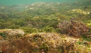
This photograph depicts a variety of algae growing in shallow water.


Differences from Chemoautotrophs


Chemoautotrophs are another type of autotroph. Like photoautotrophs, they make their own food, but they use energy from chemical reactions instead of light energy to do so. This allows them to survive in places where there is no sunlight, such as the deep ocean floor. Some examples of chemoautotrophs are nitrogen-fixing bacteria and iron-oxidizing bacteria.


Related Biology Terms


  • Autotroph – an organism that produces its own food.

  • Heterotroph – an organism that cannot produce its own food and must rely on consuming other organisms to survive.

  • Photoheterotroph – an organism that makes energy from light but uses organic materials for its carbon source instead of inorganic carbon dioxide, which is used by photoautotrophs.

  • Photosynthesis – the process by which photoautotrophs absorb light and convert its energy into chemical energy to power their cells.

Test Your Knowledge


1. Which organisms are NOT photoautotrophs?
A. Algae
B. Cyanobacteria
C. Humans
D. Apple trees

Answer to Question #1

2. What molecule is responsible for absorbing light in the cells of photoautotrophs?
A. Oxygen
B. Hemoglobin
C. Chlorophyll
D. Sodium chloride

Answer to Question #2

3. What cell organelle carries out photosynthesis?
A. Endoplasmic reticulum
B. Ribosome
C. Mitochondrion
D. Chloroplast

Answer to Question #3


Photoautotroph

Monday, February 27, 2017

Homologous Chromosomes

Homologous Chromosomes Definition


Chromosomes are the genetic information coded into human DNA. Each person has 23 pairs of chromosomes, or 46 chromosomes in total. Homologous chromosomes are essentially similar in size, and carry the same genetic information.


Examples of Homologous Chromosomes


Homologous chromosomes primarily occur in two scenarios:


  • Cell division

  • Reproduction


Example #1: A Divisive Matter


Homologous chromosomes exist in one person during mitosis, or cell division. When in mitosis, cells replicate themselves – DNA included – to regenerate human tissue. Although these copies of DNA are identical, the chromosomes they carry are about the same size and have the same genetic information as their predecessors. Therefore, technically speaking, the chromosomes produced during mitosis are homologous to an extreme level.


Example #2: Females and Males


Single-chromosome gametes, or sex cells, are more common examples of homologous chromosomes in action. When the body undergoes meiosis, or the creation of sex cells, chromosomes do not divide from replicated pairs. Instead, the original chromosome pair and its copy split in two. What results are four, single-chromosome gametes. They are stored in male sperm and the female egg, respectively.


When a female becomes pregnant, she combines one of her eggs with a male sperm. On a genetic level, her 22-chromosome set combines with her male partner’s 22-chromosome set. Since each chromosome from the female’s 22-chromosome set is similar in size to, and carries similar genetic information as, the male’s 22-chromosome set, they are homologous.


Homologous chromosomes

Homologous chromosomes


Related Biology Terms


  • Mitosis – Cell division where DNA is replicated, and one new cell divides from the original.

  • Meiosis – Cell division where DNA is replicated, and individual chromosomes do not form pairs. Four new cells divide from the original, each containing only one set of genetic information.

  • Gamete – A male or female reproductive cell that contains only one set of human genetic information.

Test Your Knowledge


1. This process allows for homologous chromosomes to come into contact without sexual intercourse.
A. Meandering
B. Cell division
C. Cell divisiveness
D. Cell direction

Answer to Question #1

2. When a female becomes pregnant, the homologous chromosomes necessary for fetal formation are located in these.
A. Grametes
B. Gromits
C. Gametes
D. Gamers

Answer to Question #2

3. Homologous chromosomes are about the same ______ and carry similar ________.
A. age, credit scores
B. width, DNA
C. beast, fake IDs
D. size, genetic information

Answer to Question #3


Homologous Chromosomes

Sunday, February 26, 2017

Phospholipid

Phospholipid Definition


A phospholipid is a type of lipid molecule that is the main component of the cell membrane. Lipids are molecules that include fats, waxes, and some vitamins, among others. Each phospholipid is made up of two fatty acids, a phosphate group, and a glycerol molecule. When many phospholipids line up, they form a double layer that is characteristic of all cell membranes.


Phospholipid Structure


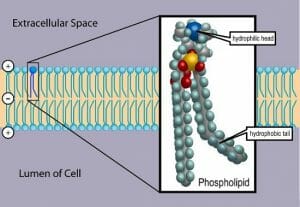
A phospholipid is made up of two fatty acid tails and a phosphate group head. Fatty acids are long chains that are mostly made up of hydrogen and carbon, while phosphate groups consist of a phosphorus molecule with four oxygen molecules attached. These two components of the phospholipid are connected via a third molecule, glycerol.


Phospholipids are able to form cell membranes because the phosphate group head is hydrophilic (water-loving) while the fatty acid tails are hydrophobic (water-hating). They automatically arrange themselves in a certain pattern in water because of these properties, and form cell membranes. To form membranes, phospholipids line up next to each other with their heads on the outside of the cell and their tails on the inside. A second layer of phospholipids also forms with heads facing the inside of the cell and tails facing away. In this way, a double layer is formed with phosphate group heads on the outside, and fatty acid tails on the inside. This double layer, called a lipid bilayer, forms the main part of the cell membrane. The nuclear envelope, a membrane surrounding a cell’s nucleus, is also made up of phospholipids arranged in a lipid bilayer, as is the membrane of mitochondria, the part of the cell that produces energy.


This figure depicts the lipid bilayer and the structure of a phospholipid:


Phospholipid TvanBrussel


Functions of Phospholipids


As membrane components, phospholipids are selectively permeable (also called semi-permeable), meaning that only certain molecules can pass through them to enter or exit the cell. Molecules that dissolve in fat can pass through easily, while molecules that dissolve in water cannot. Oxygen, carbon dioxide, and urea are some molecules that can pass through the cell membrane easily. Large molecules like glucose or ions like sodium and potassium cannot pass through easily. This helps keep the contents of the cell working properly and separates the inside of the cell from the surrounding environment.


Phospholipids can be broken down in the cell and used for energy. They can also be split into smaller molecules called chemokines, which regulate a variety of activities in the cell such as production of certain proteins and migration of cells to different areas of the body. Additionally, they are found in areas such as the lung and in joints, where they help lubricate cells.

In pharmaceuticals, phospholipids are used as part of drug delivery systems, which are systems that help transport a drug throughout the body to the area that it is meant to affect. They have high bioavailability, meaning that they are easy for the body to absorb. Valium is an example of a medication that uses a phospholipid-based drug delivery system.


In the food industry, phospholipids can act as emulsifiers, which are substances that disperse oil droplets in water so that the oil and water do not form separate layers. For example, egg yolks contain phospholipids, and are used in mayonnaise to keep it from separating. Phospholipids are found in high concentrations in many other animal and plant sources, such as soybeans, sunflowers, cotton seeds, corn, and even cow brains.


Related Biology Terms


  • Lipid – a class of molecules that includes fats, waxes, and some vitamins, among other molecules.

  • Hydrophilic – a molecule that “loves water”; it is attracted to water molecules and can usually dissolve in water.

  • Hydrophobic – a molecule that “hates water”; it is not attracted to water, but will usually dissolve in oils or fats.

  • Lipid bilayer – a double layer of phospholipids that makes up the cell membrane and other membranes, like the nuclear envelope and the outside of mitochondria.

Test Your Knowledge


1. Which is NOT a component of a phospholipid?
A. Glycerol
B. Fatty acids
C. Deoxyribose
D. Phosphate group

Answer to Question #1

2. Which molecule is hydrophobic?
A. Fatty acid
B. Phosphate group
C. Glucose
D. Carboxylate group

Answer to Question #2

3. What is a function of phospholipids?
A. Being part of a drug delivery system in some pharmaceuticals
B. Regulating cellular activities such as cell migration
C. Forming the cell membrane and the membranes of other organelles in the cell
D. All of the above

Answer to Question #3


Phospholipid

Thursday, February 23, 2017

Biotic Factors

Biotic Factors Definition


Biotic factors are the living parts of an ecosystem.


Because of the way ecosystems work – as complex systems of competition and cooperation, where the action of every life form can effect all the others – any living thing within an ecosystem can be considered a biotic factor.


Biotic factors such as soil bacteria, plant life, top predators, and polluters can all profoundly shape which organisms can live in an ecosystems and what survival strategies they use.


Biotic factors, together with non-living abiotic factors such as temperature, sunlight, geography, and chemistry, determine what ecosystems look like and what ecological niches are available.


Types of Biotic Factors


Biotic factors are grouped by scientists into three major groups, which define their role in the flow of energy which all living things in the ecosystem need to survive. These groups are producers or autotrophs, consumers or heterotrophs, and decomposers or detritivores.


Producers


Producers – also known as autotrophs, from the Greek words “auto” for “self” and “troph” for “food” – are organisms that make their own food using inorganic materials and energy sources.


Producers are extremely important: without them, no life could exist at all!


The very first life forms on Earth had to learn to make fuel and building materials to make more cells out of non-living materials. That’s because when the first life forms appeared, there were no other life forms to feed on! So the first life forms had to be producers. Producers remain vital today as the life forms that can harness inorganic energy to be used as fuel for life.


There are two major classes of producers:


1. Photoautotrophs are by far the most common type of producer on Earth today. These producers harness energy from sunlight to power their life functions. Green plants, green algae, and some bacteria are photoautotrophs.


Most photoautotrophs use a pigment, such as chlorophyll, to catch photons from the Sun and harvest their energy. They then package that energy into a form that all life forms can use, and use it to create proteins, sugars, lipids, and more essential materials for life.


In most ecosystems, plants – which are producers that are multicellular, highly complex, and very efficient at turning sunlight into fuel for living organisms – form the bottom of the energy pyramid. All other organisms depend on the energy plants harvest from the Sun to survive.


2. Chemoautotrophs are fairly rare in most ecosystems. They obtain energy from chemicals such as hydrogen, iron, and sulfur, which are not common in most environments. Nonetheless, they can still play an important role in ecosystems because of their unusual biochemistry.


Some methanogens – microorganisms that make methane – are chemoautotrophs. Methane, a greenhouse gas which is much more powerful than carbon dioxide, may play a major role in regulating the planet’s temperature. Other chemoautotrophs can produce similarly powerful chemicals with their unique metabolisms.


It is actually not known whether the first forms of life on Earth were photoautotrophs or chemoautotrophs. Photoautotrophs are more common today, but that may simply be because sunlight is more plentiful than the chemicals chemoautotrophs use as their energy source.


Consumers


Consumers, also called “heterotrophs,” are organisms that eat other living organisms in order to obtain energy. Their name comes from the Greek “hetero” for “other” and “troph” for “food.”


Herbivores who eat plants, carnivores who eat animals, and omnivores who eat both plants and animals, are all heterotrophs.


Heterotrophy probably evolved when some organisms discovered that they could eat autotrophs as a source of energy, instead of creating their own energy and organic materials.


Some autotrophs subsequently evolved symbiotic relationships with consumers, such as angiosperms – plants which produce nectars and fruits to attract animals, who ultimately help them to reproduce.


Most levels of most ecosystems’ energy pyramids consist of consumers – herbivores, minor predators, and top predators who eat other organisms.


Decomposers


Decomposers, or detritovores, are organisms that use organic compounds from producers and consumers as their source of energy. They are important to ecosystems because they break down materials from other living things into simpler forms, which can then be used again by other organisms.


Decomposers include soil bacteria, fungi, worms, flies, and other organisms that break down dead materials or waste products from other life forms. They are distinct from consumers, because consumers usually consume other organisms while they are still alive.


Decomposers, on the other hand, metabolize waste products that might not be of interest to consumers, such as rotting fruit and dead animals. In the process they break down these dead things into simpler chemicals that can be used by heterotrophs to thrive and produce more energy for the ecosystem as a whole.


This is the principle behind the practice of composting – where waste scraps of plants and animal products are put into a pile, where decomposers such as bacteria, worms, and flies are allowed to thrive. These decomposers turn the waste products into rich fertilizer for the composter’s garden, which then grows bigger and healthier thanks to the decomposers breaking down the waste products in the compost.


Decomposers are the link between the bottom of an ecosystem’s energy pyramid and the other levels. Decomposers can take energy and raw materials from dead plants, herbivores, lesser carnivores, and even top carnivores, and break it down into a form that can be used by the ecosystem’s producers to make it easier for them to harness sunlight. In this way, the ecosystem’s energy cycle is preserved.


Examples of Biotic Factors


Example #1: Cyanobacteria and Life on Earth


Scientists believe that the earliest widespread form of life on Earth was cyanobacteria. These fairly simple cells, which made food and organic materials from sunlight, played a massively important role in creating all of Earth’s modern ecosystems.


Prior to the success of cyanobacteria, Earth did not have an oxygen atmosphere. That meant that aerobic respiration was not possible – and also meant that it was impossible, or very difficult, for any organisms to live on land because of the DNA-destroying ultraviolet radiation from our sun.


However, cyanobacteria developed a method for storing the energy of sunlight in organic molecules. For this they needed to take molecules of carbon from inorganic sources, such as carbon dioxide in the air, and turn them into carbon-based organic compounds such as sugars, proteins, and lipids.


To achieve this, cyanobacteria took in the inorganic gas CO2, and released a new gas, O2.


O2, or molecular oxygen turned out to be the perfect fuel for the most powerful type of heterotroph metabolism: aerobic respiration. Molecules of O2 also reacted with ultraviolet light in the upper atmosphere to form, O3 – a molecule also known as ozone, which absorbed ultraviolet light in the upper atmosphere and made it safe for life forms to colonize land.


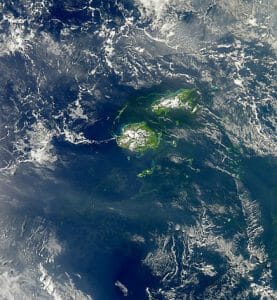
In the billions of years to come, cyanobacteria would be mostly replaced by its more sophisticated descendants such as trees, grass, and algaes who would take over its role as Earth’s primary oxygen producers. However, cyanobacteria itself still appears in blooms which can sometimes be seen from space!


2010 Filamentous Cyanobacteria Bloom near Fiji

2010 Filamentous Cyanobacteria Bloom near Fiji


As biotic factors, cyanobacteria and its modern descendants supplied not only energy and organic compounds, but also oxygen, to all of Earth’s ecosystems!


Example #2: Wolves in North America


When European colonists arrived in North America, wolves were common in many of the continent’s ecosystems. These large carnivores were the top predators in many places, using a combination of their large size and teamwork to take down large prey animals.


The colonists and their descendants hunted wolves fiercely, due to safety concerns over the fact that wolves could eat sheep that farmers depended on for food, and could even eat human children.


However, the disappearance of wolves eventually started to cause new problems for the humans of North America. Without their top predator, deer and other herbivore species multiplied to unprecedented numbers.


This might have seemed nice for human hunters who ate deer meat and sold deer skins at first, but the problem became serious when the deer started eating so many plants that crops, gardens, and wild plant species became endangered. Humans began to have to hunt deer themselves, not just for meat and skin, but to prevent serious damage to their ecosystems.


Humans didn’t realize the full extent of the roll of wolves until a ban on wolf hunting was introduced, and wolves bred in captivity were released back into the wild to repopulate the wolf species in some areas.


The areas where wolves were re-introduced underwent startling transformations. Numbers of deer and other large prey species went down, sure enough – which led to populations of many plant species increasing.


To the surprise of human scientists studying the ecosystems, even land forms began to change: it turned out that deer had been eating grass and other small plants whose root systems held soil in place against erosion. With wolves keeping the deer population in check, the plant populations began to come back – and erosion decreased, and the courses of rivers changed! Fish were also effected by a decrease in loose soil washing into the river.


This is an excellent example of how complex and interconnected ecosystems are – and how the removal of one element of the ecosystem, even if its only role is to eat other animals, can cause big changes for all other organisms living in the ecosystem.


Example #3: Humans


In 2016, biologists around the world decided to declare that the Earth had entered a new geologic era: the Anthropocene.


The name “Anthropocene” comes from the Greek words “anthropo” for “human” and “cene” meaning “new” or “recent.”


This era is defined by the effects of human technology, which has caused massive changes to the global ecosystem on par with the effects of past major climate change events and even asteroid impacts.


Human activity has drastically changed the carbon cycle of Earth, with the burning of wood, coal, and oil releasing millions of years worth of carbon dioxide into the atmosphere in the space of just a couple of centuries. Over the same time scale, humans have cut down about half of all Earth’s forests, which had previously acted to take carbon dioxide out of the air and incorporate it back into plant life.


In addition, humans have begun to release many new substances into Earth’s land, air, and oceans, including plastics, heavy metals, and radioactive materials, none of which exist in nature.


The result has been the beginning of alarmingly rapid climate change and a mass extinction, in which species are disappearing faster than they have been since the asteroid impact that killed the dinosaurs and made way for the rise of mammals 65 million years ago.


Humans are therefore maybe the most powerful example of how the living factors in an ecosystem can change it since cyanobacteria.


This has led some environmentalists to suggest that humans are “evil,” and “bad for the Earth. But the truth is that the Earth always survives ecological upheavals. It’s just a question of whether the species that exist at their beginning survive to their end.


That’s why many scientists say humans should be concerned about their effect on the planet. Not because changing the planet is in itself morally wrong, but because humans themselves rely on the complicated ecological interplay of thousands of species for their food.


Scientists are already beginning to raise alarms that the pollinators on which many human food crops depend appear to be dying off due to new chemicals that humans have released to the environment.


Human food crops are also threatened by climate change caused by the carbon dioxide humans have released into the air, which has brought severe drought to many areas with dense human populations who require large amounts of food to survive.


Medical scientists also caution that man-made climate change is allowing dangerous insect-borne diseases, which used to be restricted to the regions near the equator, to spread to new areas.


As the dominant species on Earth, it is important that humans learn about the ecosystems upon which they depend for well-being and survival. We have the power to seriously disrupt these ecosystems – and as living things which rely on other life forms for our own survival, we may set in motion events that could lead to our own extinction if we are not careful.


Related Biology Terms


  • Ecosystem – A community of organisms, and their physical environment.

  • Energy Pyramid – A diagram which shows the flow of energy through organisms in an ecosystem.

Test Your Knowledge


1. Which of the following is not an example of a producer, or autotroph?
A. Cyanobacteria
B. A daisy
C. A wolf
D. A chemoautotroph

Answer to Question #1

2. Which of the following is not an example of decomposers in action?
A. A fruit fly laying eggs in a rotting fruit
B. A compost pile turning food scraps into fertilizer
C. Mushrooms growing on a piece of dead wood
D. A venus fly trap consuming a fly

Answer to Question #2

3. Which of the following is an example of major ecosystem change that was NOT caused by biotic factors?
A. The release of oxygen into the atmosphere, allowing aerobic respiration and the colonization of dry land.
B. The rise in global temperatures during the 20th century, caused by the release of large quantities of carbon dioxide.
C. The extinction of the dinosaurs following the impact of an asteroid in the Yucatan peninsula.
D. All of the above.

Answer to Question #3


Biotic Factors

Wednesday, February 22, 2017

Autotroph

Autotroph Definition


Autotrophs are organisms that can produce their own food, using materials from inorganic sources. The word “autotroph” comes from the root words “auto” for “self” and “troph” for “food.” An autotroph is an organism that feeds itself, without the assistance of any other organisms.


Autotrophs are extremely important because without them, no other forms of life can exist. Without plants that create sugars from carbon dioxide gas and sunlight via the process of photosynthesis, for example, no herbivorous animals could exist, and no carnivorous animals that eat herbivores could exist.


For this reason, autotrophs are often called “producers.” They form the base of an ecosystem’s energy pyramid, and provide the fuel that all the heterotrophs (organisms that must get their food from others) need to exist.


The first life forms on Earth would have had to be autotrophs, in order to exist and make energy and biological materials in a previously non-living environment. Heterotrophs most likely evolved as autotrophs became more common, and some life forms discovered that it was easier to simply eat the autotrophs than to make energy and organic materials for themselves.


Types of Autotrophs


Scientists classify autotrophs according to how they obtain their energy. Types of autotrophs include photoautotrophs, and chemoautotrophs.


Photoautotrophs


Photoautotrophs are organisms who get the energy to make organic materials from sunlight. Photoautotrophs include all plants, green algaes, and bacteria which perform photosynthesis.


All photoautotrophs perform photosynthesis – a word that comes from the root words “light” and “to make.” Photoautotrophs capture photons from the Sun and harvest their energy, using it to perform important biochemical processes such as making ATP.


Photoautotrophs make more than just fuel and organic compounds for heterotrophs like ourselves!


Many photoautotrophs take carbon from the atmosphere and use it to make sugars and other molecules that store the Sun’s energy in their molecular bonds. To do this, they take in molecules of CO2, which is created by nonliving geological processes, and release molecules of O2 – also known as the oxygen we need to breathe!


It is thought that free oxygen was not present in Earth’s atmosphere until after photoautotrophs became common in Earth’s seas. Then, they produced so much free oxygen that large amounts of iron that had previously been dissolved in ocean water reacted with the oxygen and turned into rust!


This process created rocks called banded iron formations, which we can still look at today to see this record of our Earth’s history. The release of large amounts of free oxygen into Earth’s atmosphere by photoautotrophs paved the way for large animals, like ourselves, who need the highly efficient process of aerobic respiration to survive.


It is thought that some of the oxygen produced by photoautotrophs also created the Earth’s ozone layer, which allowed life to move onto dry land without fear of DNA damage from the Sun’s UV light.


Chemoautotrophs


Chemoautotrophs are organisms that obtain energy from inorganic chemical processes. Today, chemoautotrophs are most commonly found in deep water environments which receive no sunlight. Many need to live around deep sea volcanic vents, which produce enough heat to allow metabolism to occur at a high rate.


Chemoautotrophs use volatile chemicals such as molecular hydrogen, hydrogen sulfide, elemental sulfur, ferrous iron, and ammonia as their energy sources. This makes them well-suited to live in places that would be toxic to many other organisms, as well as places without sunlight. Chemoautotrophs are usually bacteria or archaebacteria, as their metabolisms are usually not efficient enough to support multicellularity.


Scientists have speculated that life might be able to exist in dark, chemically volatile environments such as the seas of Jupiter’s moon Titan by using similar metabolisms to those seen in chemoautotrophs on Earth. No proof of such life has yet been found, but some scientists believe that the range of metabolic options offered by chemosynthesis drastically expands the range of places in the universe where we can expect to find life.


It is actually unknown whether photoautotrophs or chemoautotrophs were the first life forms on Earth. Many favor the idea that the first cells were photosynthetic, since the Sun’s light shines on the entire surface of the Earth. But some scientists believe that volcanic sites in the deep sea or on the surface of the Earth could have supplied more concentrated energy and more volatile chemicals, potentially leading to the creation of the first cells.


These scientists speculate that these cells could then have evolved photosynthesis as an energy source that would work anywhere on the Earth’s surface they spread further from their volcanic points of origin.


Because single cells and their biochemistry do not fossilize well, we may never know whether chemoautotrophs or photoautotrophs were the first ever forms of life on Earth.


Examples of Autotrophs


Example #1: Plants


Plants, with very few exceptions (such as the venus fly trap which can eat insects) are photoautotrophs. They produce sugars and other essential ingredients for life by using their pigments, such as chlorophyll, to capture photons and harness their energy. When plants are consumed by animals, animals are then able to use that energy and those organic materials for themselves.


Example #2: Green Algae


Green algaes, which may be familiar to you as pond scum, are also photoautotrophs. Green algae may in fact bear a great resemblance to the first common life form on Earth – cyanobacteria, a green bacteria that grew in mats and began the process of turning Earth into a world with an oxygen atmosphere.


Example #3: ”Iron Bacteria” – Acidithiobacillus ferrooxidans


The bacterium Acidithiobacillus ferrooxidans obtains energy from ferrous iron. In the process, it converts the iron atoms from a molecular form where they cannot be dissolved in water to a molecular form where they can.


As a result, Acidithiobacillus ferrooxidans has been used to extract iron from ores that could not be extracted through conventional means.


The field of biohydrometallurgy is the study of using living organisms to obtain metals by dissolving them in water, where they can be further processed.


Related Biology Terms


  • Energy pyramid – A structure that shows the flow of energy through an ecosystem.

  • Heterotroph – An organism that relies on other organisms, such as plants or prey animals, for food.

  • Photosynthesis – The process used by phototrophs to extract energy from sunlight.

Test Your Knowledge


1. Which of the following statements is true of chemoautotrophs?
A. They harness energy from sunlight to make food.
B. They rely on other organisms, such as plants and prey animals, for food.
C. They harness energy from chemicals such as hydrogen, sulfur, and iron to make food.
D. None of the above.

Answer to Question #1

2. Which of the following is NOT an example of a photoautotroph?
A. Daisies
B. Iron bacteria
C. Cyanobacteria
D. None of the above.

Answer to Question #2

3. The first form of life on Earth was likely…
A. A photoautotroph.
B. A chemoautotroph.
C. Neither of the above.
D. No one knows.

Answer to Question #3


Autotroph

Tuesday, February 21, 2017

Punnett Square

Punnett Square Definition


A Punnett square is a graphical representation of the possible genotypes of an offspring arising from a particular cross or breeding event. Creating a Punnett square requires knowledge of the genetic composition of the parents. The various possible combinations of their gametes are encapsulated in a tabular format. Therefore, each box in the table represents one fertilization event.


The inherent assumption is that each trait is determined by a single gene locus and that various traits assort independently from one another. Though this is true for many useful traits, especially when choosing characters for plant or animal breeding, there are many exceptions.


This tool was created in the twentieth century, much after Mendel’s seminal experiments on genetics. However, they are now commonly used to explain the results that Mendel obtained, especially when combined with our current knowledge of DNA, genes and chromosomes.


Common Terms in Genetics


Some terms are often used in the study of genetics and these are particularly useful in understanding the function of Punnett squares. Among these is the term ‘allele’ and is used to denote a variant of a gene. For example, a pea plant can have red or white flowers and the gene variants coding for each of these is called an allele.


When an organism contains two copies of the same allele, its genetic composition or genotype is said to be homozygous. These are also called true-breeding specimens. For instance, plants with white flowers are homozygous at the genetic loci coding for flower color.


Individuals who have two different alleles are said to be heterozygous at that locus. Many plants that have red flowers can have one allele for red color and another for white color. The externally observed characteristic of an individual is called the phenotype. The phenotype in a heterozygous individual is said to be the ‘dominant’ form of the gene and the trait that is suppressed is considered as the ‘recessive’ allele. In the example of flower color, the allele coding for red color is dominant over the one for white.


In a cross between a dominant homozygote and a recessive homozygote, all the offspring will have a heterozygous genotype and a dominant phenotype.


Some gene loci are on sex chromosomes and are called sex-linked traits, while all the others are said to be autosomal.


Functions of Punnett Squares


In large-scale experiments, such as those conducted by Mendel, Punnett squares can accurately predict the ratios of various observable traits as well as their underlying genetic composition. For instance, when a true-breeding tall pea plant is cross fertilized with pollen from a true-breeding short pea plant, the Punnett square can predict that all the offspring will be tall, and all of them will be heterozygous with both the allele for shortness and tallness. It can further predict that if these heterozygous plants are allowed to self-fertilize, approximately seventy-five percent of the second generation plants will be tall, and the remaining twenty-five percent will be short. Among the tall plants, one-third will remain true-breeding while the remaining two-thirds will be heterozygous. This tool is therefore used by plant and animal breeders to choose appropriate specimens in order to obtain offspring carrying a desired trait.


They are also used in genetic counseling to help couples make the decision about having children. For example, in cases where both parents are carriers for an autosomal recessive disease such as cystic fibrosis, there is a twenty-five percent chance of their child suffering from the illness and a fifty-percent chance that their offspring will be carriers. However, if one parent has the disease and the other is neither a carrier nor suffering from the illness, the couple can be reassured that their child will not develop cystic fibrosis since she will carry only one copy of the abnormal gene.


Types of Punnett Squares


Two types of Punnett squares are commonly used. The first is relevant when a single trait determined by one genetic locus is being observed. This is called a monohybrid cross and examples include some of Mendel’s original experiments, where he chose true-breeders for a single trait and crossed them with members carrying a different allele. For a monohybrid cross, these are 2X2 squares with four boxes, each representing one fertilization event between the parent gametes.


The second type is used to predict the outcome of breeding experiments where two traits are being followed and the Punnett square is larger, with sixteen boxes. The 4X4 square is necessary since each of the parents can produce four types of gametes, based on the distribution of the alleles of the two genes.


When more than two traits are being observed, a Punnett square becomes unwieldy and other tools are used to predict the outcomes of such crosses.


Examples of Punnett Squares


Most people are introduced to Punnett squares through the experiments of Mendel. Among the various traits of the common pea plant that he observed, one was the color of the peas. Other common examples used to elucidate the predictive power of this tool are the inheritance of blood types and eye color in humans.


Example #1: Seed Color in Common Pea Plant Pisum sativum


Mendel created true-breeding homozygous plants for both the alleles – yellow and green color seeds. When he cross pollinated these homozygotes, he found that all the offspring had yellow seeds. When he allowed these yellow offspring to undergo self pollination, he was surprised to find that nearly twenty-five percent of the second generation of pea plants contained green seeds. He concluded that the yellow allele was dominant over the green one. In order to better understand this phenomenon, he crossed some of the first generation plants with yellow seeds with a true-breeding green plant. This would later be known as a test cross.


In every Punnett square, an allele is represented by the first letter of the dominant phenotype. In this case, the dominant yellow color allele is denoted by the capital letter ‘Y’ and the recessive allele by the small letter ‘y’. Each allele is allowed to segregate independently into a gamete and the gametes are represented just outside the 2X2 table.


Punnett Square


Each of the boxes shows one possible genotype for the offspring. In this test cross, half the offspring have yellow seeds and are genotypically heterozygous. The other half are homozygous and have green seeds.


Example #2: Tail And Hair Color in Cats


When a homozygous short-tailed, white haired cat is mated with a long-tailed brown haired cat, all the offspring appear to inherit one trait from each parent. They all have short tails and brown hair, showing that brown color is dominant over white and the allele for a short tail is dominant over the one for a long tail.


Dihybrid cross


When members of this first generation mate with each other, a large majority of their offspring will have short tails and brown hair. Additionally, there is a three-in-sixteen probability that the parental combinations will reappear: short tail with white hair or long tail with brown hair. Finally there is a one-in-sixteen probability that a new combination could appear – long-tailed and white colored.


If an animal breeder was looking for a long-tailed, white-haired specimen, he would know that it would only appear in the second generation.


Limitations of Punnett Squares


While Punnett squares are a convenient tool to understand Mendelian genetics, they cannot be used in many situations involving complex genetic inheritance. For example, they are not effective in estimating the distribution of genotypes and phenotypes when there is linkage between two genes. Genetic linkage is a phenomenon where two genes exist close to each other on the same chromosome. Therefore, during gamete formation, the chances of these two traits being inherited together, in the same combination as that found in the parent, is high. One instance of this is the linkage between the locus of the gene causing Nail-patella Syndrome (NPS) and the one determining blood group. Analysis of one family whose members suffer from NPS found that it was often inherited along with a B-type blood group. These linkages will change the random distribution of the two traits among offspring, therefore making the Punnett square unreliable as a predictive device.


In addition, when a single trait is determined by multiple genes and the effect of each of these genes is graded, Punnett squares cannot accurately predict the distribution of phenotypes in the offspring. Human height is determined by over four hundred genes distributed across the genome. In addition, this trait is also influenced by environmental factors such as nutrition.


Finally, genes that are inherited completely from one parent, such as those in the mitochondria or on the Y-chromosome, as well as genotypes that are lethal to the foetus, confound the results from a Punnett square.


Related Biology Terms


  • Codominance – A situation where two alleles are neither dominant nor recessive towards each other and both are expressed as phenotype.

  • Diploid – A cell containing two sets of chromosomes, one set from each parent. Diploid cells contain two copies of nearly every gene.

  • Gametes – Mature, haploid germ cells from the male and female that can fuse with one another to form a zygote.

  • Haploid – A cell containing a single set of chromosomes.

Test Your Knowledge


1. Which of these is inherited completely from the mother?
A. Genes for eye color
B. Genes for cystic fibrosis
C. Genes from the Y-chromosome
D. Mitochondrial genes

Answer to Question #1

2. Which of these are assumptions in creating a Punnett square?
A. The alleles for each trait segregate during meiosis
B. Each trait assorts independently of the others
C. Only one gene locus is involved in a particular trait
D. All of the above

Answer to Question #2

3. How many rows and columns would be needed to create a Punnett square for a trihybrid cross?
A. 3X3
B. 6X6
C. 8X8
D. 9X9

Answer to Question #3


Punnett Square

Autosomes

Autosome Definition


An autosome is a chromosome in a eukaryotic cell that is not a sex chromosome.


Unlike prokaryotic cells, eukaryotic cells have many chromosomes in which they package their DNA. This allows eukaryotes to store much more genetic information.


Most eukaryotic organisms reproduce through sexual reproduction – meaning that each individual has two copies of each chromosome. One copy is inherited from one parent, while the other is inherited from the other parent.


This system enhances genetic diversity and protects against some diseases, since it enables individuals to inherit immune system genes from two different parents, and having two copies of a gene often enables a healthy copy inherited from one parent to “cover for” a copy of a gene that has been corrupted through harmful mutation.


It’s normal for diploid eukaryotic organisms (those which have a full set of chromosomes inherited through sexual reproduction) to have two copies of each autosome.


Sex chromosomes are considered separately from autosomes, since their inheritance pattern works differently. In humans, the sex chromosomes are referred to as the X chromosome and the Y chromosome. Other animals, like birds, use a different system of sex chromosomes.


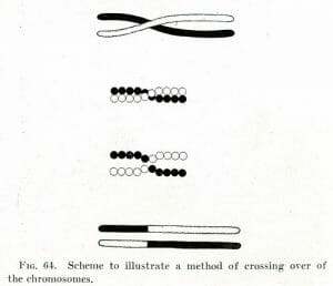
During the process of meiosis which creates eukaryotic sex cells, the sex cells “remix” DNA between their two copies of each autosome in the process of crossing over. The result is a unique set of chromosomes which has a mix of material from both of the individual’s parents. This process is illustrated below.


Morgan crossover


The sex cell then discards one of each of the resulting remixed autosomes, resulting in a gamete cell that has only one copy of each autosome.


When two gametes combine, they produce a cell which will grow into a new individual which will possess a copy of each chromosome from each parent. The individual’s unique genetic profile will include DNA from each of its four grandparents.


During the growth of a multicellular organism, it’s normal for a cell to make a full copy of each of its chromosomes, and give one copy to each daughter cell.


When errors are made in distributing chromosomes during meiosis or early in embryonic development, serious diseases can result due to many cells in an individual’s body having the wrong number of chromosomes.


Because each chromosome contains thousands of genes, having too many or too few chromosomes can result in serious imbalances in gene expression. In humans, many pregnancies that do not survive the first trimester are cases where the embryo inherited the wrong number of chromosomes and was not able to survive.


Other errors in chromosome replication can cause more mild syndromes such as Down syndrome, which is caused by inheriting an extra copy of chromosome 21 from one parent.


Function of Autosomes


Each autosome stores many thousands genes, each of which performs a unique function in the organism’s cells.


Under normal circumstances, each chromosome follows a “map” that is shared across individuals in the species. This allows cells to “know” where to start gene expression when they want to express a certain gene. It is thought that factors which effect gene expression use this “map” to accurately respond to a cell’s needs.


When autosomes are healthy, this enables cells to perform an awesome array of functions. Each of hundreds of subtly differing cell types in a eukaryotic organism express a different combination of genes in the right place at the right time, enabling the huge array of cellular functions we see in eukaryotic organisms like ourselves.


Each of our cells contain the necessary compliment of genes to reproduce our whole bodies. Differences between brain cells, skin cells, and muscle cells are made by cells transcribing the right genes in the right places at the right times.


Our bodies get it right almost all the time! But biologists often learn how something works by watching cases where it breaks, and seeing what happens when the mechanism is not working properly.


In the case of autosomes and their carefully arranged “map” that allows for the complexity of our bodies, problems can arise when chromosomes break and their pieces end up in the wrong place.


This event, called “translocation,” can cause genes to the expression of the wrong genes at the wrong times. Some types of cancer may be caused by translocations leading to errors in cell development and reproduction.


Examples of Autosomal Disorders


Example #1: Trisomy 21 (Down Syndrome)


Down syndrome occurs when a person inherits all or part of an extra copy of chromosome 21 from one parent. This usually occurs due to a one-time error in meiosis and is not passed down through the generations.


People with Down syndrome have a variety of unusual traits and symptoms related to skeletal tissue (unusual skeletal shape, weak ligaments), nerve tissue (cognitive disabilities, poor muscle tone), and have a higher risk of some diseases due to extra expression of material from chromosome 21.


Due to the range of symptoms seen in Down syndrome, some people with Down syndrome can complete regular schooling and have independent careers, while others may need special education classes and may not be able to function independently in the workplace.


The only known risk factor for Down syndrome is having older parents, which can increase the chance that parents’ bodies will incorrectly sort chromosomes during meiosis.


Example #2: Cri du Chat


Cri du chat, also known as “chromosome 5p deletion syndrome” or “Lejeune’s syndrome,” happens when a person inherits just one copy of part of chromosome 5. Some people with cri du chat also have extra copies of other parts of chromosome 5.


As with Down syndrome, cri du chat usually occurs due to an error in the sorting of the parents’ chromosomes during meiosis.


The syndrome’s name comes from French for “cry of the cat,” in reference to the unusual catlike cry that babies with cri du chat have due to their unusual skeletal and neurological traits.


Like people with Down syndrome, people with cri du chat can have unusual skeletons, weak muscles, and cognitive impairment due to the under-expression of the 5p chromosomal section.


People with cri du chat may also have hearing loss, heart problems, and microcephaly (a small head).


Example #3: Philadelphia Chromosome


The Philadelphia chromosome is a chromosome found in many leukemia cancer cells, which may give a clue as to how the cancer gets started.


In the Philadelphia chromosome, chromosome 9 and chromosome 22 have swapped some genetic material. The specific place where the two are joined creates a fusion protein – that is, a protein coded for by a fusion of two different genes, one from chromosome 9 and one from chromosome 22.


This gene turns cell replication to “always on,” and as a result leads to uncontrolled replication of cells which never mature and become properly functional. Leukemia occurs when these non-functioning cells multiply out of control and destroy healthy, functioning tissue.


Some scientists believe that chromosomal translocations are a common cause of cancer. At least fifteen different kinds of cancer have been found to frequently involve chromosomal translocation, often resulting in the creation of fusion proteins.


Related Biology Terms


  • Gametes – The sex cells used by sexually reproducing species to produce offspring with new gene combinations, created by remixing and combining genetic material from each parent.

  • Gene balance – The theory that genes need to be expressed in the right amount within cells. In the theory of gene balance, too much or too little expression of a given gene compared to others results in cellular problems.

  • Gene expression – The process by which genes are turned into proteins, which perform important functions in a cell.

Test Your Knowledge


1. Which of the following is NOT an autosome?
A. Chromosome 21
B. Chromosome 9
C. X Chromosome
D. Chromosome 22

Answer to Question #1

2. Which of the following is NOT a function of autosomes?
A. To store large amounts of genetic information needed to make a complex multicellular organism.
B. To ensure that genes are expressed in the proper amounts to create healthy tissues.
C. To pass on genetic information to daughter cells.
D. To determine an individual’s sex.

Answer to Question #2

3. Which of the following is not true of gene balance?
A. Expressing too many copies of the same gene can cause problems for a cell.
B. Expressing too few copies of the same gene can cause problems for a cell.
C. Even if the right number of copies of a gene are present, it can still be expressed too much or too little if it is in the wrong place on the chromosomal “map.”
D. A cell cannot survive with the wrong number of copies of a gene.

Answer to Question #3


Autosomes

Monday, February 20, 2017

Asexual Reproduction

Asexual Reproduction Definition


Asexual reproduction occurs when an organism makes more of itself without exchanging genetic information with another organism through sex.


In sexually reproducing organisms, the genomes of two parents are combined to create offspring with unique genetic profiles. This is beneficial to the population because genetically diverse populations have a higher chance of withstanding survival challenges such as disease and environmental changes.


Asexually reproducing organisms can suffer a dangerous lack of diversity – but they can also reproduce faster than sexually reproducing organisms, and a single individual can found a new population without the need for a mate.


Some organisms that practice asexual reproduction can exchange genetic information to promote diversity using forms of horizontal gene transfer such as bacteria who use plasmids to pass around small bits of DNA. However this method results in fewer unique genotypes than sexual reproduction.


Some species of plants, animals, and fungi are capable of both sexual and asexual reproduction, depending on the demands of the environment.


Asexual reproduction is practiced by most single-celled organisms including bacteria, archaebacteria, and protists. It is also practiced by some plants, animals, and fungi.


Evolution and animal life

Evolution and animal life


Advantages of Asexual Reproduction


Important advantages of asexual reproduction include:


1. Rapid population growth. This is especially useful for species whose survival strategy is to reproduce very fast.


Many species of bacteria, for example, can completely rebuild a population from just a single mutant individual in a matter of days if most members are wiped out by a virus.


2. No mate is needed to found a new population.


This is useful for species whose members may find themselves isolated, such as fungi that grow from wind-blown spores, plants that rely on pollinators for sexual reproduction, and animals inhabiting environments with low population density.


3. Lower resource investment. Asexual reproduction, which can often be accomplished just by having part of the parent organism split off and take on a life of its own, takes fewer resources than nurturing a new baby organism.


Many plants and sea creatures, for example, can simply cut a part of themselves off from the parent organism and have that part survive on its own.


Only offspring that are genetically identical to the parent can be produced in this way: nurturing the creation of a new organism whose tissue is different from the parents’ tissue takes more time, energy, and resources.


This ability to simply split in two is one reason why asexual reproduction is faster than sexual reproduction.


Disadvantages of Asexual Reproduction


The biggest disadvantage of asexual reproduction is lack of diversity. Because members of an asexually reproducing population are genetically identical except for rare mutants, they are all susceptible to the same diseases, nutrition deficits, and other types of environmental hardships.


The Irish Potato Famine was one example of the down side of asexual reproduction: Ireland’s potatoes, which had mainly reproduced through asexual reproduction, were all vulnerable when a potato-killing plague swept the island. As a result, almost all crops failed, and many people starved.


The near-extinction of the Gros-Michel banana is another example – one of two major cultivars of bananas, it became impossible to grow commercially in the 20th century after the emergence of a disease to which it was genetically vulnerable.


On the other hand, many species of bacteria actually take advantage of their high mutation rate to create some genetic diversity while using asexual reproduction to grow their colonies very rapidly. Bacteria have a higher rate of errors in copying genetic sequences, which sometimes leads to the creation of useful new traits even in the absence of sexual reproduction.


Types of Asexual Reproduction


There are many different ways to reproduce asexually. These include:


1. Binary fission. This method, in which a cell simply copies its DNA and then splits in two, giving a copy of its DNA to each “daughter cell,” is used by bacteria and archaebacteria.


2. Budding. Some organisms split off a small part of themselves to grow into a new organism. This is practiced by many plants and sea creatures, and some single-celled eukaryotes such as yeast.


3. Vegetative propagation. Much like budding, this process involves a plant growing a new shoot which is capable of becoming a whole new organism. Strawberries are an example of plants that reproduce using “runners,” which grow outward from a parent plant and later become separate, independent plants.


4. Sporogenesis. Sporogenesis is the production of reproductive cells, called spores, which can grow into a new organism.


Spores often use similar strategies to those of seeds. But unlike seeds, spores can be created without fertilization by a sexual partner. Spores are also more likely to spread autonomously, such as via wind, than to rely on other organisms such as animal carriers to spread.


5. Fragmentation. In fragmentation, a “parent” organism is split into multiple parts, each of which grows to become a complete, independent “offspring” organism. This process resembles budding and vegetative propagation, but with some differences.


For one, fragmentation may not be voluntary on the part of the “parent” organism. Earthworms and many plants and sea creatures are capable of regenerating whole organisms from fragments following injuries that split them into multiple pieces.


When fragmentation does occur voluntarily, the same parent organism may split into many roughly equal parts in order to form many offspring. This is different from the processes of budding and vegetative propagation, where an organism grows new parts which are small compared to the parent and which are intended to become offspring organisms.


6. Agamenogenesis. Agamenogenesis is the reproduction of normally sexual organisms without the need for fertilization. There are several ways in which this can happen.


In parthenogenesis, an unfertilized egg begins to develop into a new organism, which by necessity possesses only genes from its mother.


This occurs in a few species of all-female animals, and in females of some animal species when there are no males present to fertilize eggs.


In apomoxis, a normally sexually reproducing plant reproduces asexually, producing offspring that are identical to the parent plant, due to lack of availability of a male plant to fertilize female gametes.


In nucellar embryony, an embryo is formed from a parents’ own tissue without meiosis or the use of reproductive cells. This is primarily known to occur in citrus fruit, which may produce seeds in this way in the absence of male fertilization.


Examples of Asexual Reproduction


Example #1: Bacteria


All bacteria reproduce through asexual reproduction, by splitting into two “daughter” cells that are genetically identical to their parents.


Some bacteria can undergo horizontal gene transfer – in which genetic material is passed “horizontally” from one organism to another, instead of “vertically” from parent to child. Because they have only one cell, bacteria are able to change their genetic material as mature organisms.


The process of genetic exchange between bacterial cells is sometimes referred to as “sex,” although it is performed to change the genotype of a mature bacterium, not as a means of reproduction.


Bacteria can afford to use this survival strategy because their extremely rapid reproduction makes harmful genetic mutations – such as copying errors or horizontal gene transfer gone wrong – inconsequential to the whole population. As long as a few individuals survive mutation and calamity, those individuals will be able to rebuild the bacterial population quickly.


This strategy of “reproduce fast, mutate often” is a major reason why bacteria are so quick to develop antibiotic resistance. They have also been seen to “invent” whole new biochemistries in the lab, such as one species of bacteria that spontaneously acquired the ability to perform anaerobic respiration.


This strategy would not work well for an organism that invests highly in the survival of individuals, such as multicellular organisms.


Example #2: Slime Molds


Slime molds are a fascinating organism that sometimes behave like a multicellular organism, and sometimes behave like a colony of single-celled organisms.


Unlike animals, plants, and fungi, the cells in a slime mold are not bound together in a fixed shape and dependent on each other for survival. The cells that make up a slime mold are capable of living individually and may spread or separate when food is abundant, much like individuals in a colony of bacteria.


But slime mold cells are eukaryotic, and can display a high degree of cooperation to the point of creating a temporary extracellular matrix and a “body” which may become large and complex. Slime molds whose cells are working cooperatively can be mistaken for fungi, and can perform locomotion.


Slime molds can produce spores much like a fungus, and they can also reproduce through fragmentation. Environmental causes or injury may cause a slime mold to disperse into many parts, and units as small as a single cell may grow into a whole new slime mold colony/organism.


Example #3: New Mexico Whiptail Lizards


This species of lizard was created by the hybridization of two neighboring species. Genetic incompatibility between the hybrid parents made it impossible for healthy males to be born: however, the female hybrids were capable of parthenogenesis, making them a reproductively independent population.


All New Mexico whiptail lizards are female. New members of the species can be created through hybridization of the parent species, or through parthenogenesis by female New Mexico whiptails.


Possibly as a remnant of their sexually reproducing past, New Mexico whiptail lizards do have a “mating” behavior which they must go through to reproduce. Members of this species are “mated with” by other members, and the lizard playing the female role will go onto lay eggs.


It is thought that the mating behavior stimulates ovulation, which can then result in a parthenogenic pregnancy. The lizard playing the “male” role in the courtship does not lay eggs.


Related Biology Terms


  • Gamete – Sexual reproductive cells, which contain half of the parent organism’s genetic material.

  • Reproductive strategy – A strategy that describes how a given population uses its resources to produce offspring.

  • Sexual Reproduction – A means of reproduction in which the genetic material of two parents is combined to produce offspring with a unique genetic profile.

Test Your Knowledge


1. Which of the following is NOT an advantage of asexual reproduction?
A. Rapid reproduction.
B. High genetic diversity.
C. No need for a mate.
D. Low resource investment in offspring.

Answer to Question #1

2. Which of the following events was NOT caused by low genetic diversity due to asexual reproduction?
A. The Irish Potato Famine
B. The disappearance of the Gros-Michel banana
C. The Black Death in England
D. A and B

Answer to Question #2

3. Which of the following is NOT true of asexual reproduction?
A. Some organisms can only perform asexual reproduction because their genetics does not allow for the existence of healthy males.
B. Some organisms can perform both sexual and asexual reproduction.
C. It is used by a variety of organisms, including all bacteria and some plants, animals,and fungi.
D. It is used only by single-celled organisms.

Answer to Question #3


Asexual Reproduction

Friday, February 17, 2017

Archaebacteria

Archaebacteria Definition


Archaebacteria are a type of single-cell organism which are so different from other modern life-forms that they have challenged the way scientists classify life.


Until the advent of sophisticated genetic and molecular biology studies allowed scientists to see the major biochemical differences between archaebacteria and “normal” bacteria, both were considered to be part of the same kingdom of single-celled organisms. “Kingdoms,” a way of organizing life forms based on their cell structure, traditionally included Animalia, Planitia, Fungi, Protista (for single-celled eukaryotes), and Monera (which was once considered to hold all forms of prokaryotes).


However, genetic and biochemical studies of bacteria soon showed that one class of prokaryotes was very different from “modern” bacteria, and indeed from all other modern life forms. Eventually named “archaebacteria” from “archae” for “ancient,” these unique cells are thought to be modern descendants of a very ancient lineage of bacteria that evolved around sulfur-rich deep sea vents.


Sophisticated genetic and biochemical analysis has led to a new “phylogenetic tree of life,” which makes use of the concept of “domains” to describe divisions of life that are bigger and more basic than that of “kingdom.”


The most modern version of this system shows all eukaryotes – animals, plants, fungi, and protists – constituting the domain of “Eukaryota,” while the more common and modern branching of bacteria constitutes “Prokarya,” and archaebacteria constitute their own domain altogether – the domain of “Archaea.”


Phylogenetic tree


The discovery of Archaea and its unique differences is exciting for scientists, because it’s believed that archaebacteria’s unique biochemistry might give us insight into the workings of very ancient life. Some scientists propose that the archaebacteria Thermoplasma may in fact be ancestors of the nuclei of our own eukaryotic cells, which are believed to have developed through the process of endosymbiosis.


Another remarkable trait of archaebacteria is their ability to survive in extreme environments, including very salty, very acidic, and very hot surroundings. Archaebacteria have been recorded surviving temperatures as high as 190° Fahrenheit, which is only twenty-two degrees shy of the boiling point of water, and acidities as high as 0.9 pH.


Archaebacteria have even challenged scientist’s ideas about how to define a species, since they practice a lot of horizontal gene transfer – where genes are transferred from one individual to another during their lifetimes – making it difficult to determine how closely different cells are related, or even if archaebacteria cells have the sort of stable combinations of traits that scientists typically use to define a species.


The domain of Archaea include both aerobic and anaerobic species, and can be found living in common environments such as soil as well as in extreme environments.


So what biochemical characteristics make scientists so excited about archaebacteria? Well…


Archaebacteria Characteristics


Archaebacteria have a number of characteristics not seen in more “modern” cell types. These include:


1. Unique cell membrane chemistry.


Archaebacteria have cell membranes made of ether-linked phospholipids, while bacteria and eukaryotes both make their cell membranes out of ester-linked phospholipids


Archaebacteria use a sugar that is similar to, but not not the same as, the peptidoglycan sugar used in bacteria cell membranes.


2. Unique gene transcription.


Archaebacteria have a single, round chromosome like bacteria, but their gene transcription is similar to that which occurs in the nuclei of eukaryotic cells.


This leads to the strange situation that most genes involving most life functions, such as production of the cell membrane, are more closely shared by Eukarya and Bacteria – but genes involved in the process of gene transcription are most closely shared by Eukarya and Archaea.


This has led some scientists to propose that eukaryotic cells arose from a fusion of archaebacteria with bacteria, possibly when an archaebacteria began living endosymbiotically inside a bacterial cell.


Other scientists believe that eukaryotes descended directly from archaebacteria, based on the findings of archaebacteria species, Lokiarcheota, which contains some found only in eukaryotes, which in eukaryotes code for genes with uniquely eukaryotic abilities.


It is thought that Lokiarcheota may be a transitional form between Archaea and Eukaryota.


3. Only archaebacteria are capable of methanogenesis – a form of anaerobic respiration that produces methane.


Archaebacteria who use other forms of cellular respiration also exist, but methane-producing cells are not found in Bacteria or Eukarya.


4. Differences in ribosomal RNA that suggest they diverged from both Bacteria and Eukarya at a point in the distant past


Types of Archaebacteria


There are three main types of archaebacteria. These are classified based on their phylogenetic relationship (how closely related they are to each other), and members of each type tend to have certain characteristics. The major types are:


1. CrenarchaeotaCrenarchaeota are extremely heat-tolerant.


They have special proteins and other biochemistry that can continue to function at temperatures as high as 230° Fahrenheit! Many Chrenarchaeota can also survive in very acidic environments.


Many species of Crenarchaeota have been discovered living in hot springs and around deep sea vents, where water has been superheated by magma beneath the Earth’s surface.


One theory of the origin of life suggests that life may have originally started around deep sea vents, where high temperatures and unusual chemistries could have led to the formation of the first cells.


2. Euryarchaeota are able to survive in very salty habitats. They are also able to produce methane, which no other life form on Earth is able to do!


Euryarchaeota are the only form of life known to be able to perform cellular respiration using carbon as their electron acceptor.


This gives them an important ecological niche because the breakdown of complex carbon compounds into the simple molecule of methane is the final step in the decomposition of most life forms. Without methanogens, the Earth’s carbon cycle would be impaired.


Wherever methane gas is produced by life, Euryarchaeota are responsible.


Methanogen archaebacteria can be found in marshes and wetlands, where they are responsible for “swamp gas” and part of the marsh’s distinctive smell, and in the stomachs of ruminants such as cows, where they break down sugars found in grass that are undigestible to eukaryotes by themselves. Some methanogens live in the human gut and assist us in the same way.


They can also be found in deep sea sediments, where they produce pockets of methane beneath the ocean floor.


3. Korarchaeota are the least-understood, and thought to be the oldest lineage of archaebacteria. This makes them possibly the oldest surviving organisms on Earth!


Korarchaeota can be found in hydrothermal environments much like Crenarchaeota. However, Korarchaeota have many genes found in both Crenarchaeota and Euryarcheaota, and also genes which are different from both groups. To scientists, this suggests that both other types of archaebacteria may have descended from a common ancestor similar to Korarchaeota.


Korarchaeota are rare in nature, perhaps because other, newer forms of life are better adapted to survive in modern environments than they are. Still, Korearchaeota can be found in hot springs, around deep sea vents.


Examples of Archaebacteria


Example #1: Lokiarcheota


Lokiarcheota is a hyperthermophile discovered at the deep sea vent called Loki’s Castle, which some scientists think has unique evolutionary significance.


It has a highly unique genome, consisting of roughly 26% proteins that are known to be found in other archaebacteria, 29% proteins that are known to be found in bacteria, 32% genes that do not correspond to any known protein, and – 3.3% genes that correspond to those only found in eukaryotes.


The eukaryotic genes are particularly exciting for scientists, because they are genes that appear to code for proteins that eukaryotes use to actively control the shape of their cell, including proteins for cytoskeletons, the motor protein actin, and several proteins that in eukaryotes are involved in changing cell membrane shape.


Some of these genes are involved in phagocytosis, which is exciting because the process of phagocytosis could have been used by eukaryotic ancestors to “swallow” other cells – which may have gone on to become endosymbiotes, leading to the endosymbiotic relationships between eukaryotic cells and their mitochondria, chloroplasts, and nuclei.


Lokiarchaeota’s unique genome makes it possibly our closest relative among prokaryotes, and possibly a transitional form in the extremely important jump from prokaryotic to eukaryotic life, which made the evolution of the animal, plant, fungi, and protist kingdoms possible. Scientists think that Lokiarchaeota and ourselves probably shared a common ancestor around 2 billion years ago.


It is unknown whether this means that eukaryotes likely evolved around deep sea vents, or whether Lokiarchaeota’s relatives may once have been common in other environments before they were outcompeted and driven to extinction by their more advanced descendants, the eukaryotes.


Example #2: Methanobrevibacter Smithii


Methanobrevibacter smithii is a methane-producing archaebacteria that lives in the human gut. This member of Euryarchaeota helps us to break down complex plant sugars and extract extra energy from the food we eat.


The microorganisms in our guts – including members of Euryarchaeota – also have a complex relationship with our health. While some studies show that many people with obesity and colon cancer have above-average levels of Euryarchaeota in their guts, Euryarchaeota also help people who don’t have enough food to produce more energy, and some types of these archaebacteria appear to protect against colon cancer.


Related Biology Terms


  • Domain – The highest level of classification currently recognized by biologists, refers to the broad differences between the cells of eukaryotes, bacteria, and archaebacteria.

  • Kingdom – The level of classification below “domain,” kingdoms separate eukaryotes, such as plants and animals, into groups based on their phylogenetic relationships.

  • Phylogenetics – The study of evolutionary relationships between life forms.

Test Your Knowledge


1. Which of the following is NOT a domain of life?
A. Animalia
B. Archaea
C. Bacteria
D. Eukarya

Answer to Question #1

2. Which of the following is NOT a difference between archaea and other forms of life?
A. Archaebacteria use different lipids in their cell membranes.
B. Archaebacteria have a circular chromosome like bacteria, but also a nuclear envelope like eukaryotes.
C. Archaebacteria have a circular chromosome like bacteria, but their gene transcription is more similar to that of eukaryotes.
D. Only archaebacteria can perform methanogenesis.

Answer to Question #2

3. Which of the following is not true about the major types of archaebacteria?
A. Crenarchaeota can live in temperatures as high as 230° Fahrenheit.
B. Euryarchaeota includes both methanogens who produce methane, and halophiles who prefer salty environments.
C. Lokiarchaeota is a methanogen that lives in the digestive tracts of cows.
D. Korarchaeota may be related to the common ancestor of Crenarchaeota and Euryarchaeota.

Answer to Question #3


Archaebacteria